Ribs
Historical document, translated for reference. It reflects medical knowledge of the 1920s–30s and is not medical advice.
Summary
The article discusses the comparative anatomy of ribs in vertebrates, their embryological origin, development, and variations across different species. It details the structure, classification, and functional significance of ribs in the skeletal system of various vertebrates.
Encyclopedia article (1928–1936)
Ribs (costae) of vertebrate animals are skeletal arches connected to the spine that more or completely encircle the body from the sides. Comparative anatomy. In their origin, ribs are derivatives of the axial skeleton g embryonically developing from the same mass of skeletogenic mesenchyme from which the entire spine also develops. Whether ribs are products of separation from the lateral processes, i.e., from the arches of vertebrae (Gegenbaur, Goppert), or products of independent differentiation within the system of connective tissue partitions connected to the spine (Rabl, Emelyanov), is not yet fully clarified. In any case, in vertebrates, two systems of ribs should be distinguished: lower ribs clearly connected to the arches of vertebrae, directly surrounding the abdominal cavity and located in segmental myosepta under the entire mass of trunk musculature, and upper ribs also located in myosepta but higher—in the very thickness of the trunk musculature, in the horizontal partition between its dorsal and abdominal sections. Fish as a rule have lower ribs, while terrestrial vertebrates only have upper ones. In terrestrial vertebrates, instead of the simple articulation of the rib with the vertebra, as found in fish, a double articulation generally develops, at least in the anterior half of the body. This double articulation is effected through the double lateral processes of vertebrae—one retaining the original position on the vertebra (parapophysis), and the other developing on its arch (the true transverse process, diapophysis). Accordingly, the proximal end of the rib forms a fork. Such double articulation is clearly expressed in the oldest stegocephalians and reptiles, as well as in modern tailed amphibians and crocodiles, and in modified form has been passed on to birds and mammals. In higher vertebrates, the lateral process (parapophysis) is reduced, and the rib then attaches by its head (capitulum) directly to the body of the vertebra (or, more precisely, between two adjacent bodies). Additionally, the upper branch of the proximal end of the rib is reduced to the degree of a small tubercle (tuberculum) that usually articulates with a well-developed transverse process. The double connection of ribs with the spine is explained by their function in terrestrial vertebrates—attachment of ribs in two points lying one above the other gives them the necessary support for holding the internal organs (in the horizontal position of the animal's body) and at the same time does not interfere with respiratory movements, during which the ribs alternately deviate forward and backward. Therefore, the double connection disappears in crawling reptiles (lizards and snakes), whose bellies rest on the ground, and in aquatic reptiles (ichthyosaurs) and mammals (cetaceans). Similarly, the double articulation disappears also with the shortening of ribs in the posterior part of the thoracic region. In the oldest fossil reptiles and amphibians, there were rather long ribs, the ends of which were probably cartilaginous. Undoubtedly, many ribs of the right and left sides converged and connected with each other in the abdominal wall, and through their fusion the sternum developed. In higher vertebrates, the sternum develops for the most part as a paired formation through the fusion of the abdominal ends of ribs. A more or less significant number of ribs of the thoracic region retain this connection even in the adult animal—these are the so-called "true" ribs; others connect with the sternum only through the intermediary of the ribs in front—these are "false" ribs; finally, some lose this connection altogether, shorten (posterior ribs of the thoracic region), reduce, and fuse with the transverse processes (in the lumbar region). In front of the sternum, with the development of the cervical region of the spine, ribs also shorten, reduce, and fuse with the vertebrae, but here they retain their characteristic double connection with them (hence—the perforated transverse processes of the cervical vertebrae of birds and mammals). In crocodiles, there are still well-developed independent ribs even on the first cervical vertebrae (atlas and axis). In the sacral region, there are still independent ribs in tailed amphibians—they are shortened and widened and serve for the attachment of the iliac bones of the pelvis. In all higher vertebrates, these sacral ribs (1-2 true sacral vertebrae) fuse with the transverse processes, which are correspondingly distinguished by their strength. In the caudal region, small free ribs were present only in the oldest fossil stegocephalians and reptiles. In the earliest terrestrial vertebrates, ribs were long but undivided. In all modern forms, the progressive development of the thoracic cage and its more perfect adaptation for respiratory movements was accompanied by the progressive development and division of ribs in the thoracic region into a proximal bony part and a distal, usually cartilaginous, part that becomes ossified but sometimes also ossifies (in birds and some lower mammals) part, directly or indirectly connected with the sternum. Both parts form between themselves a certain angle, the magnitude of which changes during respiratory movements. Figure 1. Sometimes the vertebral part of the rib bears a backward-directed process that overlaps the next rib behind—the processus uncinatus of crocodiles and birds, serving to strengthen the thoracic cage. The shape and position of ribs also determine the shape of the thoracic cage—flattened in crawling reptiles, high in running, and barrel-shaped in jumping and climbing birds and mammals. I. Schmalhausen. Anatomy. Ribs belong to the category of flat bones, connecting at the posterior end with the spine, at the anterior end with the sternum. In humans, there are 12 pairs of ribs (figs. 1-2). Their numbering goes from top to bottom: I, II, etc. up to the XII rib. Of the 12 pairs of ribs, seven, and sometimes eight, are attached to the sternum (costae sternales, s. verae)—sternal ribs or true ribs. Cases where the VIII rib reaches the sternum on one or both sides constitute, according to Cunningham and Robinson, 20%. If the rib reaches the sternum only on one side, then in 8 out of 9 cases it is the right one; Cunningham assumes that this is connected with the greater use of the right hand. Five pairs of lower ribs, abdominal or false (costae abdominales, s. spuriae), do not reach the sternum and are divided into two subgroups; VIII, IX, and X ribs connect at their anterior ends with the lower edge of the cartilage above. They form the costal arch (arcus costarum) and are also called connected ribs (costae conjunctae). Usually these include VIII-X ribs. Sometimes the X rib belongs to the next subdivision, consisting of XI and XII ribs, which freely project into the abdominal wall—floating ribs (costae fluctuantes). Except for the I rib, which is covered by the clavicle, all others can be palpated through the skin. The longest of the 12 ribs is the VII, the shortest are the I and XII. The length of the XII rib varies sharply: it may be absent altogether, or, in the presence of a lumbar rib, it equals the XI rib (11.5-15.5 cm, Braus). Here we encounter the same process of reduction as in the spine: due to the reduction of the last ribs, the spine gains greater mobility in the lumbar region: the absence of a direct connection of abdominal ribs with the sternum gives the same gain in the range of motion of the spine. Consequently, an increase in the number of ribs should be regarded as an atavistic phenomenon (regressive, imperfect form), while their decrease—as a progressive, perfect form (form of the future, Braus). Each of the ribs connecting the spine and sternum consists of two parts: the posterior bony (os costale, costa vertebralis) and the anterior, cartilaginous (cartilago costae, costa sternalis). Rib.
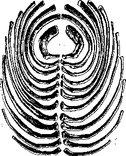
Figure 1.
Figure 2. Sometimes the vertebral part of the rib bears a backward-directed process that overlaps the next rib behind—the processus uncinatus of crocodiles and birds, serving to strengthen the thoracic cage. The shape and position of ribs also determine the shape of the thoracic cage—flattened in crawling reptiles, high in running, and barrel-shaped in jumping and climbing birds and mammals. I. Schmalhausen. Anatomy. Ribs belong to the category of flat bones, connecting at the posterior end with the spine, at the anterior end with the sternum. In humans, there are 12 pairs of ribs (figs. 1-2). Their numbering goes from top to bottom: I, II, etc. up to the XII rib. Of the 12 pairs of ribs, seven, and sometimes eight, are attached to the sternum (costae sternales, s. verae)—sternal ribs or true ribs. Cases where the VIII rib reaches the sternum on one or both sides constitute, according to Cunningham and Robinson, 20%. If the rib reaches the sternum only on one side, then in 8 out of 9 cases it is the right one; Cunningham assumes that this is connected with the greater use of the right hand. Five pairs of lower ribs, abdominal or false (costae abdominales, s. spuriae), do not reach the sternum and are divided into two subgroups; VIII, IX, and X ribs connect at their anterior ends with the lower edge of the cartilage above. They form the costal arch (arcus costarum) and are also called connected ribs (costae conjunctae). Usually these include VIII-X ribs. Sometimes the X rib belongs to the next subdivision, consisting of XI and XII ribs, which freely project into the abdominal wall—floating ribs (costae fluctuantes). Except for the I rib, which is covered by the clavicle, all others can be palpated through the skin. The longest of the 12 ribs is the VII, the shortest are the I and XII. The length of the XII rib varies sharply: it may be absent altogether, or, in the presence of a lumbar rib, it equals the XI rib (11.5-15.5 cm, Braus). Here we encounter the same process of reduction as in the spine: due to the reduction of the last ribs, the spine gains greater mobility in the lumbar region: the absence of a direct connection of abdominal ribs with the sternum gives the same gain in the range of motion of the spine. Consequently, an increase in the number of ribs should be regarded as an atavistic phenomenon (regressive, imperfect form), while their decrease—as a progressive, perfect form (form of the future, Braus). Each of the ribs connecting the spine and sternum consists of two parts: the posterior bony (os costale, costa vertebralis) and the anterior, cartilaginous (cartilago costae, costa sternalis). Rib.
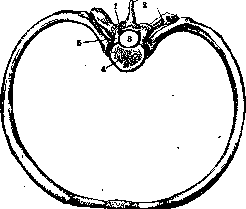
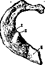
Figure 4. First rib from above: 1-sulcus subclaviae; 2 - tuberculum scaleni (Lisfranc); 3-cartilago costalis.
Figure 3. Skeleton of the thoracic segment: 1-articular apophysis; 2-muscular apophyses; 3-spinous process; 4-body of vertebra; 5-arch of vertebra. It presents a band-like bony strip, curved in three directions: in the plane, at the edge, and along the long axis. Due to the curvature in the plane, the ribs encircle the contents of the thoracic cavity, and due to the curvature at the edge, the anterior end of the rib, when fixed to the spine, is lowered below the posterior end to such an extent that in a person standing upright, it lies in the same plane as the head of a rib located 2-3 segments below; from this position, the rib is brought out by respiratory movements. The most oblique right position is occupied by the IX rib; due to the twist around the long axis (torsion), the plane of the lateral surface of the sternal end stands at a more oblique angle, the closer it is to the posterior end. The I rib constitutes an exception. The ribs are fixed to the spine at an angle open downward (angulus costo-vertebralis); the lower the rib, the sharper this angle. From the vertebrae, the ribs go outward, attaching to their transverse processes; somewhat outward from the latter, the ribs sharply turn forward, forming angles of the ribs (anguli costarum), and before connecting with the corresponding costal cartilage, they again slightly change direction and go from outside inward (fig. 3). Each rib consists of a body (corpus costae) and two ends-anterior and posterior (fig. 4-8). The body of the rib is flattened; its lateral surface, convex in the middle ribs, stands vertically, in the upper ones it is turned upward, in the lower ones more downward. On the lateral surface, away from the tubercle of the rib (see below), as mentioned, a blunt angle is formed-angulus costae. The angle in the I rib is in the area of the tubercle, in the lower ribs it moves outward and is completely absent on the last two. On the median surface of the body at the lower edge passes the costal groove (sulcus costae) for the intercostal vessels; it is expressed in the posterior part, disappears forward; in the first and last ribs, the groove is either absent or weakly expressed.
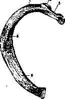
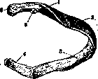
In TRUE RIBS, the anterior end is widened behind: it has a rough, more often cup-shaped surface for connection with the costal cartilage, while in false ribs it is usually smooth (Batu). At the posterior end of the rib, the following are distinguished: the head (capitulum costae), the neck (collum costae), and the tubercle (tuberculum costae). With the head, i.e., the thickened end, the rib articulates with the body of the vertebra; on the head there is a cartilage-covered surface-the articular surface of the head (facies articularis capituli), divided from II to IX ribs by a crest (crista capituli) into two parts. The costal neck-the next part after the head, somewhat narrowed; along its upper edge passes the longitudinal crest of the neck (crista colli costae), continuing onto the body of the rib and absent in I and XII ribs. The costal tubercle is located on the lower-posterior surface of the neck, on it there is a small cartilage surface-the articular surface of the tubercle (facies articularis tuberculi) for articulation with the transverse process of the vertebra; the tubercle is largest in the I rib, and in XI and XII ribs (sometimes also in X) it is completely absent.
Figure 6. Fourth right rib from behind: 1-tuberculum costae; 2-angulus costae; 3-facies lateralis; 4-cartilago costalis; 5-facies articularis tuberculi; 6-facies articularis capituli.
Figure 7. Seventh right rib from within: 1-corpus costae; 2-crista colli costae; 3-crista capituli; 4-facies articularis capituli; 5-sulcus costae.

Of the 12 ribs, four (I, II, XI and XII) have some peculiarities in their shape. The first rib is curved only at the edge, one of its surfaces is turned upward and slightly outward, the other-downward inward. In the middle of the upper surface there are two flat grooves: the anterior one-for the passage of the subclavian vein and the posterior one, deeper (sulcus subclaviae)-for the subclavian artery; between them is located the scalene tubercle of Lisfranc (tuberculum scaleni Lisfranci), to which the anterior scalene muscle is attached. On the upper surface of the anterior end of the first rib there is a roughness for the attachment of lig. costo-clavicularis. The posterior end of the rib stands at a right angle to the body; the head with one articular facet, the neck is very thin, the tubercle protrudes strongly. Both surfaces of the II rib are oblique; like the I, II rib,
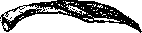
Figure 8. Twelfth right rib from behind.
not twisted along its axis and lacks the costal groove; on its upper-lateral surface there is a roughness (tuberositas costae II) for the attachment of the anterior serratus muscle. Each of the last two ribs articulates with only one vertebra; tubercles are absent on them. These ribs are only slightly curved. The costal groove is often absent on the XII rib. Ribs are flat bones and consist of two compact plates connected at the edges and interspersed with spongy tissue. The periosteum of the ribs is so strong that when a rib fractures, it often remains intact and holds the fragments from displacement; it is easier to remove from the outer surface than from the inner. The costal cartilage (cartilago costalis) generally has the shape of the rib and continues it forward, for the first seven ribs to the attachment to the sternum, for the next three it adjoins the cartilage of the rib above, and for the XI and XII the anterior ends of the cartilages are lost in the abdominal muscles. Costal cartilages differ from each other in length. According to Sappey, the I costal cartilage has a length of 2 cm; III and IV-2-3 cm. The following costal cartilages lengthen up to VII, which equals 12-14 cm; VIII-10 cm; IX-7 cm; X-1 cm; XI-2 cm; XII-6-8 mm. Costal cartilages also differ from each other in their direction and some other details: thus, the I costal cartilage is directed straight obliquely downward inward, approaching the sternum at an obtuse angle, open downward. The II costal cartilage is located almost horizontally. III-VII rise upward and inward, forming with the sternum an acute angle, open downward. V, VI and VII costal cartilages pass so close to each other that they often touch each other (articulate) at their edges. VIII, IX and X costal cartilages articulate with each other, carrying on their edges horizontal articular facets. The ends of XI and XII costal cartilages are thin, pointed, and curved. Costal cartilages consist of hyaline (glassy) cartilage, the ground substance of which has a homogeneous structure. Cartilage cells are enclosed in it, lying in cartilage cavities. The cartilage is covered by perichondrium, which on one side passes into the periosteum of the rib, and on the other into the periosteum of the sternum. In adults and elderly people, the ground substance of the cartilage becomes fibrous and lime is often deposited in it (it ossifies), which reduces the elasticity of the costal cartilage and hinders respiratory movements. Costal cartilages anatomically undergo age-related changes and have typical vascularization in all ages. Up to 10 years of age, vascular channels in large numbers penetrate from the perichondrium into the depth in the form of conical channels, especially many of them in the area of the rib-cartilage boundary; by 10 years, longitudinal vascular channels are already observed, located in the central parts of the cartilage, and in each channel three thin-walled vessels are distinguished (1 artery and 2 veins); by 20 years, bone marrow elements are found in the longitudinal vascular channels; by 30 years, the vascular system of the cartilages reaches full development, and in the central longitudinal channels there are in large quantities fat cells and blood vessels (bone marrow elements). After 50 years, the marrow elements in the central channels atrophy and are replaced by smooth-walled cavities (Popov).-The blood supply to the ribs goes through the intercostal arteries and veins, and the ribs are innervated by the intercostal nerves. The lymphatic circulation of bones, in particular the ribs, is a question still far from resolution. Testut writes that true lymphatic vessels do not exist in either the compact or spongy part of the ribs. Streltsov (1873), Rauber (1876) and others describe in Haversian channels perivascular passages covered with endothelium; these authors consider these passages as lymphatic pathways of the bone. Lymphatic outflow is carried out by intercostal lymphatic vessels into the corresponding regional glands. Development of the ribs. Each rib has 4 points of ossification: one primary and 3 secondary. The primary point of ossification (the earliest) appears on the 40-50th day of intrauterine life. The three additional, otherwise epiphyseal points, are late points: from the first of them develops tuberculum costae, from the second-facies articularis tuberculi costae and from the 3rd - the articular surface of the head of the rib. Various anatomical changes in the ribs can concern their shape, development and number. The width of the rib sometimes reaches double the norm. The ribs are normally fixed at their ends to the spine and sternum, but are shortened in length (straight ribs), as a result of which the corresponding half of the thoracic cage is proportionally reduced (narrowed thoracic cage). There are observed ribs fused together with ceremiches or with a joint in the area of the tubercle or along the course, as well as in the area of the costal cartilages, as is normally found in birds and turtles.-The costal cartilage sometimes does not reach the sternum, to which it is attached in normal conditions, or else the rib cartilage, which normally does not reach the sternum, is fixed directly to the latter. With the first anomaly, the number of false ribs increases, with the second it decreases by one rib. Sometimes a rib is interrupted in the middle of its course by unossified cartilage.-The number of ribs can be reduced or increased (11 or 13). The anomaly can be unilateral and bilateral. With a reduction in the number of ribs, the XII rib is always absent. There are frequent cases when the I rib, remaining rudimentary, connects to the sternum by means of a ligament or does not reach it at all. In the absence of fusion of the I rib with the sternum, it remains mobile in the soft tissues of the neck or its anterior end fuses with the II rib, then the latter at its vertebral end is forked, which is normal for many cetaceans (P. J. Beneden). There is the first rib, as if consisting of two segments: the posterior-vertebral and the anterior-sternal, connected to each other either by a ligament or by a joint. Forked ribs are common, and their anterior ends (the fork) are connected to one or even two costal cartilages. Sometimes a forked rib becomes whole again (perforated rib) with an oval opening, which can be located in either the bony or the cartilaginous part of it. This opening is closed by a fibrous or muscular plate. Ribs are articulated with the spine and their cartilages; costal cartilages-with the sternum and with each other. Ribs articulate with vertebrae at two points: 1) with the bodies of the vertebrae, forming the costovertebral joint (articulatio costovertebralis) and 2) with the transverse process of the vertebra-the joint of ribs and transverse processes (articulatio costo-transversaria) (fig. 9). Both connections of the ribs form mechanically one joint. The heads of II-X ribs articulate with the corresponding articular fossae of the bodies of two neighboring vertebrae and with the depression in the intermediate intervertebral fibrocartilage. From the crest of the head of these ribs, the interarticular ligament of the head (lig. capituli costae interarticulare) (fig. 10) goes to the mentioned cartilage, which divides the joint into two cavities. I, XI and XII ribs connect only with the body of their own vertebra, so here there is a single joint. On the anterior surface of the joint of the rib heads, there is a diverging fan-like from the rib to the body of the vertebra flat radiate 8 ligament of the head of the rib (lig. capituli costae radiatum) (fig. 10), partially covered by the longitudinal ligament of the spine. At the point of contact of the tubercles of the upper 10 ribs with the transverse processes of the corresponding vertebrae, there are joints of ribs and transverse processes-articulatio costo-transversariae. The joint capsules of these joints are reinforced by ligaments: the anterior ligament of the rib and transverse process (lig. costo-transversarium anterius) (fig. 10), more%strong, going from the lower surface of the overlying transverse process downward and inward to the crest of the neck of the rib, and the posterior (lig. costo-transversarium posterius), going from the base of the overlying
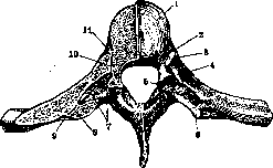
Figure 9. Articulations of rib heads and transverse processes: 1-fibrocartilago intervertebralis; 2-lig. capituli costae interarticulare; 3-facies articularis capituli costae VII; 4-crista colli costae; 5-proc. articularis sup.; 6-proc. transversus; 7-lig. colli costae; 8-articulatio costo-transversaria; 9-lig. tuberculi costae; 10-articulatio capituli costae; 11 - lig. capituli costae radiatum.
vertebral bodies and with the depression in the intermediate intervertebral fibrocartilage. From the crest of the head of these ribs, the interarticular ligament of the head (lig. capituli costae interarticulare) (fig. 10) goes to the mentioned cartilage, which divides the joint into two cavities. I, XI and XII ribs connect only with the body of their own vertebra, so here there is a single joint. On the anterior surface of the joint of the rib heads, there is a diverging fan-like from the rib to the body of the vertebra flat radiate 8 ligament of the head of the rib (lig. capituli costae radiatum) (fig. 10), partially covered by the longitudinal ligament of the spine. At the point of contact of the tubercles of the upper 10 ribs with the transverse processes of the corresponding vertebrae, there are joints of ribs and transverse processes-articulatio costo-transversariae. The joint capsules of these joints are reinforced by ligaments: the anterior ligament of the rib and transverse process (lig. costo-transversarium anterius) (fig. 10), more%strong, going from the lower surface of the overlying transverse process downward and inward to the crest of the neck of the rib, and the posterior (lig. costo-transversarium posterius), going from the base of the overlying
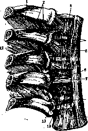
Figure 10. Articulations of rib heads: ^-lig. intercostale internum; 2-lig. costo-transversarium ant.; 3- vertebra thoracalis III; 4-lig. capituli costae radiatum; 5-lig. longitudinale ant.; 6-lig. capituli costae interarticulare; 7-fibrocartilago intervertebralis; 8- fovea costalis inf.; 9-vertebra thoracalis VII; 10-fovea costalis sup.; 11-capitulum costae VII; 12-foramen costo-transversarium.
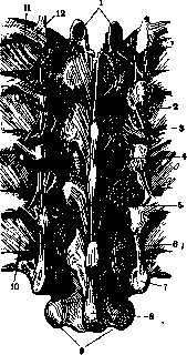
Figure 11. Ligaments of thoracic vertebrae and ribs on the posterior side: 1-processus articulares sup-
of the transverse process downward to the posterior surface of the neck of the R. and to the costal tubercle (fig. 11). The named joints are additionally reinforced by the ligament of the costal tubercle (lig. tuberculi costae), stretched between the apex of the transverse process and the posterior surface of the costal tubercle, and by the ligament of the costal neck (lig. colli costae), a short, broad, horizontal ligament stretched between the posterior surface of the neck of the rib and the anterior surface of the transverse process, almost completely filling the space between them, leaving only small openings in the anterior and posterior parts - foramen costo-transversarium. In the XI and XII R., there is no joint with the transverse processes. The joints of the ribs with the vertebrae receive blood from the intercostal arteries of the VI thoracic vertebra; 2-lig. radiatum; 3-lig. costotransversarium post.; 4-lig. supraspinale; 5-capsula articulationis intervertebralis; 6-lig. intertransversarium; 7-proc. transversus; 8-proc. spinosus of the X thoracic vertebra; 9-processus articulares inf.; 10 and 11-lig. intercostale int.; 12-lig. tuberculi with branches of the intercostal arteries
costae. The nerves of the ribs. The costosternal joints (articulations sterno-costales) are arthrodies similar to those between the ribs and the spine. The cartilage of the first rib, with very rare exceptions, directly passes into the sternum without forming a joint. The cartilages of ribs II-VII usually join with the sternum through a joint, in which the joint cavity of the second cartilage and sternum is divided into two by an interarticular ligament (lig. sterno-costale interarticulare); similar fiber bundles are found in the joints of the lower cartilages, but they do not completely separate the joint cavity. In front and behind, the joint capsules are reinforced by costosternal radiate ligaments (lig. sterno-costalia radiata), which extend fan-like from the ends of the cartilages to the sternum; meeting with similar ligaments of the opposite side and interweaving with the longitudinal fibers on the sternum, they form the anterior and posterior sternum membranes (membrana sterni). The joint of the seventh cartilage with the sternum is additionally strengthened by a strong broad ligament, the costoxiphoid ligament (lig. costo-xiphoide-um). Between the lower edge of the cartilage and the anterior surface of the xiphoid process, at the points where the cartilages of the false ribs join each other, there are small joint cavities with joint capsules, the interchondral joints (art. interchondrales).-The arteries of the costosternal joints are the anterior branches of the internal mammary arteries. Nerves are intercostal. The arteries of the interchondral joints are the musculophrenic artery, a branch of the internal mammary artery. The intercostal spaces are filled with intercostal muscles (see Muscles) and shiny fiber bundles (lig. intercostalia ext., otherwise lig. coruscantia and lig. intercostalia interna). The former continue the external intercostal muscles in the area of the rib cartilages, the latter partially cover them from the inside, then continue the layer of internal intercostal muscles from the angles of the ribs to the vertebrae. Between the external and internal intercostal muscles are located (from top to bottom) the v., a., and n. intercostales. From the upper edge of the first rib to the tubercle of the clavicle goes the costoclavicular ligament (lig. costo-clavicular). From the I and II lumbar vertebrae to the XII rib go strong, arched, fiber bundles - the lumbar costal ligament (lig. lumbo-costale). Movements of the ribs. The ribs rise and fall. When rising, the angle between the rib and vertebra increases, when falling it decreases. In its movement, the rib represents a lever of the third kind with a fulcrum in the costovertebral joint, with resistance at the anterior end and the point of application of force in its middle part, at the points of attachment to the rib of numerous muscles. The movements of the ribs are very complex; when rising, each rib performs a triple movement: 1) forward translation, 2) outward, and 3) rotation around an imaginary axis passing through both its joints with the vertebra, parallel to the axis of the neck of the rib; at this time, its inner surface turns slightly downward; when falling, the rib performs opposite movements. The sternum, being intimately connected with the rib, necessarily follows the ribs in all their movements. ;- If we examine the movements of the entire thoracic cage, we will notice that the elevation of the ribs leads to an increase in its transverse and anteroposterior diameters, while the depression of the ribs leads to a decrease in them. Consequently, all muscles that elevate the ribs increase the thoracic cavity and are inspiratory musculature, while those that depress them are expiratory. The muscles moving the ribs are divided into elevators and depressors. The first: diaphragm, sternocostalis, scaleni, subclavius, levatores costarum, pectoralis major and minor, latissimus dorsi, serratus posterior superior, upper and lower bundles of serratus anterior; the second: rectus abdominis, external and internal oblique abdominis, transverse abdominis, serratus posterior inferior, and middle bundles of serratus anterior, quadratus lumborum. Pathology. Acute inflammatory diseases of the ribs and cartilages are rare and most often develop as a complication of a purulent process in adjacent layers of the chest wall (in phlegmon of the wall, in empyema of the pleura, especially in empyema necessitatis) or as metastatic lesions in parasitic typhoids (typhus and relapsing fever) and in typhoid fever, in scarlet fever, measles, trauma, and other unclear causes. In post-typhoid cases, the cartilage is more often affected: acute purulent chondritis develops, characterized by all inflammatory phenomena of the cartilage, in which the lesion occurs inside the cartilage in the vascular canal in the form of an intra-cartilaginous abscess, which increases by destroying the cartilage tissue incapable of inflammatory reaction, and leads to the formation of an intra-cartilaginous cavity. The intra-cartilaginous abscess ruptures into the surrounding soft tissues, where an abscess forms; the latter, breaking through outward, leads to an unhealing fistula. The inflammatory process often spreads to adjacent cartilages; such chondrites and perichondrites become chronic and last for months. Salvarsan therapy has been proposed, and in case of failure, surgical intervention with removal of the entire affected area. Severe acute purulent diseases of the ribs include acute hematogenous osteomyelitis - a not common disease: according to Trendel, out of 1252 cases of acute osteomyelitis, there were only 11 cases (0.87%) of rib osteomyelitis, while later Parcelier collected in the literature 94 cases of rib osteomyelitis out of 1924 cases, with men being twice as many as women. In children, according to Vyshgorodskaya, out of 118 cases there were 7 cases of rib osteomyelitis. The ribs are either the primary and only focus of osteomyelitis or a metastatic focus in the presence of an osteomyelitic process elsewhere. Most often one rib is affected; the involvement of several ribs is exceptional. - Anatomical changes consist of early, very small subperiosteal abscess with involvement of adjacent soft tissues of the chest wall; then the periosteum proliferates, bone destruction is usually focal, and only in 1/10 of cases does a sequestrum form later (Parcelier). The clinical picture - see Osteomyelitis. - Treatment of acute osteomyelitis is only surgical - incision of tissues penetrating to the bone, without drainage of the wound, so that after the severe condition subsides, one can proceed to the operation of opening the focus already in the bone itself, striving to remove only obviously necrotic tissue; one should be more radical in chronic osteomyelitis, where it is necessary to remove all affected tissue; after thoroughly cleaning the purulent area of bone, one sometimes (carefully with the pleura!) resorts to resection of the entire affected area of the rib. Tuberculosis of the ribs, in contrast to osteomyelitis, is observed very often, lesions of the ribs constitute up to 10% of all diseases of the ribs; men are affected somewhat more often. Tuberculosis of the ribs is a disease of middle and later decades of life (Schubert). Most often the middle ribs (IV-VIII) are affected, and primarily the anterior part of the ribs, especially frequently at the border of the rib with the cartilage, in which the primary focus is more often observed in the rib cartilage (Riedel). One or many ribs are affected. The process is localized either in the periosteum (external and internal) - periosteal form or in the bone itself - osteomyelitic form. In the first case, bone destruction occurs on the surface, in the second - central bone destruction occurs with the formation of a central sequestrum followed by perforation of the bone. When the cartilage is affected, the process may be limited to only the cartilage or may also involve the bone. Tuberculosis of the ribs and cartilages gives as complications fractures of the ribs and cartilages (rarely) and spread of the inflammatory process to the soft tissues, but nevertheless, ruptures of pus into the pleural cavity, the pericardium, or the mediastinum are extremely rarely observed, as the soft tissues surrounding the tuberculous focus manage to resist the movement of pus inward by inflammatory thickening. This does not prevent the movement of pus in the plane of the chest wall and the formation of distant cold abscesses. The connection of trauma with tuberculosis of the ribs is difficult to establish, because if in traumatic fracture of the rib, tuberculosis of the rib is diagnosed after several days or weeks, it is almost impossible to establish the cause of the appearance of the tuberculous process in the rib in connection with the trauma, due to the need for a long period - 2-3 months - for the development of the tuberculosis itself (Kaufmann). Recognition of tuberculosis of the ribs at the beginning of the development of the process is difficult: as long as there are only small flat, chronic, painless tumors on the chest wall, they can be confused with true tumors; when the tuberculous foci become adherent to the skin, in most cases the diagnosis of inflammatory tumor is made - the nature of the inflammation can be established bacteriologically by obtaining pus by puncture. At this time, an open tuberculous abscess does not yet indicate the site of rib involvement. This may be an abscess far from the affected focus. After emptying the abscess, it should be filled with a contrast medium (25% sodium bromide, Lipiodol), the patient should be positioned so that the latter, due to its weight, can descend to the focus, and a radiograph (preferably stereoscopic) should be taken; then the site of rib involvement can be determined by the cavity of the abscess and the course of the fistulous tracts leading to it.
In the case of a fistula opening onto the skin, it is preferable to use the same contrast medium than a probe, and only with short fistulous tracts, especially for confirming a sequestrum, in individual cases a probe may be good. The appearance of the fistula opening, the shape of its undermined and cyanotic edges speak in favor of tbc. The differential diagnosis between tuberculosis and osteomyelitis of the R. in the acute stage can be made on the basis of the entire clinical picture, which is completely different. In chronic osteomyelitis, in the absence of specific pus, the diagnosis is decided by the history (acute onset in osteomyelitis) as well as the X-ray picture: in osteomyelitis the bone is dense (the shadow of the affected area of R. is intense), in tbc there is a picture of bone atrophy (the shadow is weakly expressed). At the same time, it should be taken into account that the specific X-ray picture in tbc appears late, in contrast to the early appearance of such in osteomyelitis, so that a negative X-ray finding does not yet speak against tbc (Kisch). In chronic tbc and syphilis, diagnostic errors are not uncommon, the history, the presence of other signs of syphilis (RW) and tissue biopsy help to clarify. Sometimes tuberculous abscesses of R. are mixed with soft tumors, e.g. lipomas; diagnostic puncture decides the issue. Treatment of tbc of the ribs is conservative and operative; in most cases the question is decided in favor of the latter. In a closed tuberculous focus, radical will be the surgical removal of the entire affected area, healing as a rule by first intention. If the process is present not only in R. but also in the lungs (in middle-aged people), removal of one focus helps in the fight against other foci, therefore it is absolutely indicated. In widespread tbc in weakened persons, it is better to refrain from radical intervention. In children, it is also better to use conservative treatment and to resort to surgery only if the first is unsuccessful. The conservative method consists in the use of sun (heliotherapy), air, quartz lamps, small doses of iodine internally, puncture of the abscess with the injection of iodoform-glycerin into the cavity, strengthened rational nutrition. In the presence of fistulas, it is necessary, by outlining the opening of the fistula, to isolate the entire fistulous tract in healthy tissues and remove it, if possible, with the affected area of the rib; if due to extensive damage this cannot be done entirely, one must try to remove the maximum, resorting to a knife, scissors and a sharp curet. If it is impossible to close the wound tightly, it has to be tamponed with iodoform. Congenital syphilis manifests itself mainly in the form of osteochondritis syphilitica, almost exclusively at the junction of the cartilage with the R. and on many R. simultaneously. The diagnosis is made on the basis of general clinical data characteristic of syphilitic lesions of the entire skeleton (softness of the skull, numerous thickenings of the long tubular bones; these changes, confirmed radiologically, are located in the area of the epiphyses), on the basis of the history and blood examination. Since syphilis of R. is only part of the general lesion of the body, treatment cannot be only local. Acquired syphilis of R. manifests itself in two forms: gummatous periostitis and primary gummatous osteomyelitis; at the same time, centrally located gummatous nodes are joined by central bone necroses (Kaufmann), the latter form more often occurs in the sternum. Gummatous periostitis can turn into osteomyelitis and vice versa. Syphilis of R. is very similar to chronic osteomyelitis and tbc in its clinical picture. In making the diagnosis, one must be guided by the general changes, blood examination and other specific manifestations of the disease: multiple star-shaped scars, fused with the bone, on the leg, skull and sternum. The multiplicity of these symptoms with great probability speaks against a tumor. In the case of a single focus, in which despite positive RW and conducted anti-syphilitic treatment there remains suspicion of a malignant tumor, the question is decided by biopsy of the tumor. If on microscopy of non-characteristic granulation tissue strong changes in blood vessels and small foci of necrosis are found, then syphilis can be diagnosed with probability. Treatment is general anti-syphilitic; only if this therapy is unsuccessful is surgery for removal of the affected area (resection) indicated. Actinomycosis of R. is a complication of the same lesion of the neck or lung. According to Tikhov, in Russian literature out of 158 cases of actinomycosis, 38 cases of lung lesions were registered, and in most cases the process spread to the bony wall of the chest cavity, simulating a tuberculous lesion. Actinomycosis, starting as bronchopneumonia, spreads to the neighboring serous membranes and then to the chest wall (to the ribs, etc.). Actinomycosis manifests itself as an extensive, dense, painful swelling with a chronic course; over months this infiltrate softens, fluctuation appears and the thinned skin of a blue-red color ruptures. Long fistulous tracts are formed. The lymph glands do not swell. Pus flows out with grains visible to the naked eye (drusen). With this picture, actinomycosis differs from tbc, the question is decided by microscopic examination of the pus. The prognosis in actinomycosis is clouded in the presence of simultaneous lung lesions, if this is not the case, then the prognosis, as in the local form, is favorable. Treatment: first place is given to therapy with radiant energy (X-rays); internally large doses of potassium iodide. Widely open all fistulous tracts, clean out the affected area with a sharp curet. In cases not complicated by extensive damage to neighboring tissues, one should try to resect the affected area of R.; if after resection it turns out that complete removal of the focus is impossible, then it is recommended to tampon the wound with gauze soaked in 1-2% Cupr. sulfur. (Baracz). Echinococcus of R. is encountered much less frequently than echinococcus of internal organs, and most often as a secondary lesion as a result of spread from the lung. Primary localization of echinococcus in R. is extremely rare, and moreover, echinococcus is often sharply demarcated multilocular. On palpation of such a tumor, a parchment-like crackle of the thinned R. is heard, nevertheless, spontaneous fracture of such R. is observed much less frequently than in the long tubular bones. The diagnosis of echinococcus of R. is made on the basis of a combination of data: palpation of the tumor, X-ray, Casoni reaction (see Echinococcus), eosinophilia and puncture of the tumor, but directly after puncture surgery should follow. If the degree of damage allows, it is necessary to perform resection of the affected area of R.; in case of impossibility of such a radical procedure, the cyst must be opened, its cavity freed from the chitinous shell, treated with 2% formalin in glycerin, and sutured tightly, resorting to repeated suction punctures if there is content in it. Lesions of R. in systemic diseases. In rickets in 69.4% of cases (according to Quisling) changes of the chest skeleton are found, the earliest age is mainly affected (see Chest cage - deformities of the chest cage, Rickets). On the disease of R. in infantile scurvy - see Barlow's disease. Beading on R. and angular curvature between the handle and the body of the sternum are observed in chondrodystrophia foetalis. Osteogenesis imperfecta (see) on the ribs manifests as multiple fracture of one or many R.; at the same time, alongside fresh fractures there are thickenings (calluses) at the site of former fractures. As a result of multiple fractures, a deformation of the chest cage can occur. Softness of R. is observed in osteomalacia, at the same time depressions are formed on them along the line of contact with the chest cage by the hands (Kaufmann). Tumors. Among tumors originating from the chest skeleton, chondromas and sarcomas prevail numerically. In 213 cases of these tumors (according to the data of the Mayo clinic) there were 61.4% sarcomas, 18.7% chondromas; 78.8% of these tumors affect R. and only 21.3% fall on the sternum. Chondromas stand on the border between malignant and benign tumors. Chondromas are anatomically diverse, as in them elements of predominantly sarcomatous character are often found; they often undergo myxomatous degeneration; sometimes chondromas are observed in the form of numerous, limited, small, rarely reaching the size of a walnut tumors, growing on the costal cartilages; this form of chondromas does not present special clinical interest; due to their multitude and small size they rarely serve as an object of surgical intervention. Another form of chondroma is located on the bony part of R. and mostly consists of hyaline cartilage; they grow slowly, reaching large sizes, appear between the ages of 20-40 years. Such a tumor is dense, lobular and immobile; these tumors sometimes give metastases; therefore the prognosis is doubtful. Treatment consists in removing the tumor, which is sometimes very difficult to perform due to ingrowth and fusion with important anatomical structures. - Sarcoma of R. is more often observed in the periosteal form, denser varieties predominate; myelogenous sarcoma of R. is rare. The size of sarcomas of R. is usually insignificant; the tumor is closely fused with the bone, more or less dense in consistency, at first not painful, later causes severe pains. The tumor quickly causes general phenomena of exhaustion of the patient.
Prognosis unfavorable. Treatment: for small sizes, surgical removal followed by X-ray irradiation; for large, difficult or completely inoperable tumors, X-ray therapy is indicated, often as a preliminary act before surgical intervention. Cancer of the Ribs usually manifests as a secondary tumor as a result of growth from neighboring organs and tissues or as a metastasis (for example from the prostate gland). Metastases of hypernephromas are observed in the ribs. Among benign tumors of the Ribs, exostoses occur; it is often difficult to determine the origin of these tumors, sometimes they appear after bruises, in syphilis, osteomyelitis, etc. (Tikhov). Exostoses are observed as independently developing primary tumors. Exostoses appear as a bony tumor on the outer side of the Ribs in the form of an uneven bony mass of small size. Clinically, the tumor manifests with insignificant subjective sensations, often without any sensations, it is a bony mass immovably fused with the Ribs. Prognosis favorable. If the tumor is of inflammatory nature, it is treated conservatively, in other cases surgical removal is indicated. Osteomas of the Ribs originate from the periosteum or from the bone itself; they are of benign character, grow very slowly (Tikhov's case, where the tumor was 50 years old), reach significant sizes without causing any subjective disorders; the tumor is knobby, hard, immovable, painless. Recognition presents no difficulty. Prognosis favorable. Treatment only surgical--removal of the tumor. The Ribs, like the clavicle, are a favorite location for the development of ostitis fibrosa (see Fibrous osteitis). Multiple myelomas are very rarely localized in the ribs, closely related to aleukemic tumors; they are most often found in the form of sharply demarcated tumors reaching the size of a chicken egg. The rib is destroyed to such an extent that spontaneous fractures occur. Clinical course and symptoms of tumors. Tumors of the Ribs originating from their outer surface reach significant size, but sometimes they barely rise above the outer surface of the chest wall, while the main mass of the tumor grows inward into the chest cavity; clinical symptoms with inward growth of the tumor appear late; suddenly appearing intercostal pains already indicate infiltration of the tumor into the intercostal space. Bloody exudate indicates tumor growth into the pleura. Respiratory disorders are rare, if they occur, they indicate large size of the tumor. For visible dense (of bony consistency) tumors, diagnosis is easy. The differential diagnosis between benign and malignant tumor plays no practical role, since with the always doubtful nature of tumors and without it, the question can only be about their complete removal, therefore, one should not resort to trial excision of the tumor. Significant development of the subcutaneous venous network is always suspicious in terms of intracavity growth of the tumor. Percussion and auscultation do not always help in diagnosis, as there are known cases of large tumors that remained unnoticed (Schubert). X-ray examination, especially stereography, is decisive, as it not only indicates the presence of a tumor but also its location and the possibility of its removal (scale of tissue and organ capture). If with the presence of a tumor, exudative fluid is obtained by puncture, it should be examined for the presence of blood and tumor cells. Key advises to induce pneumothorax to determine lung metastases, which will also be useful for the operation--the lung gets used to the state of collapse. Fractures of the Ribs are common in adults and very rare in children, as their ribs are very flexible. Most often the middle ribs break and rarely the upper and lower ones: the upper ones are well protected by the shoulder girdle, and the lower ones by their great mobility, helping them to escape the effect of trauma. With age, the fragility of the ribs increases, and at the same time their predisposition to fractures increases. Most of these injuries are observed in men engaged in physical labor between the ages of 30 and 60. The cause of a rib fracture is trauma, which acts as a direct force violating the integrity of the bone at the point of application, or as indirect violence, expressed in excessive bending of the ribs, for example when caught between buffers of railway cars (in railway couplers), or the fracture is caused by excessive muscle tension, e.g. when lifting a heavy weight, with excessive and sharp bending of the trunk to the side, in old people during coughing attacks. In addition to the listed causes, there are other causes of rib fractures: pathological fractures of ribs weakened in their strength by various diseases of the ribs (see above), in patients in psychiatric departments, in persons with senile dementia and progressive paralysis. Fractures are distinguished as simple and complicated (in particular gunshot wounds), single and multiple, in which either one rib breaks in several places or several ribs break at once. Any part of the rib can be the site of fracture, but most often it is in the middle part, near the angle of the rib. The most severe forms can be contrasted with the mildest, namely fractures (infractio) of the Ribs, in which only the cortical plate is broken, while the periosteum remains intact. In fractures in several places of one rib, fragments may not be displaced and are held by intact ribs and intercostal muscles; in fractures of several ribs, fragments are more often displaced and overlap each other. Fractures of the Ribs are often complicated by rupture of the pleura and lungs either by a rib fragment or simultaneously and independently of the rib fracture as a result of the trauma. The clinical picture of a rib fracture is usually characteristic; the patient at the moment of injury hears a bone crunch, difficulty in breathing appears. On examination of the patient, his position is striking; he stands tensely, holds his hand on the sore side, speaks with difficulty, breathes superficially, conversation with pauses. On palpation, a point of greatest tenderness can be found right at the fracture site. On auscultation, weakened breathing and often a crunch (crepitus) of fragments are noted. It is more difficult to recognize a fracture of the vertebral part of the rib; X-ray helps to clarify this. With intrapleural complications, there are signs characteristic of them; percussion and auscultatory changes (dullness and muffled sound), subcutaneous emphysema, etc. Prognosis is generally favorable for uncomplicated fractures, for complicated ones it can be doubtful. The fracture usually heals in 3-4 weeks. There are healings of two or more adjacent ribs with a large callus fusing several ribs into one bony mass. As with any other fracture, here it is necessary to limit the mobility of the chest wall, achieved by applying a narrow (10 cm) adhesive plaster bandage. It is applied after several forced respiratory movements at the moment of maximum expiration. The strip is applied to the lower part of the chest wall due to the greatest respiratory excursions in this department. For fractures of the upper ribs, a strip is also applied over the shoulder joint to deprive or at least sharply limit movements in this joint and thus give rest to the fracture. The ends of the adhesive strips must necessarily go to the healthy half of the chest wall, but not be circular. On the first day with severe pain, morphine should be given for pain relief, and then codeine to reduce irritating cough. Over the adhesive plaster to hold it, several passes of a gauze bandage are recommended. When the strips weaken after 5-6 days (usually), they should be changed and the patient kept this way for 3 weeks. Beeler recommends prophylactically against bronchitis to inject 1 cm3 of a mixture of ether with olive oil. Subcutaneous emphysema does not require special treatment. For pneumothorax with increased pressure, aspiration of air by puncture is indicated. Bleeding into the chest cavity, if it is not increasing, does not require any intervention, if increasing--puncture, but not earlier than five days, otherwise blood will accumulate again. For open fractures, primary wound treatment is necessary; excise all bruised and any tissues questionable in terms of viability, carefully suture the skin. If there is suppuration in the pleura, puncture is performed and it is treated like any pleural empyema. Fractures of the costal cartilages are very rare due to the elasticity of cartilaginous tissue. The mechanism of injury is the same as in fractures of the Ribs. Cartilage fractures are observed almost exclusively in men. *In Souligoux's 45 years, there was not a single woman with such fractures. Fractures of costal cartilages are also observed in children. Most often the VII-IX ribs break (especially VIII). The clinical picture is the same as in fracture of the Ribs, except for crepitus, instead of which characteristic sliding of fragments is observed. The cartilages heal with a scar. Treatment is the same as for fractures of the Ribs. Dislocations of the Ribs (luxatio costarum) are not common; they are observed in the costovertebral, costosternal, and interchondral joints; the first are extremely rare. Such a dislocation requires great violence.
The clinical picture is not well developed due to the small number of observations; pain at the site of dislocation, tenderness during breathing, sometimes a depression forms at the site of the costovertebral joint. X-ray examination resolves the diagnosis. Dislocations in the sternocostal joints are not common, the displaced end of the cartilage is pushed either forward or backward from the sternum, or the cartilage overlaps the adjacent cartilage. Due to the accessibility of the cartilages, the clinical picture is clear from the anatomy of the injury. The prognosis is favorable. If it is not possible to reduce the dislocation by a bloodless method, in cases of severe pain and significant deformity, one should proceed to the operative method, and if reduction fails, resect the displaced portion of the cartilage. Operations on Ribs. Resection of Ribs (resectio costae) is performed 1) for diseases: tumors of the ribs, inflammatory processes (tuberculosis, syphilis, diseases of the cartilages), deformities of the chest; 2) for access to organs of the chest and upper abdomen [pleura, lungs, heart, diaphragm, thoracic part of the esophagus, stomach (cardia), spleen, liver, adrenal gland]; 3) with the aim of causing artificial deformation of the chest wall - thoracoplasty (diseases of the pleura, lungs); 4) for free bone plastic surgery: a) for the restoration of defects (vault of the skull, lower jaw, nose); b) as a fixing plastic material (operation of Albee and others). For anesthesia during this operation, local anesthesia is used (as a rule). In thoracoplasty (see), due to the extensive nature of the intervention, conduction anesthesia, paravertebral anesthesia, and anesthesia along the nerve in the intercostal spaces are used. The operation is performed either in the lying position on the healthy side or in a semi-sitting position. Technique of rib resection [see vol. XXV (articles 390 and 391), figs. 4-8]; skin incision, linear along the rib or arc-shaped with convexity downward when resecting several ribs. The muscles are incised along the rib, spread apart with hooks, the periosteum is cut in the middle of the rib when it is resected for access to the depth, or along its edges when the removed segment of the rib is used as plastic material. The periosteum is separated with a periosteal elevator up to the edges, a curved elevator is inserted onto the inner surface of the rib, a special Duval elevator is introduced and the periosteum is stripped from the inner surface of the rib, without removing the elevator, the blunt branch of rib shears is brought behind the rib and a piece of rib is bitten off; after this, the bottom of the wound is lined with periosteum; it together with the pleura is carefully opened so as not to cause displacement of the mediastinal organs by disturbing the pressure; the air entering the pleural cavity causes the strongest cough reflex, and the contents of the cavity with a noise fly out of the wound, therefore, to avoid splashing pus over a large distance, it is recommended to cover the wound with a sterile towel. To approach the lateral surface of the vertebral bodies and to expose the posterior part of the mediastinum, Menard proposed the operation of subperiosteal resection of the transverse process and the adjoining part of the rib (head, neck and part of the body - costo-transversectomia). The incision is made along the rib from the spinous process for a distance of 8-10 cm. The musculature is incised layer by layer to the bone; the periosteum of the transverse process is incised and detached first from behind and then from the front; the pleura should not be damaged. The transverse process, head of the rib and its neck up to the tubercle or a little further are removed in parts with Luer forceps. The pleura is carefully pushed aside to approach the vertebral body. The defect in the chest wall resulting from rib resection, especially in the cartilaginous part, can be closed by adjacent costal cartilages; in such cases, the upper and lower cartilages are split along the plane and bent one downward (upper) and the other upward (lower) and fixed with silk to the remainder of the sternal end of the resected costal cartilage. Extensive resection of all or many ribs, having the aim of making the chest wall mobile and thereby compressing the organs of the pleural cavity, is called thoracoplasty (see). Additional ribs. In case of an increase in the number of ribs, additional ribs are located either above the first or below the twelfth thoracic rib. On the neck, additional ribs are usually attached to the transverse process of the seventh and rarely the sixth vertebra. Lumbar additional ribs are attached to the transverse processes of the first lumbar vertebra (more often) and very rarely to the second, third and fourth (Cruveilhier) and - to this day the only observation by Ebstein - to the fifth lumbar vertebra. Heise describes in 1,050 patients with complaints of back pain 80 cases of lumbar ribs (x-ray examination). The first lumbar additional rib resembles the twelfth thoracic, while the cervical rib is often in the form of a simple bony plate with a free anterior end, or sometimes it is a true (and then the first in the series of ribs) rib, attached to the spine and sternum. Sometimes the cartilage of an additional rib fuses with the cartilage of the first thoracic rib; sometimes this rib is interrupted in the middle and has no bony part, only fibrous, or it is interrupted and consists of two free parts (sternal and vertebral); sometimes this rib is represented only by a small bony, rounded at the end plate, connected by a fibrous cord to the first rib (extremely rarely). This (cervical) rib is reduced to a small bony peristernal plate. This anomaly is not uncommon; Adson and Coffey provide material on 303 cases of cervical ribs, of which 84 in men and 219 in women. Cervical ribs are more often unilateral and less often bilateral. The interval between a fully developed cervical rib and the first thoracic rib is closed by intercostal muscles, the anterior scalene muscle is attached to the cervical rib, while the middle one is attached partly to it and partly to the first thoracic. The subclavian artery and vein and the brachial plexus most often pass over the cervical rib; the dome of the pleura is adjacent to it. Vessels stretched over the cervical rib, like strings of a violin over the 'bridge', give rise to disorders consisting in impaired circulation, which sometimes leads to occlusion of the subclavian artery. These disorders manifest as paleness, cooling of the corresponding limb, weakening or loss of pulse in the radial artery and even gangrene of the fingers. Edema and profuse sweating of the limb (primary disorders), pain throughout the arm, sometimes there are paralyses or atrophy of muscles (atrophy of the thenar eminence, Mouchet) can be observed. A paradoxical fact is observed - short ribs more often give rise to stronger nervous disorders in the arm. Adson and Coffey explain these vascular disorders not by mechanical causes, but see in them paralyses of the periarterial sympathetic system. The appearance of the listed vascular and nervous disorders, which usually occur between the ages of 20 and 60, is often caused by trauma (acute or chronic - carrying weights on the shoulder or in the hands) or diseases of the true ribs (periostitis, osteomyelitis). The diagnosis of cervical ribs is based on the detection in the supraclavicular fossa of a painless, hard, bony tumor, sometimes in the form of a plate resembling a rib; pulsation of a vessel can be palpated over the tumor if the artery passes over the cervical rib and is not occluded. It can be confused with an exostosis of the first thoracic rib. X-ray examination resolves the question. The prognosis is generally favorable, as cervical ribs in many cases do not cause any disorders at all, and serious complications are extremely rare, and if they have not caused irreparable consequences - gangrene of the fingers, etc. - much can be improved and even cured by operative means, but functional disorders decrease slowly. Treatment. Preventive measures: do not carry weights with the corresponding hand, rest for the arm; conservative methods: massage, galvanic current. The operative method of treatment should be resorted to in cases of significant vascular or nervous disorders, and in persons doing physical labor, indications for the operation should be expanded. Either the entire rib is removed or a portion of it in the posterior part is excised, in both cases with the periosteum to prevent regeneration of the rib. Partial resection of the rib, giving sufficient mobilization of it, completely eliminates pressure on the neurovascular bundle. This operation should be preferred to complete removal of the rib, which is associated with the danger of damaging vessels (mainly large veins), the plexus and the pleura. There are two approaches to the cervical rib: anterior - by an incision for ligation of the subclavian artery, the posterior end of the incision is bent upward along the anterior edge of the trapezius muscle. It is difficult to approach the posterior part of the rib with this incision, as the nerve plexus is an obstacle; it is more advantageous to use the posterior incision according to Sauerbruch-Yesipov: a vertical, parallel to the spine skin-muscle incision, starting from the level of the sixth-fifth cervical vertebra; such an incision increases the field of vision of the deep part of the wound above. The dangers of the operation are reduced by the use of an apparatus for increased pressure.
a. Sirotkin
Related articles
Mentioned in
Cite this page
“Ribs.” Soviet Medical Encyclopedia. English translation of Bolshaya Meditsinskaya Entsiklopediya, 1st ed. (Moscow, 1928–1936), ed. N. A. Semashko. https://sovietmedicalencyclopedia.pages.dev/article/ribs/