Dysostosis
Historical document, translated for reference. It reflects medical knowledge of the 1920s–30s and is not medical advice.
Summary
This article from the 1928–1936 Great Medical Encyclopedia discusses dysostosis, a rare, congenital bone formation disorder primarily affecting the cranium and clavicles. It details the clinical manifestations, including cranial deformities, delayed fontanelle closure, facial bone hypoplasia, and clavicular defects.
Encyclopedia article (1928–1936)
DYSOSTOSIS (from Greek dys-, a prefix denoting bad quality, and Latin os, bone); literally bone disorder; a rare, congenital disorder of bone formation that originates in the intrauterine period of life and is inherited, predominantly affecting those bones that are preformed in connective tissue.
qaePaltaufa. do-cranialis. This name has been retained in the literature, although subsequent observations revealed that, besides the skull and clavicles, other skeletal bones are also affected, and one can speak of a "general dysostosis" (Paltauf); however, the skull and clavicles are predominantly affected. On the part of the skull, there is an increase in its volume, chiefly in the transverse direction (brachycephaly); the superciliary, frontal, and parietal tuberosities are massive. The base of the skull is significantly flattened, and the facial angle approaches or is less than a right angle. Especially characteristic and constant is the failure of the fontanelles to close and the persistence (and sometimes separation) of the cranial sutures. Sometimes a multitude of small accessory bones, ossa Wormiana, are observed in the parietal and occipital regions. Changes in the bones of the cranial vault in many respects resemble hydrocephalus. Besides the cranial vault, the bones of the facial skeleton are also significantly altered. Compared to the vault, the facial skeleton appears small and atrophic; the upper jaw is especially atrophic, while the lower jaw may be well developed, resulting in prognathism. The nasal bones are underdeveloped, giving the nose a saddle-like appearance (Fig. 1). The vault of the hard palate is deep, teeth erupt late, they are irregular in shape and position, and dentition is significantly delayed, incomplete, and irregular (Paltauf's case).
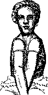
Figure 2. Case of P. Marie.
Changes in the clavicles consist of their atrophy. Complete absence is rarely observed, and usually the sternal and humeral ends are present, while the middle section is replaced by a connective tissue cord. Muscles normally attached to the clavicle (m. trapezius, subclavius, sternocleidomastoideus, etc.), as well as the middle cervical aponeurosis, attach to the bone remnants and to the connective tissue cord. Due to the absence of the clavicles, unusual mobility of the upper part of the shoulder and scapula is created: in markedly pronounced cases, the heads of the humeral bones can be shifted to complete contact on the anterior chest wall (Fig. 2, Marie's case). Changes in the rest of the skeleton are inconsistent and variable, and include: deformations of the ribs, sternum, kyphoscoliosis, deformations of the pelvic bones and limbs, atrophy of the finger phalanges, dislocations, genu valgum, etc. Along with typical forms, there are also those in which all manifestations are significantly milder, or those affecting only the skull (dysostosis cranio-facialis Crouzon). The disease usually appears spontaneously in a previously healthy family and is subsequently hereditary, transmitted in a direct line regardless of sex; not all children of affected parents fall ill. The nature and cause of the disease remain completely unexplained. No organ changes specific to this disease are observed in the internal organs.
Related articles
Cite this page
“Dysostosis.” Soviet Medical Encyclopedia. English translation of Bolshaya Meditsinskaya Entsiklopediya, 1st ed. (Moscow, 1928–1936), ed. N. A. Semashko. https://sovietmedicalencyclopedia.pages.dev/article/dysostosis/