Occipital Region
Historical document, translated for reference. It reflects medical knowledge of the 1920s–30s and is not medical advice.
Summary
This article describes the anatomical structure of the occipital region, including its boundaries, layered composition of soft tissues, and the complex arrangement of the neck musculature. It details the four layers of muscles, their origins, insertions, and functions, along with their innervation.
Encyclopedia article (1928–1936)
OCCIPITAL REGION (regio occipitalis, regio nuchae), on the head corresponds to the position of the occipital bone; in front it borders on the parietal region (regio parietalis), on the sides on the mastoid regions (regiones mastoideae), and below it passes into the occipital region of the neck—regio nuchae (nuchal region), the lower boundary of which is marked by a line drawn from the spinous process of the VII cervical vertebra on both sides to the acromion, and the lateral boundaries by lines connecting the mastoid processes with the acromion. Not differing in its layered structure from the parietal and frontal regions and having no convex boundaries, the upper section of the occipital region is usually combined in topographic-anatomical descriptions with these two regions into one—the fronto-parieto-occipital region (regio fronto-parieto-occipitalis), the posterior-lower boundary of which is the external occipital protuberance and the superior nuchal line. The soft parts of this section of the occipital region consist of skin covered with hair and containing sebaceous glands, and a layer of adipose tissue, permeated with connective tissue fibers extending from the fascia lining the outer surface of the paired occipital muscle, which constitutes the next layer. The connection between these layers is strong, which provides grounds for practically considering them as one—the "thick skin of the head" ("Kopfschwarte" of German authors), in which vascular and nerve trunks are embedded. The occipital muscle represents a flat muscle, individually developed in various ways, originating along the superior nuchal line throughout the entire extent between the bases of the mastoid processes; anteriorly it passes into a tendinous expansion—the aponeurosis (galea aponeurotica), also firmly connected by connective tissue bundles to the scalp, which explains why the hair cover of the head moves when these muscles contract. This is all the more facilitated by the fact that under the aponeurosis, between it and the deeper-lying periosteum, there is a layer of loose connective tissue, ensuring a less strong connection of these layers. The periosteum (pericranium) is also connected to the bone rather weakly due to the fact that a layer of loose tissue, although insignificantly expressed, is located under it; this layer is absent along the line of the sutures, where the connection of the periosteum to the bone is strong. Below the superior nuchal line, the skin becomes thinner, the hairiness is less abundant, and in the lower section it disappears; the connection between the skin and the superficial fascia (fascia colli superficialis) becomes less strong.
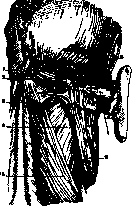
Figure 1. Muscles of the occipital region: 1 - protub. occip. ext.; 2 - m. transv. nuchae; 3 - m. semispin. capit.; 4 - m. splen. capit. et cervic.; 5 - m. trapezius; 6 - m. sterno-cleido-mast.; 7 - m. auric. post.; 8 - m. occipit. (According to Spalteholz.)
The musculature is arranged in four layers. The first layer—m. trapezius s. cucullaris (Fig. 1), lined by the split superficial fascia forming its sheath, originates from the superior nuchal line, external occipital protuberance, nuchal ligament, and from the spinous processes of the VII cervical and all thoracic vertebrae; it attaches to the acromial end of the clavicle, to the acromion, and to the spine of the scapula. It is innervated by the accessory nerve and from the cervical plexus. Upon contraction of the upper sections, it raises the scapula upward, and the lower sections lower it; upon simultaneous contraction of both sections, it brings the scapula closer to the spine; with a fixed scapula, simultaneous contraction of both muscles pulls the head backward. In the second layer are located: 1) m. levator scapulae, which originates from the transverse processes of the 4 upper cervical vertebrae and attaches to the angle of the scapula and along its inner edge; upon contraction, it raises the scapula; upon fixation of the latter, it flexes the cervical spine; 2) m. rhomboideus minor—originates from the spinous processes of Cvi-vii, attaches along the vertebral edge of the scapula in the upper section; it is innervated in the same way as the previous one, by the dorsal scapular nerve; its contraction raises and brings the scapula closer to the midline; 3) m. serratus post. superior—originates from the spinous processes of Cvi-vii and Di-ii, attaches to the II-V ribs, somewhat outward from their angles. It is innervated by the intercostal nerves I-IV; its contraction raises the indicated ribs; 4) m. splenius cervicis et capitis—takes origin from the lower two-thirds of the nuchal ligament and the spinous processes of Cvii and the first five-six thoracic vertebrae, attaches to the transverse processes of the I and II cervical vertebrae, to the outer side of the mastoid process, and to the outer third of the superior nuchal line, it is innervated by the cervical nerves II-VIII; upon contraction, it turns the head, and upon simultaneous contraction from both sides, it pulls the head backward. In the third layer are found: 1) m. ilio-costalis cervicis (m. cervicalis ascendens)—takes origin from the 6 upper ribs and attaches to the transverse processes of Civ-vi; 2) m. longissimus capitis (m. complexus minor)—originates from the transverse processes of Civ-vii and Di-iii, attaches to the mastoid process; 3) m. longissimus cervicis—from the transverse processes of Di-v to the transverse processes of Cii-v; 4) m. spinalis cervicis—from the spinous processes of Di-ii and Cvi-vii to the spinous processes of Cii-iv; all the listed muscles of the third layer are part of the m. sacrospinalis (m. erector trunci). Their contraction entails extension of the spine as a whole or its parts and extension of the head; 5) m. semispinalis capitis—from the transverse processes of Civ-vii and Di-vi, attaches to the occipital bone in the extent between the superior and inferior nuchal lines; 6) m. semispinalis cervicis—from the transverse processes of Di-vi to the spinous processes of Cii-v. In the fourth layer (Figs. 2 and 3)—two groups of muscles: the first—mm. occipito-vertebrales,
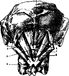
Figure 2. Muscles of the occipital region: 1 - protub. occip. ext.; 2 - proc. transv. atlant.; 3 - a. vertebr.; 4 - tuberc. post. atl.; 5 - proc. spin. epistr.; 6 - m. obliq. capit. inf.; 7 - m. rect. capit. post. major; 8 - m. rect. capit. post. min.; 9 - m. obliq. capit. sup. (According to Spalteholz.)
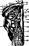
consisting of m. rectus capitis posterior major (from the spinous process of Cii to the outer part of the inferior nuchal line; rotates and pulls the head backward); m. rectus capitis posterior minor (from the posterior tubercle of Ci to the inner part of the inferior nuchal line; pulls the head backward); m. obliquus Figure 3. Deep muscle triangle of the nuchal region: 1 - m. sterno-cl.-mastoid.; 2 - m. splenius capit.; 3 - a. occipit.; 4 - a. vertebral.; 5 and 7 - m. semispin. cap.; 6 - m. trapezius; 8 - m. obliq. cap. super.; 9 - m. rect. cap. major; 10 - m. rect. cap. min.; 11 - n. suboccip.; 12 - posterior arch of the atlas; 13 - n. occip. major; 14 - m. obliq. cap. inf.; 16 - m. semisp. cervic.; 15, 17 and 18 - ram. post. n. Ciii-v; 19 - a. cervic. post. (According to Dyakonov.) capitis superior (from the transverse process of Ci to the occipital bone; pulls the head backward) and m. obliquus capitis inferior (from the spinous process of Cii to the transverse process of Ci; rotates the head); they are innervated by the suboccipital nerve. The second group consists of mm. multifidus, rotatores, intertransversarii dorsales cervicales, and interspinales.
S41
Following the muscles are: in the upper section, the squamous part of the occipital bone; in the lower section, the arches of the cervical vertebrae, connected to each other by ligamenta flava. The space between the posterior margin of the foramen magnum and the posterior arch of the atlas is filled by strong ligaments called membrana atlanto-occipitalis. The occipital region receives its blood supply from 1) the occipital artery (from the external carotid artery), which at the lateral margin of the trapezius muscle is situated quite superficially, covered by the sternocleidomastoid and splenius muscles, and goes to the upper section of the occipital region, where it appears medial to the posterior margin of the mastoid process; 2) the transverse cervical artery (from the subclavian artery), covered by the trapezius muscle, divides into ascending and descending branches; 3) the vertebral artery, upon exiting the foramen transversarium of the axis, forms a lateral bend and approaches the foramen transversarium of the atlas. Upon exiting here, it curves behind the articular surfaces of the atlas and is situated here in a triangle formed by the obliquus capitis superior and inferior muscles and the rectus capitis major muscle. Then, piercing the membrana atlanto-occipitalis, it enters the foramen magnum. The veins of the occipital region drain into the vertebral, occipital, external jugular, and deep cervical veins. On the membrana atlanto-occipitalis is situated a venous plexus (posterior cervical venous plexus), which communicates with the external venous plexus, located under the membrane, directly on the spine, and with the sinuses of the dura mater (via an emissary vein passing through the foramen condyloideum). The lymph vessels of the occipital region are directed to the nodes located along the line of attachment of the trapezius muscle (occipital lymph nodes), as well as to the nodes of the axillary cavity. Throughout the upper sections, there are no anastomoses between the lymphatic vessels of this region and those adjacent to it. One to three relatively large trunks, collecting from the occipital lymph nodes, having passed the clusters of nodes under the angles of the lower jaw and the upper half of the neck, drain into the jugular lymphatic trunk. The nerves of the occipital region are branches from the posterior branches of the cervical roots (Fig. 4). The greater occipital nerve (from CII) exits between the arch of the atlas and the axis; having curved from below around the obliquus capitis inferior muscle, it lies on the rectus capitis posterior major and minor muscles, medial to the occipital artery. Piercing then the trapezius muscle, it branches in the skin (it also supplies the longissimus capitis, semispinalis, and obliquus capitis inferior muscles). The suboccipital nerve (from CI) exits between the occipital bone and the arch of the atlas and is situated behind the vertebral artery; it gives branches to the rectus capitis posterior major and minor, semispinalis cervicis, and obliquus capitis superior and inferior muscles. Among acute inflammatory diseases in the occipital region, carbuncles are observed. The process is often not limited to the hair follicles and surrounding tissues, but spreads along the surface and into the depth. In the latter case, phlegmons form on the muscles, and in the presence of dense infiltration of the skin, it is sometimes difficult to determine fluctuation. Acute abscesses, the source of which are usually lymph nodes, are also observed quite often. Deeper are located abscesses originating from the mastoid process (in the depth behind and below the mastoid process, under the attachment of the sternocleidomastoid muscle), sometimes penetrating into the occipital region ("Bezold's abscess"); they are preceded by an inflammatory process in the middle ear. Osteomyelitis of the occipital bone can be accompanied by the formation of an abscess, which usually penetrates between the first and second or second and third layers of muscles into the lateral sections of the neck; the disease is quite rare and more often develops secondarily. Also, tuberculous involvement of the occipital lymph nodes is noted relatively rarely. Their symmetrical swelling indicates a luetic lesion. Woody phlegmon of the neck, also observed in the occipital region, represents a chronic infection.
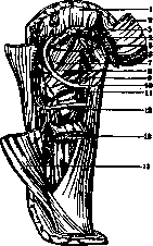
Figure 4. Posterior branches of the first three cervical nerves: 1 - cucullaris muscle; 2 and 5 - complexus major muscle; 3 - sternocleidomastoid muscle; 4 and 8 - splenius muscle; 5 - posterior branch of the I cervical nerve; 6 - obliquus capitis superior muscle; 7 - rectus capitis posterior minor muscle; 8 - rectus capitis posterior major muscle; 9 - obliquus capitis inferior muscle; 10 - posterior branch of the II cervical nerve (greater occipital nerve of Arnold); 11 - common nerve trunk for the complexus muscle; 12 - posterior branch of the III cervical nerve.
of unclear nature (diplococcus?), accompanied by diffuse, hard, pale-gray, painless infiltration without elevated temperature (phlegmon ligneux Reclus, Holzphlegmone of German authors). The occipital region is a site for the emergence of cerebral hernias, which reach large sizes here. Encephalocele (encephalomeningocele) and meningocele are observed. Based on the location and exit from the cranial cavity, upper and lower occipital cerebral hernias are distinguished, depending on whether they are located above or below the external occipital protuberance. So-called blood cysts of the sinus (sinus pericranius) also emerge here. False meningocele (cephalocele traumatica) is a consequence of trauma (usually during childbirth—forceps), accompanied by brain damage, as a result of which a communication is established between the brain ventricle and the subcutaneous tissue. Accumulation of blood between the periosteum and the bone as a consequence of birth trauma (cephalohaematoma) is rarely observed in the occipital region. Among solid tumors, fibromas, lipomas, papillomas, and angiomas are encountered. Fibromas are observed quite often and can be solitary, the size of a pea or larger, or multiple, reaching significant sizes in the form of hanging folded formations (fibroma molluscum). In their structure, they are diverse: purely fibrous, with the inclusion of pigment, vascular (fibr. angiomatodes), or nervous (fibr. neuromatodes) elements, or they have signs of a malignant neoplasm (fibrosarcomas). The starting point for them is the aponeurosis, the spine, and rarely the skin and the dura mater. Lipomas can reach large sizes and have significant adhesions, whereby the spread of the tumor into intermuscular spaces creates difficulties during their surgical removal. So-called periganglionic lipomas represent accumulations of fatty masses in the vicinity of lymph nodes and can reach large sizes. The accumulation of fat in the form of a collar encircling the neck ('fatty neck' Madelung), apparently, is a particular manifestation of a general disease (see Dercum's disease). Angiomas in the occipital region (as in general on the head) present the peculiarity, compared to other areas of the body, that they can have a direct connection with intracranial vessels (through a bone erosion, more often with the superior longitudinal sinus) and consequently present a danger during trauma, infections, and complicate surgical intervention. Among the various types of angiomas, cavernous angioma is encountered here more often. Among cystic tumors, dermoids, atheromas, and very rarely, as a consequence of trauma, epithelial cysts are observed. A special group is formed by so-called air cysts (pneumatocele)—accumulations of air between the bone and soft parts, which reach significant sizes in the occipital region and are sometimes accompanied by changes in the bone (Kadyan). Sarcoma of the soft parts as a primary disease is rarely encountered; usually, in the occipital region, a tumor originating from the bone or from the cranial cavity grows through. Cancer is a relatively frequent phenomenon: about 6% of all skin cancers occur on the scalp, mainly in the occipital and frontal regions. More often it is epithelioma, less often glandular cancer, and even less often colloid and pigment cancer (Tikhov). The neoplasm, rapidly growing through the bone, destroys it and is prone to decay with the subsequent formation of a characteristic cancerous ulcer. Metastatic adenocarcinomas are rarely observed (in cases of lesions of the thyroid gland and adrenal gland). Syphilitic lesion of the occipital bone is encountered relatively rarely (more often in the anterior part of the skull), but manifestations of all forms of bone syphilis are possible. Trauma to the occipital region can affect the soft parts and be accompanied by subperiosteal and muscular hemorrhages, and also cause fractures of the occipital bone. The latter is possible with direct violence and when falling from a height onto straightened legs. In this latter case, the skull is, as it were, impaled on the spine, and a fracture occurs in the region of the ring of the foramen magnum. Fractures of the occipital bone of the latter type belong to fractures of the base of the skull, represent severe injuries, and have a poor prognosis. Gunshot wounds of the occipital region, usually associated with damage to the cerebellum or the posterior parts of the cerebrum, according to wartime data, are observed in 7-8% of all skull wounds. However, their frequency cannot be accurately determined, because due to the severity of the injury, a large part of the wounded remain on the battlefield and cannot be accounted for. In the occipital region, operations are undertaken with the aim of exposing the cerebellum, the cerebellopontine angle, and the medulla oblongata. There are the following methods: the Krause method—an incision down to the bone, 1-2 cm above the external occipital protuberance, from the posterior edge of the mastoid process of one side to the other, slightly arcuate, with the convexity upward. From the ends of this incision, two others are made, slightly converging toward the posterior semicircle of the foramen magnum. After placing trepanation holes at the corners and above the external occipital protuberance and connecting them with Dahlgren forceps or by another method, the bone flap is incised at the base with a Doyen chisel, and the osteo-musculo-cutaneous flap is reflected downward; at the same time, the posterior semicircle of the foramen magnum is partially broken off. Cushing's method: an incision in the form of a cross-bow, in which the horizontal part is the same as in the Krause method, and the vertical part is from the middle of the horizontal one downward along the midline. The soft parts are detached to the sides together with the periosteum to the level of the foramen magnum. In the area of the nuchal plane, on both sides of the occipital crest, trepanation holes are placed, which are expanded to the necessary limits. The dura mater is incised parallel to the sinuses, 1 cm away from them. Puncture of the cerebellomedullary cistern ('Suboccipitalstich') is performed with the patient in a lying or, more often, sitting position, with the head lowered in a strictly median position. Slightly away downward from the spinous process of the axis, a needle is inserted into the thickness of the skin and muscles, in the direction of the posterior edge of the foramen magnum, down to the bone. Then, having pulled the needle back slightly, the posterior edge of the foramen magnum is felt with its tip, and, sliding along it, one penetrates into the cistern through the atlanto-occipital membrane. In order to create conditions for constant drainage into the musculature and subcutaneous tissue of the occipital region in cases of hydrocephalus, Anton and Schmieden proposed the following operation: an incision of 8-12 cm from the external occipital protuberance to the spinous process of the second cervical vertebra, strictly along the midline (through the nuchal ligament), the posterior edge of the foramen magnum and the posterior arch of the atlas are exposed. The atlanto-occipital membrane and simultaneously the cerebellomedullary cistern are opened, and the edges of the incision (at the same time a quadrangular opening can be made) are sutured to the occipital musculature.
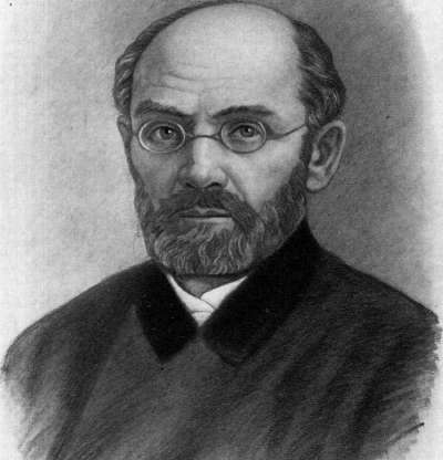
P. Kupriyanov. SAUERBRUCH, Ferdinand (born in 1875), a famous German surgeon, a student of Langerhans, Mikulicz, and

?
Friedrich. In 1910, Sauerbruch took the chair of surgery in Zurich, from where in 1918 he moved to Munich, where he gained worldwide fame for his outstanding works in the field of thoracic surgery. Capital, brilliantly illustrated—'Technik der Thoraxchirurgie' (Berlin, 1911, together with E. Schumacher) and especially the two-volume 'Die Chirurgie der Brustorgane' (Vol. I-II, Berlin, 1927-28; the latter work was published in a third edition) have no equal in world literature on this subject. Having devoted himself mainly to work in the field of thoracic surgery and especially to the surgical treatment of pulmonary tuberculosis, Sauerbruch is no less intensively engaged in the study of a number of other highly relevant chapters of general surgery, as evidenced by numerous works of both Sauerbruch and his students. Sauerbruch participates in the work of the most common surgical journals (Deutsche Zeitschrift für Chirurgie, Archiv für klinische Chirurgie, Zentralblatt für Chirurgie), of which he is currently an active editor. In 1927, after the death of Hildebrand, he took his chair at the University of Berlin and is one of the chairmen of the Berlin Surgical Society. Besides those mentioned above, the main works of Sauerbruch are: 'Vorlesungen zur operativen Behandlung schwerer Skoliosen' (Archiv für klinische Chirurgie, Vol. CXVIII, 1921); 'Blutleere Operationen am Schädel unter Überdruck nebst Beiträgen zur Hirndrucklehre' (Mitteilungen aus den Grenzgebieten der Medizin und Chirurgie, 1907, Supplement 3); 'Willkürlich bewegbare künstliche Hand' (Berlin, 1923); 'Weitere Mitteilungen über die Parabiose bei Warmblütern mit Versuchen über Ileus und Urämie' (Zeitschrift für experimentelle Pathologie und Therapie, Vol. VI, 1909).
Related articles
Mentioned in
Cite this page
“Occipital Region.” Soviet Medical Encyclopedia. English translation of Bolshaya Meditsinskaya Entsiklopediya, 1st ed. (Moscow, 1928–1936), ed. N. A. Semashko. https://sovietmedicalencyclopedia.pages.dev/article/occipital-region/