Occipital Bone
Historical document, translated for reference. It reflects medical knowledge of the 1920s–30s and is not medical advice.
Summary
This article from the 1928–1936 Soviet Great Medical Encyclopedia provides a detailed anatomical description of the occipital bone, detailing its structure, foramina, and connections to the brain and spinal cord.
Encyclopedia article (1928–1936)
OCCIPITAL BONE (os occipitale), one of the primitive bones of the cranial vault, located in the lower posterior part of the skull. It represents a curved plate of rhombic shape, thickened in the anterior-lower part, where there is a large, irregularly oval opening (foramen occipitale magnum) connecting the cranial cavity with the vertebral canal. Through this opening pass: the medulla oblongata with its membranes, the posterior arteries of the dura mater (rami meningei aa. vertebralis), the anterior and posterior spinal arteries (aa. spinales ant. et post.), vertebral arteries, basilar plexus, accessory nerves of Willis (nn. accessorii Willisii), and the roots of the first cervical spinal nerves (nn. cervicales I). The shape of this opening is subject to variations and corresponds to the shape of the skull: in dolichocephalic skulls it is elongated longitudinally, in brachycephalic skulls it has outlines close to a circle, or is even expanded in the transverse diameter. In front of the occipital foramen is the basal part of the occipital bone (pars basilaris), which adjoins the body of the sphenoid bone with its anterior surface and is connected to it by a layer of hyaline cartilage (synchondrosis spheno-occipitalis), which ossifies between the ages of 13 and 20 years (Figures 1, 2, and 3). The lower (outer) surface of the body is vaulted and participates in the formation of the Turkish saddle's vaulted plane, the clivus Blumenbachi. On it are located the vertebral and basilar arteries with their branches, the pons Varolii. The lateral edges are serrated, adjoin the petrous part of the temporal bone (synchondrosis petro-occipitalis), and together with the latter form on the upper surface a groove (sulcus petrosus inferior) in which the inferior petrosal sinus passes. To the basal part of the occipital bone on the sides adjoin the lateral parts (partes laterales os. occipit.), on the lower surface of which there are articular processes (condylus), convex in the sagittal and frontal directions, of oval shape, with long diameters converging forward; in front and behind of them are noticeable depressions (fossa praecondyloidea et postcondyloidea). At the bottom of the postcondyloid fossa, on one or both sides, the posterior condyloid canal (canalis condyloideus post.) may open, through which a vein passes connecting the lateral sinus with the plexus venosus suboccipitalis located outside the cranial cavity. In the precondyloid fossa, located outward from the anterior end of the condyles, the canal (canalis hypoglossi) for the XII pair of cranial nerves opens. Outside, to the lateral parts adjoin the petrous parts of the temporal bone of the nasal pharynx. In the middle of it there is a tubercle (tuberculum pharyngeum; Figure 1), to which the longitudinal ligament of the pharynx (lig. longitudinales ant.) and the superior constrictor muscle of the pharynx (m. constrictor pharyngis sup.) are attached; in front of it lies a depression (fossa navicularis), not always clearly expressed. On both sides of the tubercle are roughnesses (tuberositas occipitalis), to which the muscles (m. longus capitis, m. rectus capitis ant.) and the pharyngobasilar fascia are attached. The upper surface of the body of the occipital bone is concave in the transverse direction and, together with the posterior surface of the atlas, forms the fissura petro-occipitalis, in which a deep notch (incisura jugularis) is outlined, divided by a bony process (processus intrajugularis) into a front part (pars nervosa) for the IX, X, XI pairs of cranial nerves and the inferior petrosal sinus, and a back part (pars venosa-foramen jugulare) for the internal jugular vein (bulbus) and the posterior meningeal artery. The posterior part of the lateral part of the occipital bone adjoins the occipital edge of the mastoid part of the temporal bone and is connected to the latter by a suture (sutura occipito-mastoidea). On the lower surface of the lateral part, outward from the posterior half of the articular processes, there is a triangular process (proc. jugularis), limiting the sigmoid or transverse sulcus behind and outward; on the lower surface of it the muscle m. rectus capitis lateralis is attached. The posterior part of the occipital bone, the squama (squama occipitalis), with its anterior edge forms the posterior half-circle of the foramen magnum. The free posterior-upper edges of the squama are connected to the occipital edge of the parietal bones by a suture (sutura lambdoidea). Approximately in the middle of the convex posterior surface there is a process (protuberantia occipitalis externa), from which rough lines (linea nuchae superior) extend to the sides; above and below them are located two other rough, less pronounced lines (lin. nuchae suprema et inferior). Downward, towards the foramen magnum, from the occipital protuberance passes a crest (crista occipitalis externus) that bifurcates at the edge of the opening, along which the nuchal ligament is attached. The parts of the occipital bone above the occipital protuberance are called planum occipitale and serve for the attachment of the occipital muscle; the lower part (below the nuchal lines) is called planum nuchale for the attachment of the trapezius, semispinalis capitis, rectus capitis posterior (minor et major), obliquus capitis superior muscles and partially the sternocleidomastoid and splenius capitis muscles. On the inner concave surface of the squama, corresponding to the position of the occipital protuberance, lies a cruciform elevation (eminentia cruciata), in the middle of which a tubercle (protuberantia occipit. interna) protrudes and which divides four fossae: two upper, in which the posterior parts of the cerebral hemispheres are located, and two lower for the cerebellar hemispheres. Corresponding to the cruciformly located lines are grooves: sulci transversi - the right and left, a receptacle for the transverse sinus; sulcus sagittalis, usually continuing into the right transverse groove and containing the sagittal sinus, and the internal occipital crest; sometimes in the latter there is a groove in which the occipital sinus is located. In the lambdoid suture, accessory bones are often encountered, sometimes reaching significant sizes and giving occasion to confuse them with the so-called os incae, s. interparietale. The latter was described for the first time on the skull of the Incas (ancient Peruvian skulls), but is also encountered in other peoples. This bone represents the upper, triangular part of the squama (planum occipitale), originating from a membranous rudiment and separating from the rest of the squama, which develops from a cartilaginous rudiment. In this case, between the parts of the squama there is a suture (sutura transversa occipitalis). Sometimes the os interparietale itself consists of several parts (os incae tripartitum Walcker's, unilaterale, bilaterale, unicum, etc.; Stieda), which is determined by the course of ossification of the bone nuclei. At first, the occipital bone represents a cartilaginous ring limiting the large occipital foramen. Ossification usually begins in the beginning of the third month from points located in the pars basilaris (one), the lateral parts (one each), and two in the cartilaginous squama, which soon fuse. In addition, the membranous part contains two points; under normal conditions, the part of the squama of the occipital bone formed from them connects with the bony plate developing from the lower points (cartilaginous part). In lower vertebrates, the four bones developing from four ossification points remain separated by layers of cartilage (ossa occipitalia basilare, superius and lateralia); in man and mammals, their fusion occurs by the 3rd to 4th year of life.
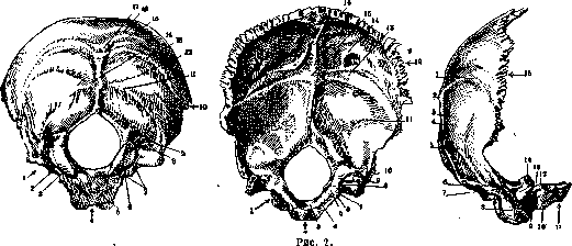
Figure 1
Figure 3. Figure I. Occipital bone from behind: 1-pars lat.; 2-proc. intrajug.; 3-can. hypogl.; 4-pars basil.; 5-tub. pharyng.; 6--cond. occip.; 7-incis. jugul.; 8-can. condyl.; 9-fossa cond.; 10-squama occip.; 11-pla-num nuchae; 12 and 13-lin. nuchae inf. et sup.; 14-crista occip. ext.; 15-lin. nucliae suprema; 16- planum occip.; 17-protub. occip. ext. Figure 2. Occipital bone from front: 1-pars lat.; 2-pars basil.; 3-clivus; 4-sulc. petr. inf.; 5-tub. jugul.; 6-proc. intrajug.; 7-can. hypogl.; 8-incis. jugul.; 9-can. condyl.; 10-sulc. transv.; 11-fossa occip. Inf.; 12-squama occip.; 13-sulc. transv.; 14-fossa occip. sup.; 15-protuber. occip. int.; 16-sulc. sagittalis. Figure 3. Occipital bone on the right: 1-lin. nuchae sup.; 2-prot. occip. ext.; 3-crista occip. ext.; 4- planum nuchae; 5-lin. nucliae inf.; 6-fossa condyl.; 7-can. condyl.; 8-condyl. occip.; 9-can. hypogl.; 10-tub. pharyng.; 11-pars basil.; 12-proc. intrajug.; 13-incis. jugul.; 14-proc. jugul.; 15-squama occip. (According to Spalteholz.)
Related articles
Mentioned in
Cite this page
“Occipital Bone.” Soviet Medical Encyclopedia. English translation of Bolshaya Meditsinskaya Entsiklopediya, 1st ed. (Moscow, 1928–1936), ed. N. A. Semashko. https://sovietmedicalencyclopedia.pages.dev/article/occipital-bone/