Temporal Bone
Historical document, translated for reference. It reflects medical knowledge of the 1920s–30s and is not medical advice.
Summary
The temporal bone is one of the most complex bones in the human body, housing the organs of hearing and balance, and containing the carotid artery and nerves. It forms the base of the cranial vault and supports the facial skeleton.
Encyclopedia article (1928–1936)
TEMPORAL BONE (os temporale), one of the most complex bones, as it serves as a container for the organs of hearing and balance and through it pass the carotid artery and nerves. This paired bone, located between the occipital and sphenoid bones, forms the base of the cranial vault and serves as support for the facial skeleton. The main part of the temporal bone is the petrous part (pars petrosa s. pyramis), which has the appearance of a four-sided pyramid and is constructed from extremely dense bone substance; an appendage to it is the mastoid part (pars mastoidea), named after the homonymous process. In childhood, the tympanic part (pars tympanica), which initially appears as a ring and then grows into a tube leading to the external auditory canal, separated from the tympanic cavity by the eardrum, and the squama, which rises almost vertically and with its beveled edge overlaps the parietal bone (sutura squamosa), remain separated by sutures, as well as the styloid process (proc. styloideus), which serves for the attachment of the ligament suspending the hyoid bone and three muscles. On the lower surface of the pyramid begins the carotid canal, arching through the pyramid and opening on the upper surface near its apex; behind it is the jugular fossa (fossa jugularis), which contains the bulbous enlargement of the homonymous vein; along its inner edge is noted a notch, which together with the homonymous notch of the occipital bone limits the jugular foramen (for.
II 12
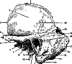
Figure 1. Temporal bone (left, external view): 1-margo parietalis (sutura squamosa); 2-squama (facies temporalis); 3-margo sphenoidalis; 4-proc. zygomaticus; 5-tuberc. articulare; 6-fossa mandibularis; 7-fissura petro-squamosa; 8-fiss. petro-tympanica (Glaseri); 9-vagina proc. styloidei; 10-proc. styloideus; 11-meatus acusticus ext.; 12-fiss. tympano-mastoidea; 13-proc. mastoideus; 14-incisura mastoidea; 15-remains of the mastoid-squamous suture; 16-for. mastoideum; 17-margo occipitalis; 18-spina suprameatum; 19-linea temporalis; 20-incisura parietalis; 21-sulcus arteriae temporalis mediae (according to Rauber). jugulare), through which passes the jugular vein and the IX-XI pairs of cranial nerves.
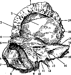
Figure 2. Temporal bone (left, internal view): 1-margo parietalis; 2-squama (facies cerebralis); 3-eminentia arcuata; 4-incisura parietalis; 5-sulcus petrosus sup.; 6-sulcus sigmoideus; 7-for. mastoideum; 8-margo occipitalis; 9-proc. mastoideus; 10-apertura externa aquaeductus vestibuli; 11-incisura jugularis; 12-proc. intrajugularis; 13-fossa subarcuata; 14-meatus acusticus int.; 15-angulus post. pyramidis; 16'-apex pyramidis; 17-angulus sup. pyramidis; 18-sulcus art. meningeae med.; 19-margo sphenoidalis (according to Rauber).
nerves. On the posterior-inner surface is seen the internal auditory canal, at a depth of 8 mm blocked by a perforated bony plate, through which branches of the auditory nerve and the facial nerve, entering the so-called Fallopian canal, penetrate into the inner ear; the latter opens on the lower surface of the bone through the stylomastoid foramen (foramen stylo-mastoideum). At the apex of the pyramid is noted the musculo-tubal canal, the lower part of which represents the bony part of the Eustachian tube, originating in the tympanic cavity and provided with a cartilaginous extension opening into the nasopharynx. On the inner surface of the mastoid process is a groove containing the transverse venous sinus of the dura mater.
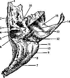
Figure 3. Temporal bone (left, inferior view): 1-apex pyramidis; 2-sulcus petrosus inf.; 3-apertura externa canaliculi cochleae; 4-proc. intrajugularis; 5-sulcus canaliculi mastoidei; 6-fossa jugularis; 7-for. mastoideum; 8-sulcus art. occipitalis; 9-incisura mastoidea; 10-proc. mastoideus; 11-fissura tympano-mastoidea; 12-for. stylo-mastoideum; 13-proc. styloideus; 14-fossa mandibularis; 15-tub. articulare; 16-canal caroticus (according to Rauber).
ki (sinus transversus). From the squama extends forward the zygomatic process (proc. zygomaticus), which abuts against the zygomatic bone, with which it forms the zygomatic arch; at its root is located the articular tubercle, which, together with the articular fossa located behind it, participates in the formation of the jaw joint. In the so-called petromastoid part there is a cavity (tympanic), which communicates via two openings (oval and round windows) with the vestibule of the auditory labyrinth (vestibulum). In the tympanic cavity is located the chain of auditory ossicles; it communicates with the cavity of the mastoid process, which appears at the time of sexual maturity (antrum mastoideum). Ossification is completed within one year, the sutures close significantly later. Some time after birth, the mastoid process is outlined. The eardrum of the newborn occupies almost a horizontal position.
p. karuapya
Related articles
Mentioned in
- Anastomoses
- Articular Menisci
- ARTICULATION
- Cholesteatoma
- Dura Mater
- Dural Sinuses
- Facial Diplegia
- Facial Nerve (facial nerve, the seventh pair of cranial)
- Gasserian Ganglion
- Hiatus
- INNER EAR
- Jaws
- Labyrinth
- Mastoid Process
- Meningitis
- Occipital Bone
- Otosclerosis
- Oxycephaly
- Parietal Bone
- Sphenoid Bone (the main bone located posteriorly - foramen)
Cite this page
“Temporal Bone.” Soviet Medical Encyclopedia. English translation of Bolshaya Meditsinskaya Entsiklopediya, 1st ed. (Moscow, 1928–1936), ed. N. A. Semashko. https://sovietmedicalencyclopedia.pages.dev/article/temporal-bone/