Parietal Bone
Historical document, translated for reference. It reflects medical knowledge of the 1920s–30s and is not medical advice.
Summary
The parietal bone is a paired, quadrangular, flat bone of the cranial vault located between the frontal, occipital, temporal, and sphenoid bones. It participates in forming the roof of the skull and has specific anatomical features including edges, angles, and surfaces with various markings.
Encyclopedia article (1928–1936)
PARIETAL BONE (os parietale), a paired quadrangular flat bone of the cranial vault, located on either side of the median line (sagittal suture) between the frontal, occipital, temporal, and sphenoid bones. Together with adjacent bones, the parietal bone participates in the formation of the roof of the skull. The parietal bone has four edges: 1) margo sagittalis ossis parietalis, s. superior, s. parietalis - the edge connecting the parietal bone of one side with the corresponding edge of the parietal bone of the other side (via the suture - sutura sagittalis) (fig. 1 and 2); 2) margo frontalis, s. coronalis - for connection with the squama of the frontal bone (sutura coronalis) (fig. 1 and 2); 3) margo occipitalis - for connection with the squama of the occipital bone (sut. lambdoidea) and 4) margo squamosus, s. sphenotemporalis, s. temporalis - for connection with the squama of the temporal bone (sut. squamosa). As a quadrangular bone, os parietale has four angles: 1) the postero-inferior (angulus mastoideus, s. margo mastoideus), inserted into the incisura parietalis of os. temporalis; 2) the antero-inferior (angulus sphenoidalis, s. margo sphenoidalis), connecting with the upper edge of the greater wing of the sphenoid bone; 3) the antero-superior (angulus frontalis) (equal to 106°) and 4) the postero-superior (angulus occipitalis). Two surfaces of the parietal bone are distinguished: the outer, convex (facies superficialis) and the inner, concave - cerebral (facies cerebralis). On the outer surface, relatively smooth, the upper and lower temporal lines (crests) (lineae temporales sup. et inf.) are visible to varying degrees, appearing as two parallel arches. The first of these is the site of attachment of the temporal fascia (fascia temporalis); the second, more sharply defined and forming the inserted part of a common crest that extends from the zygomatic process of the frontal bone to the squama of the temporal bone, serves as the site of attachment of the temporal muscle (m. temporalis) and the upper boundary of the temporal fossa (fossa temporalis). The most prominent part of the outer surface is designated by the term parietal tuber (tuber parietale); the degree of protrusion of the parietal tubers varies depending on age, sex, individuality, etc. The inner surface bears a series of impressions from adjacent anatomical structures of the cranial cavity: 1) sulcus sagittalis - a groove running along the margo parietalis of the parietal bone (fig. 2). From two half-grooves (right and left) along the line of the sagittal suture, a groove is formed, in which most of the sinus sagittalis superior is located, and to the edges of which the falx cerebri (proc. falciformis major) of the dura mater is attached. 2) Part of the sulci transversi in the region of the postero-inferior angle. 3) Sulci meningei, s. arteriosi (fig. 2) - a tree-like branching groove - a trace of the adjacent branching of art. meningeae mediae. This groove begins in the antero-inferior angle, from where it extends backward and upward, giving off smaller grooves along its path. Sometimes the initial part of the groove is represented by a canal (from 0.2 mm to 3.6 mm) (Yatsuta). 4) Foveolae granulares (Pacchioni) - for the accommodation of small protrusions of the arachnoid membrane, i.e., Pacchionian granulations. The larger ones are called Trolard's pits; they are the receptacle for expansions of the venous sinuses (parasinusoids). 5) Impressiones digitatae - depressions, as if imprints of the cerebral convolutions, and juga cerebralia - ridges between them corresponding to the brain's sulci. From the latter, a ridge corresponding to the fissura Sylvii is often distinguished, which is called crista Sylvii. The most concave part of the inner surface is marked as fossa parietalis (corresponding to tuber parietale). The parietal bone in its postero-superior quadrant (2-3 cm from sut. lambdoidea) has a small opening (foramen parietale) (fig. 1) for the passage of the emissary vein (emissarium parietale). This opening is sometimes absent, rarely greatly increased in diameter, and finally, the right and left parietal foramina may merge into a single unpaired one. Foramen parietale is a remnant of a small fontanelle (fonticulus obelicus), which closes off from the small fontanelle in the seventh month of intrauterine life. The parietal bone belongs to the covering (appositional) bones. In the primordial skull, it is represented by a connective tissue anlage and does not pass through a cartilaginous stage. On the 10th week of intrauterine life (according to Toldt) or at the end of the 2nd month (according to Mall and others), two areas of ossification appear in the center in the shape of an hourglass. The process of ossification of the parietal bone spreads from the center to the periphery (toward the edges and angles of the bone). In the newborn, ossification is not completed, so connective tissue areas - fontanelles - remain between the parietal bones and adjacent bones (fig. 3). Paired appositional parietal bones are already found in bony fishes. In Dipnoi (lungfishes), the parietal bones fuse with the frontal bones, forming the skull roof - an unpaired fronto-parietale. In the parietal region of the amphibian skull, the parietal bones fuse with the frontal bones into a pair of bones (fronto-parietale). Extinct Stegocephala have a single foramen parietale in the middle of the parietal bone for the passage of the parietal eye nerve. In reptiles (except turtles), the parietal bones are fused into one bone.
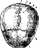
Figure 1. Roof of the skull - external view: 1-squama occipitalis; 2-sutura sagittalis; 3-sutura lambdoidea; 4-foramen parietale; 5-os parietale; 6-sutura coronalis; 7-squama frontalis.
The inner surface bears a series of impressions from adjacent anatomical structures of the cranial cavity: 1) sulcus sagittalis - a groove running along the margo parietalis of the parietal bone (fig. 2). From two half-grooves (right and left) along the line of the sagittal suture, a groove is formed, in which most of the sinus sagittalis superior is located, and to the edges of which the falx cerebri (proc. falciformis major) of the dura mater is attached. 2) Part of the sulci transversi in the region of the postero-inferior angle. 3) Sulci meningei, s. arteriosi (fig. 2) - a tree-like branching groove - a trace of the adjacent branching of art. meningeae mediae. This groove begins in the antero-inferior angle, from where it extends backward and upward, giving off smaller grooves along its path. Sometimes the initial part of the groove is represented by a canal (from 0.2 mm to 3.6 mm) (Yatsuta). 4) Foveolae granulares (Pacchioni) - for the accommodation of small protrusions of the arachnoid membrane, i.e., Pacchionian granulations. The larger ones are called Trolard's pits; they are the receptacle for expansions of the venous sinuses (parasinusoids). 5) Impressiones digitatae - depressions, as if imprints of the cerebral convolutions, and juga cerebralia - ridges between them corresponding to the brain's sulci. From the latter, a ridge corresponding to the fissura Sylvii is often distinguished, which is called crista Sylvii. The most concave part of the inner surface is marked as fossa parietalis (corresponding to tuber parietale). The parietal bone in its postero-superior quadrant (2-3 cm from sut. lambdoidea) has a small opening (foramen parietale) (fig. 1) for the passage of the emissary vein (emissarium parietale). This opening is sometimes absent, rarely greatly increased in diameter, and finally, the right and left parietal foramina may merge into a single unpaired one. Foramen parietale is a remnant of a small fontanelle (fonticulus obelicus), which closes off from the small fontanelle in the seventh month of intrauterine life. The parietal bone belongs to the covering (appositional) bones. In the primordial skull, it is represented by a connective tissue anlage and does not pass through a cartilaginous stage. On the 10th week of intrauterine life (according to Toldt) or at the end of the 2nd month (according to Mall and others), two areas of ossification appear in the center in the shape of an hourglass. The process of ossification of the parietal bone spreads from the center to the periphery (toward the edges and angles of the bone). In the newborn, ossification is not completed, so connective tissue areas - fontanelles - remain between the parietal bones and adjacent bones (fig. 3). Paired appositional parietal bones are already found in bony fishes. In Dipnoi (lungfishes), the parietal bones fuse with the frontal bones, forming the skull roof - an unpaired fronto-parietale. In the parietal region of the amphibian skull, the parietal bones fuse with the frontal bones into a pair of bones (fronto-parietale). Extinct Stegocephala have a single foramen parietale in the middle of the parietal bone for the passage of the parietal eye nerve. In reptiles (except turtles), the parietal bones are fused into one bone.
Figure 2. Roof of the skull - internal view: 1-squama occipitalis; 2-sutura sagittalis; 3-sutura lambdoidea; 4-os parietale; 5-sulci arteriosi; 6-sutura frontalis; 7-sulcus sagittalis.
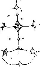
Figure 3. Diagram (Scammon's) of the location of fontanelles (permanent and accessory): a-os frontale; b-os parietale; c-os occipitalis; 1-fonticulus glabel-laris; 2-fonticulus metopicus; 3-fonticulus frontalis; 4-fonticulus sphenoidalis; 5-fonticulus parietalis; 6'-fonticulus mastoideus; 7-fonticulus occipitalis; 8-fonticulus cerebellaris.
The parietal bones are fused into one bone.
The parietal bones of various representatives of mammals differ only in size, the external and internal relief of the surfaces, and in the presence, as for example in primates, carnivores, etc., of a bone that fuses with the occipital bone, and in other cases remains free as os interparietale. Such a bone on human skulls is called os Incae. The splitting of the ossification center of the parietal bone can lead to the appearance of sutura interparietalis, dividing the bone into separate parts in the longitudinal or vertical direction. This gives rise to variations of the parietal bone: for example, os parietale bipartitum (fig. 4), extremely rarely tri-partitum and with greater division. Double parietal bones are not found in lower vertebrates and are a progressive formation (N. Braus). Among the variations of the parietal bone should be included the defect of ossification of the posterior part of the upper edge of the right and left parietal bones (close to lor. pariet.), as a result of which fibrous tissue remains in the form of Gerdy's parietal fontanelle (font. parietalis Gerdy) (fig. 3); on the postero-inferior angle, at the junction with the temporal bone, the presence of a tubercle (tub. asteriacum) is sometimes observed.
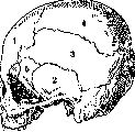
Figure 4. Double parietal bone (os parietale bipartitum): 1-frontal bone; 2-temporal bone; 3-lower parietal bone; 4-upper parietal bone; 5-greater wing of the sphenoid bone.
As a flat bone, the parietal bone consists of outer and inner plates of compact substance (the inner one is called lamina vitrea due to its brittleness when struck from the outside) and a layer of spongy substance between them, the so-called diploe. The latter is permeated with channels for veins (vv. diploicae).- The pathology of the parietal bone-see Skull.
Related articles
Mentioned in
Cite this page
“Parietal Bone.” Soviet Medical Encyclopedia. English translation of Bolshaya Meditsinskaya Entsiklopediya, 1st ed. (Moscow, 1928–1936), ed. N. A. Semashko. https://sovietmedicalencyclopedia.pages.dev/article/parietal-bone/