Caput Succedaneum
Historical document, translated for reference. It reflects medical knowledge of the 1920s–30s and is not medical advice.
Summary
Caput succedaneum is a type of swelling that forms on the presenting part of the fetus's head during childbirth due to pressure from the birth canal. This condition causes venous and lymphatic congestion in the tissues, leading to a temporary swelling that typically resolves within two days after birth without requiring treatment.
Encyclopedia article (1928–1936)
CAPUT SUCCEDANEUM (Latin caput-head and succedaneus-substitute), a head tumor, one of the types of tumors that form during childbirth. It develops on the presenting part of the fetus and arises due to pressure from the soft tissues of the birth canal (cervix and pelvic floor muscles) on the advancing head. This ring-shaped compression impedes the outflow of venous blood and lymph, thereby causing congestion in the tissues of the head lying below the compression site. Additionally, there is a difference in pressure experienced by parts of the head lying above and below the circular 'belt of contact' (Gruzdev, Sellheim). The part of the skull below this belt is under atmospheric pressure, while the part of the head lying above it, like the entire fetal body, is under significantly higher intrauterine pressure. Thus, conditions are created analogous to those in a Birn-Hood. The amniotic sac plays a prominent role here. The more
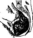
Formation of caput succedaneum on the right parietal bone in occipital position.
amniotic fluid, the more difficult the formation of S. s. The size and extent of S. s. depend on the degree of resistance of the compressing maternal tissues, the strength of contractions, the duration of labor after rupture of the sac, the size of the presenting head, and the characteristics of the latter. S. s. in most cases develops after rupture of the amniotic sac on the most deeply situated part of the head (see figure). Its skin bulges dome-shaped into the vaginal canal and is cyanotic in color. All layers of the skin, subcutaneous tissue, and periosteum are impregnated with serosanguineous gelatinous fluid. Ruptures of small vessels lead to the formation of numerous hemorrhages, ranging in size from pinhead to pea-sized. Hemorrhages are found not only in the S. s. itself but also above the ring-shaped compression. Due to the significant displacement of soft tissues on the flat bones of the skull, ruptures of vessels located between the periosteum and bone may occur. The resulting bleeding detaches the periosteum from the bone, forming the so-called subperiosteal haematoma, which, according to Sellheim, represents a pathological intensification of the physiological phenomenon itself, S. s. Venous hyperemia with hemorrhages, not limited to the coverings of the skull, may involve the spongy layer of bone, the dura mater, and even the brain (Schwartz). Therefore, the presence of the described phenomena in an infant corpse should in no way serve as a basis for assuming that violence was committed against the child. Even mild and rapidly progressing births can be accompanied by the formation of a markedly expressed S. s. with all its features. To the examining finger, S. s. appears as a doughy tumor, due to which it is impossible to detect the sutures and fontanelles without prolonged and fairly strong pressure. S. s. usually almost completely disappears by the end of the second day of extrauterine life and requires no therapeutic measures. The mechanism of labor determines the location of S. s. Thus, for example, in first occipital anterior positions, S. s. is localized on the upper posterior third of the right parietal bone, often crosses the sagittal suture, involving adjacent parts of the left parietal bone, and also extending backward to the occipital bone. In cephalic presentations, subconjunctival hemorrhages are often observed. Congestion in the retinal veins and bluish discoloration of the optic nerve papilla are frequently observed, probably due to hemorrhage into the sheath of this nerve. In 25% of all newborns, retinal hemorrhages were observed. If the head remained engaged at the pelvic outlet in its anteroposterior diameter or close to it, compressed by the pelvic floor muscles, a new tumor may arise, located almost symmetrically and usually completely covering the initially formed one. This new tumor is located mainly on the occipital bone. In anterior cephalic presentations, S. s., although mostly localized on the parietal bone, extends forward and reaches the anterior fontanelle. In first facial presentation, S. s. is located on the right cheek, in second presentation on the left, involving the area of the mouth and eye. The latter are deformed to such an extent that the face resembles the profile of a Negro, the nasolabial fold is smoothed, and the line of the mouth sometimes acquires a vertical direction (Spiegelberg); the child cannot open its eyes. In rarer cases, retrobulbar edema or hemorrhage into the eyeball cause symptoms of lagophthalmus. The thyroid gland, due to compression of the pharyngeal region by the lower edge of the symphysis, may also swell, simulating a congenital goiter. A birth tumor can of course only be observed in a child who was alive during the corresponding period of the birth process.
Related articles
Mentioned in
Cite this page
“Caput Succedaneum.” Soviet Medical Encyclopedia. English translation of Bolshaya Meditsinskaya Entsiklopediya, 1st ed. (Moscow, 1928–1936), ed. N. A. Semashko. https://sovietmedicalencyclopedia.pages.dev/article/caput-succedaneum/