Cellulitis (a1332)
Historical document, translated for reference. It reflects medical knowledge of the 1920s–30s and is not medical advice.
Summary
Cellulitis is a loose fibrous connective tissue located between organs and between the skin and superficial fascia. It contains numerous irregular cavities that can easily fill with fluid or gas, and contains fat lobes, blood vessels, and lymphatic vessels.
Encyclopedia article (1928–1936)
Cellulitis (in anatomy), a loose fibrous connective tissue located between organs and between the skin and superficial fascia. The term C. originates from the fact that when stretched and inflated with air, numerous irregularly shaped and variously sized cavities-cells can be easily seen with the naked eye; these cavities collapse just as easily and are just as easily filled with gas or liquid. The walls of the cavities consist of bundles and plates made of bundles of collagen fibers, loosely connected to each other and easily movable. In the normal state, these cavities contain a small amount of interstitial fluid and during congestion can easily fill with a larger or smaller amount of edematous fluid; they can also be easily filled artificially by injecting fluid under slight pressure. In many places, larger or smaller amounts of fat lobules are embedded in C.: then one speaks of fatty C. In places, homogeneous accumulations of C. are pierced by dense connective tissue plates, fasciae, the arrangement of which, having great importance in medicine, is considered in topographic anatomy. Blood vessels and lymphatic vessels pass through C. The latter form a closed network here, which can optionally connect with the interstitial spaces ('cells') containing metabolic products and sometimes cellular decay. Due to the contractility of endothelial cells, in the blind flask-like outgrowths of the lymphatic capillary network, a kind of fine dense sieve is formed, through which not only colloidal fluid but also particles and grains of decay easily enter the lymphatic current and are carried away from the tissue.
V. Fomin. C. in pathology. In loose C., circulatory disorders have significance in the form of hemorrhages, which here due to the looseness of the tissue have a tendency to spread significantly; on the other hand, this same property of the tissue is the cause of rapid absorption of the extravasated blood. Edema manifests as accumulation of watery fluid in the cell-spaces between the bundles of fibers of C. and the pushing apart of these latter; subsequently, dissociation of collagen fibrils and their swelling occur. In addition to ordinary edema, in rare cases there is a mucoid, mucus-like edema, which is a little-studied change in C., consisting of its impregnation with a semi-liquid mass that shows basophilia and metachromasia on staining; usually swelling is noted, and sometimes as if dissolution and disappearance of collagen fibers. Such not entirely clear pathological changes also include the fibrinoid transformation of C., consisting in the transformation of C. fibers into a substance resembling fibrin. Inflammatory processes are very often observed in C., which may be serous, fibrinous, and purulent inflammations with their usual morphological pictures, course, and outcomes. Tumors of C. belong to neoplasms of the connective tissue group (fibromas, lipomas, myxomas, sarcomas). Specifically in relation to fatty C., its changes deserve separate mention, which are connected with fluctuations in the amount of fat in it. An increase in the amount of fat is observed in obesity and manifests as thickening of the layer of fatty C. under the skin and generally an increase in the entire mass of fatty C., sometimes to colossal sizes. A decrease in the amount of fat leads to the disappearance of fatty C. through the atrophy of individual fat cells with the absorption of the fat droplets contained in them. The decrease in volume of fatty C. often observed in this case is accompanied by darkening of its color, which becomes ochre-yellow (it is thought, due to the condensation of the fat pigment lipochrome); in some places (epicardium, bone marrow) during atrophy of fatty C., its sequential edema occurs, which gives the picture of so-called serous atrophy of fatty C., externally resembling mucous degeneration of the tissue. In traumatic and inflammatory injuries of fatty C., in its necrosis of various origins, splitting of the freed fat occurs and a special granulation inflammatory reaction develops, which in general gives what is called fat granuloma or oleogranuloma (see). (See also Fatty tissue, Connective tissue.) A. Abricosov. Plant cellulose - see Cellulose. MITES (Acarina), an order of the class Arachnoidea, type Arthropoda. They are characterized by a fused body, absence of antennae, two pairs of mouth appendages (upper and lower jaws, of which the latter may merge into a single part), four pairs of legs in adults, and metamorphosis. The mouth organs of C. are gnawing or sucking. The legs may be equipped with claws and in addition sometimes with suction devices. The legs of parasitic C. are often greatly shortened or partially reduced. C. are air-breathing, breathe with tracheae, sometimes without them. They reproduce by egg-laying, more rarely - by viviparity. The larvae of C. are six-legged, nymphs have eight legs but no genital opening, which appears in imago. The transition to each stage of metamorphosis is associated with the molting of C. They are free-living or parasitize on plants or animals. They are ecto- or endoparasites. Body sizes range from microscopic to 2-3 cm. Suborder Vermiformia with family Demodicidae--see Demodex folliculorum (fig. 16).-Suborder Tetrapoda; family Eriophyidae (Phytoptidae) with microscopic small representatives that cause the formation of outgrowths on the leaves of various plants, called galls, in which the corresponding mites live.- Suborder Sarcoptoidea; family Sarcoptidae, or itch mites. Microscopically small C., whose skin forms numerous transverse stripes. Tracheae have been found in some species. The mouth organs consist of a proboscis (formed by the lower jaws) and a pair of short claw-like jaws; palpi are three-segmented. The legs are short, five-segmented with stalked suckers or long bristles. The true itch mites (Sarcoptes) are parasites of mammals; they live in the epidermis of their hosts, where they make tunnels, causing scabies (see). In humans, Sarcoptes scabiei parasitizes. Other mammals have their own itch mites, considered in taxonomy as varieties; these are: S. scabiei var. equi- the horse itch mite, S. sc. var. canis- dog, S. sc. var. suis- pig, S. sc. var. caprae- goat, etc. These itch mites can also pass to humans and other animal hosts, causing scabies in them as well. Itch mites also occur in some wild animals. Representatives of other genera of the same family are parasites only of animals and do not occur in humans; these are: Psoroptes communis (fig. 1 and 2)-parasite of horses, cows, sheep and other animals; Ps. com. var. cimiculi usually parasitizes in the ear of rabbits. Psoroptes, or skin mites, do not make tunnels in the epidermis, as they live on the surface of the skin; Otodectes cynotis lives in the ear of dogs and cats, causing in them a disease similar to epilepsy; Notoedrus muris (fig. 10) affects the ears and other parts of the body of rats; Cnemidocoptes mutans (fig. 11)-parasite of domestic birds, living under the scales of the feet; Cytoleichus nudus and Laminosioptes cysticola are internal parasites of birds; the first inhabits the air sacs and respiratory tract, the second-lives in the subcutaneous tissue, where it often becomes encapsulated. - Family Analgesidae - feather mites-live between the barbs of feathers or penetrate into the shaft; they feed on skin secretions. Nymphs of the genus Falculifer penetrate into the subcutaneous tissue and even into the thyroid gland.-Family Tyroglyphidae-microscopically small mites with smooth covers; legs are quite long with a sessile blister sucker and a curved claw. They feed on decaying organic substances, as well as animal and plant products. They live in
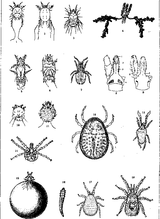
Figure 1 and 2. Psoroptes communis-male and female. Figure 3. Female Carpoglyphus from below. Figure 4. Salivary glands of female Ixodes ricinus. Figure 5. Female Pediculoides. Figure 6. Tarsonemus hominis. Figure 7. Liponyssus bacoti. Figure 8. Ixodes ricinus from the back (mouth parts): 1-hypostome; 2-chelicera; 3-sheath for chelicerae; 4-porous area. Figure 9. Ixodes ricinus (mouth parts) from the ventral side: 1-hypostome; 2-palp. Figure 10. Notoedrus muris. Figure 11. Cnemidocoptes mutans. Figure 12. Argas persicus from the back (female). Figure 13. Microtrombidium pusillum. Figure 14. Six-legged larva of Argas persicus. Figure 15. Female Pediculoides before laying eggs. Figure 16. Demodex. Figure 17. Laelaps stabularis (female). Figure 18. Dermanyssus gallinae (female).
In houses, warehouses, barns (see Barn pests), in hay, flour, fruits, corpses, etc. They can get on human skin or into the intestines. As pseudoparasites, they sometimes cause harm to humans. Tyroglyphus siro - the cheese mite; lives in the rind of old cheese, as well as in flour (found in dust in cellars, cellars, stables, haylofts, storerooms, barns, etc.). After all edible substances are used up, larvae and adults die, and nymphs turn into a special hypopial stage, so-called migratory pupae, which attach to the hairs of various insects and other animals for the purpose of moving to another place where there may be food substances. The cheese mite is considered the cause of vanillism (a professional disease of vanilla sorters); it can also cause gastric and intestinal catarrh; it has been found in urine and in the external genitalia of women. T. longior causes coconut sorters skin lesions - "coprah itch". Rhizoglyphus hyacinthi lives in decaying onions: when it gets on the skin, as shown by Stein and Pavlovsky, it causes inflammation and is the cause of skin lesions in peasants who grow large quantities of onions. Carpoglyphus passulorum (fig. 3) lives in dried fruits and causes dermatitis when sorting them. (See also Aleurobius farinae, Glyciphagus, Tyrogly-phaster.) Suborder Prostigmata. Family Trom-bidiidae, red-bodied mites, bright red in color, predatory in adult stage and parasitic in larval stage. Their larvae were described under the general name Leptus (rouget of the French); they have eyes. The spiracles are located at the base of the claw-like upper jaws. The well-known larva Leptus autumnalis belongs to the species Trombicula (Neotrombicula) autumnalis. Larvae hatch in the second half of summer and crawl onto grass and bushes, attacking humans; when they bite into the skin at the base of hairs, they cause acute itching, burning sensation, and fever. Papules with a purple edge appear on the skin. This disease is seasonal, and in Germany it is timed to coincide with the ripening of gooseberries. L. autumnalis also attacks horses, cows, sheep, dogs, and other animals, causing various disease symptoms in them (in cows - "feu d'herbe"). The remedy for humans is Peru balsam or sulfur ointment. Microtrombidium thalzahuatl in Mexico attaches to the eyelids, armpits, and other places, also causing severe local irritation. Microtrombidium akamushi (Trombicula aka-mushi - Trombidicula akamushi) - see Akamushi. Microtrombidium pusillum (fig. 13) causes autumn erythema. Family Tetranychidae - pests of various cultivated plants. With their bristle-like jaws, they pierce human skin and cause severe itching with papule formation (Tetranychus telarius, living on plane trees and grapevines). - Family Bdellidae. Tydeus molestus was apparently introduced to Belgium with shipments of guano from Peru; it settled in gardens; it attacks humans and animals, causing unbearable itching. - Family Hydrachnidae - water mites - a species-rich family. Larvae parasitize various aquatic insects (especially mosquitoes) and mollusks. Suborder Heterostigmata. Family Tarsonemidae. Pediculoides ventricosus (figs. 5 and 15) - the pot-bellied mite; f 0.2 mm, d 0.1 mm in length. Predator. It devours larvae of isopods, grain moths, and various barn pests; therefore, large batches of grain or straw can be infested with it. When loading sacks with such grain or when sleeping on straw, P. ventr. attacks humans and causes mass diseases accompanied by skin rashes, itching, urticaria, and insomnia. These mites can be carried by wind onto humans. Nephrophagus was found in urine. Tarsonemus hominis (fig. 6) and other species were sometimes found in human and animal tumors. Suborder Mesostigmata. Family Gamasidae (Parasitidae). Dermanyssus gallinae - a parasite of chickens; during the day it hides in nests and in crevices of the roost, at night it attacks chickens and sucks their blood. With massive parasitism, it causes fatal anemia in chickens, probably caused by poisonous saliva. The body of D. gallinae (fig. 18) is pear-shaped, whitish or reddish in color; f 0.7-0.75 mm in length and 0.4 mm in width, m 0.6 mm × 0.32 mm. D. gallinae easily transfers to humans. When sucking blood, it causes unbearable itching and even papular rash. In poultry farmers, it causes a professional skin disease. In some cases, D. gallinae was found in skin tumors. Besides humans, D. gallinae can attack horses, donkeys, rabbits, dogs, cats, and other animals. In horses, it causes dermatosis. Another species - D. hirundinis - is brownish, 1.2-1.4 mm in length, lives in swallow nests and also easily attacks humans, causing the appearance of polymorphic exanthemas with distressing subjective sensations. Laelaps stabularis Koch (fig. 17) is found in livestock stalls; it can transfer to humans. Some Gamasidae parasitize rats [Dermanyssus muris, Liponyssus bacoti (fig. 7), etc.], and can transfer to humans. Various species of the same family are parasites of insects (e.g. Gamasus coleopteratorum). - The superfamily Ixodoidea consists of two families - Argasidae (see) and Ixodidae. Argasidae do not have a hard chitinous shield on the back, and their mouthparts are on the ventral side of the body, set back from the anterior end. Sexual dimorphism is not strongly expressed. They suck blood repeatedly in different stages of metamorphosis. Argas persicus (figs. 12 and 14) - a parasite of chickens (lives in chicken coops); it also attacks humans. A. vespertilionis - a parasite of bats. Another genus - Ornithodorus (see). - Family Ixodidae. On the back of the mite - a hard chitinous shield. The mouthparts are terminal. They consist of a pair of upper jaws with a movable "finger" bearing chitinous spines, and the hypostoma - a chitinous plate covered with spines, the tips of which are directed toward the base of the mouthparts (figs. 8 and 9). Between them opens a pair of powerfully developed cluster-shaped salivary glands (fig. 4). When sucking blood, the mite plunges the mouthparts into the host's skin, thereby firmly holding onto the skin. In each stage of development, Ixodidae suck blood only once; this procedure lasts several days. The metamorphosis cycle of Ixodidae consists of the following stages: eggs, larvae, nymphs, and imago. They are distinguished as one-host, two-host, and three-host mites depending on how many hosts the mite lives on throughout its life. In one-host mites (Boophilus), after hatching from the egg, the larva attaches to a cow, molts into a nymph on it, and then into imago; the fed female falls to the ground for subsequent egg laying. In two-host mites, the larva and nymph live on the same host; the fed nymph falls to the ground, molts into imago; the adult mite attaches to another host; egg laying occurs on the ground (Nuazotta). In three-host mites, the larva feeds on the first host, falls off and molts into a nymph; the latter drinks blood from the second host, detaches and molts into imago; the adult mite attacks the third host and, after feeding, falls to the ground for egg laying (Ixodes ricinus). The mite's saliva is toxic; with massive parasitism on livestock, mites reduce milk yield and significantly slow down the weight gain of the animal. Many mites are carriers of pathogens of various forms of piroplasmosis of domestic animals. Ixodes ricinus (figs. 8 and 9) - the dog tick; carrier of Piroplasma bovis: it also parasitizes humans, lives on birds and mammals. Its life cycle is long: larvae hatch from eggs in 49-252 days, harden and attack the host 10-570 days after hatching; the larva feeds for 3-6 days, lives 28-140 days after feeding and molts into a nymph. The nymph attacks the host 10-540 days later; it feeds on the host for 3-5 days; the nymph lives 56-360 days; females attack the host 10-810 days after molting; they feed for 6-14 days. To destroy mites, cows are bathed in special baths with arsenic solutions. Ixodes holocyclus (Australia) causes tick paralysis in humans, dogs, and cats with its bite, which can lead to the death of the host. The typical species of the genus Dermacentor is D. reticulatus (parasite of horses, dogs). Besides it, in the fauna of the USSR are known - D. niveus (horse, horned cattle, sheep, human), D. Pavlovskii (mountain sheep), D. variegatus (deer), D. Birulai and D. Nuttalli. The three-host tick, D. reticulatus, is a carrier of Babesia caballi, the causative agent of equine piroplasmosis; in France it carries canine piroplasmosis. D. variabilis in North America parasitizes dogs, livestock, and humans. Under experimental conditions, this tick transmits the causative agent of "Rocky Mountain fever". The actual carrier of the latter is D. Andersoni venustus. The causative agent is considered to be Dermacentroxenus rickettsi. D. Andersoni also spreads tularemia - septicemia of some wild rodents with the causative agent Bacterium tularense (California and other states of North America). This disease is also transmitted to humans through the same carrier (and also the deer fly - Chrysops discalis) (North America and Japan).
Many cases of laboratory-acquired tularemia are known (London and others). Tularemia has also been detected in the USSR (Urals, Moscow region, Lower Volga), but nothing is yet known about its vectors in the USSR. Nogushi (Noguchi) was able to transmit 'oroya' fever from monkey to monkey by means of D. Andersoni. The causative agent of this disease in Peru is considered to be Bartonella (see.) bacilliformis. Finally, D. Andersoni can cause tick paralysis in humans and some domestic animals, which often ends fatally if the attached tick is not noticed and removed in time. Rhipicephalus bursa- is the vector of sheep piroplasmosis; Rh. sanguineus- is the vector of Piroplasma canis. Rh. simus (Africa)- is a parasite of cattle, transmits Theileria parva to cows. In humans, this parasite causes a short-term fever with adenitis after a 3-day incubation- 'tick fever', or 'Tick-bite fever'. Rhipicephalus- is a three-host tick. Haemaphysalis punctata- a parasite of mammals, birds, and reptiles- can cause tick paralysis in children and sheep. Three-host tick. Aponomma, parasitizes only reptiles.(See Ixodes, Boophilus, Hyalomma.) Most ticks of the family Ixodidae have a wide range of hosts, including humans.
Related articles
Mentioned in
Cite this page
“Cellulitis (a1332).” Soviet Medical Encyclopedia. English translation of Bolshaya Meditsinskaya Entsiklopediya, 1st ed. (Moscow, 1928–1936), ed. N. A. Semashko. https://sovietmedicalencyclopedia.pages.dev/article/cellulitis-2/