Amputation
Historical document, translated for reference. It reflects medical knowledge of the 1920s–30s and is not medical advice.
Summary
Amputation is the surgical removal of a peripheral body part, most commonly a limb. This article covers the historical development of amputation techniques, indications, and methods including flap, subperiosteal, and bone-plastic approaches.
Encyclopedia article (1928–1936)
Amputation (from Latin amputare - to cut off, to sever), the surgical removal of a peripheral part of an organ, for example, a limb, breast, or penis. From the standpoint of operative technique and subsequent function, amputation of limbs is of particular interest. This operation is what is primarily meant when speaking of amputation. In most cases, it is performed only for vital indications as a last resort, when it is impossible by other means to save not only the diseased limb but often the patient himself. Absolute indications for amputation at present are considered: 1) extensive comminution of bones with rupture of large vessels and nerves; 2) crushing of all the muscles of the limb; 3) necrosis of a part of the limb of various origins (senile, spontaneous, sclerotic gangrene, etc.). Tumors, prolonged, severe, extensive purulent processes, and tuberculosis of bones and joints, not susceptible to other methods of treatment, are considered relative indications for performing amputation. Pirogov said that "in determining the indications for amputation, medical logic is most clearly revealed: no operation requires as much consideration, common sense, and attention from the physician as the rational, clear formulation of indications for amputation." The operation of limb amputation is one of the oldest surgical interventions that has come down to us from the most distant times. All the main points in the modern understanding of this operation were already known to Hippocrates and were clearly indicated and substantiated by Celsus and Archigenes (II century), who gave rules for controlling bleeding by ligation of vessels and tightening of the limb above the amputation line. However, over 15 centuries there is complete oblivion of the rules of Celsus and Archigenes. During this entire time, amputation was performed as primitively as in the time of Hippocrates: limbs were severed within the necrotic tissues, physicians had forgotten how to stop and prevent operative bleeding. Only in the XVI century did the famous "surgeon from the barbershop" A. Pare introduce the ligation of vessels severed during amputation. With this proposal by Pare, the first, most difficult, stage in the development of amputation technique was completed. Surgeons were placed in even better conditions when it became possible to prevent operative bleeding during amputation. At the suggestion of Morrel in France and Young in England (1674), a special loop applied to the limb above the site of amputation began to be used. This loop, modified by Petit (I. L. Petit), in the form of a tourniquet, became universally used until the introduction of the modern Esmarch tourniquet. With this innovation in technique, surgeons significantly improved the outcomes of amputation in terms of eliminating the immediate danger of losing the patient from operative bleeding. The second stage in the doctrine of amputation is marked by the development of methodology for stump treatment. Until the XVII century, amputations were performed almost as recommended by Celsus, i.e., in the form of circular incision of soft tissues made in one plane. It is now completely clear to us that making an incision of soft tissues at once - in one blow, "en saucisson" - in the plane of the bone saw predetermined the fate of the future stump. The protrusion of the bone from the mass of soft tissues due to the latter's ability of retraction, i.e., what is now called "conicity of the stump," was the inevitable outcome of such an amputation. Surgeons of that time could not fail to notice this and therefore they sought in every way to modify this method of amputation. Thus, the aforementioned Petit and the Englishman Cheselden (Cheselden) in 1720 proposed to incise the soft tissues in two stages: first - only the skin and subcutaneous tissue, and then - muscles and bone in one plane, at the height of the pulled-up skin coverings. A number of subsequent authors - Alanson, Dupuytren, Desault, Pirogov and others - with the same purpose proposed to sever muscles by double, triple, or multiple incisions. However, despite all the variety of these modifications of soft tissue severance in amputation, the conicity of the stump was still the usual outcome of these operations. That is why from the end of the XVII century we see a significant innovation in the technique of amputation in the form of the so-called flap, or oblique, method of limb amputation. The creators of the flap method are Lowdham, Verduin, and Sabourin. Essentially this method is a circular method, to which the authors added a flap on one or two opposite surfaces. Depending on the number, shape, and composition of the flaps, single-flap and double-flap methods are distinguished. According to composition, the flaps can be skin flaps (Lowdham) and skin-muscle flaps (Langenbeck). In the XIX century, the oval method with its modifications - elliptical and rocket-shaped - was introduced into amputation technique, which constitute a transition from the circular method of soft tissue incision to the flap method. Subsequently, as methods of wound treatment improved, surgeons increasingly sought to obtain a well-functioning stump. That is why, since the time of Lister and thanks to him, surgeons began to consider the ultimate goal of any amputation to be the creation of a good (at least in terms of support) stump that would benefit the patient rather than serve as a source of continuous suffering. Not observing significant results from the application of one or another method of soft tissue incision, surgeons shifted their attention to the treatment of deeper tissues - bone, periosteum, and bone marrow. Subperiosteal and subperiosteal-free methods of amputation. Thanks mainly to the classic works of Ollier on the plastic properties of the periosteum, the proposal made earlier by Walther (1813) and Brunninghausen (1818) to cover the bone saw with periosteum received wide distribution in amputation technique. The theoretical premises made it possible to create a fully scientifically grounded subperiosteal method of amputation; its technique consists in taking an excess of periosteum in the form of a cuff, or one or two flaps, with which the bone saw is carefully covered. Due to the bone-forming ability of the periosteum, the bone saw and bone marrow cavity should quickly close with a bony plate with this method. After subperiosteal treatment of the bone saw, excessive production of bone tissue in the form of osteophytes or bone spurs of the most bizarre shape often results, which with their tips extend toward the operative scar, causing painful sensations, bedsores, and ulcers (see Figure 1 and 2). This circumstance gave
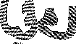
Figure 2.
Bone stump after amputation by the subperiosteal method. rise to Bunge to propose a method completely opposite to that of Walther and Brunninghausen. Bunge's method (subperiosteal-free) consists in exposing the end of the sawn bone from the periosteum for a distance of 1 cm, and the bone marrow cavity for the same distance - from the bone marrow. The soft tissues are directly applied to the exposed bone saw. This method is now widely used: it also does not prevent the formation of osteophytes, yet these osteophytes, originating from the periosteum, severed above the bone saw, are not in the plane of the operative scar but above it (see Figure 3). Sometimes with this method, sequestration of the bone saw in the form of a bony crown within the bone stump, which is deprived of periosteum and bone marrow by this method, is also possible. Bone-plastic method of amputation. Its essence is clear from the name itself. This method was introduced into amputation technique by N. I. Pirogov in 1852. His bone-plastic amputation of the shin served as the prototype for many other operations of the same type, for example, the operations of Gritti, v. m. e. t. I.
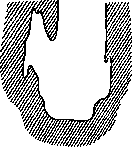
Sabanev, Delitsin, Abrazhanov, Shimanovsky, Levshin-Spasokukotsky, Tauber, Mikulich-Vladimirov, etc. The main idea of Pirogov was the desire to create a painless stump with a strong supporting surface. The richest clinical experience fully confirmed the validity of Pirogov's brilliant idea; its further development must be recognized as the proposal made by Bier (1892) to apply the bone-plastic method of amputation on the diaphyses of long bones Figure 3. Bone stump in their diaphyseal part
after amputation according to Bunge. To cover the sawn surface of the bone with a thin bone plate, connected to the maternal soil only by means of the periosteum. However, Pirogov's idea could not yet be considered as finally implemented by Vir's operation, and the path of its development was far from completed. -In 1912, the idea of free bone plasty in A. was first implemented in the clinic of A. V. Martynov (Sitkovsky). At present, this method is being applied by many authors (Bogoraz, Lukyanov, Albrecht, etc.), and for covering the bone saw, a bone plate covered with periosteum is taken either from the removable peripheral part of the limb (Martynov) or (in amputation of the thigh) the removed patella is used, from which a nail with a head (Vogoraz) or a "champagne cork" (Albrecht) is sawn out, inserted into the bone marrow cavity of the amputated bone. This method, like plasty on a pedicle (Pirogov, Bier), gives us the possibility to immediately close the bone marrow cavity of the stump with bone, covered by tissues in their natural combination, thanks to which the period of stump formation is significantly shortened. Unfortunately, the described method is limited in its application, since it requires, like any plastic operation, the most careful asepsis and good technique. And even with this method, the development of osteophytes is possible, usually originating both from the transplanted plate and from its periosteum (see Figure 4). Fascial plasty. Similar to free bone plasty, free fascial plasty (Ritter) was also introduced into the technique of amputation. This method in recent years has undergone experimental and clinical verification (Lukyanov), and it turned out: 1) due to its rapid fusion with the bone saw, the transplanted fascial flap protects the bone and its contents from ascending infection from the soft tissues of the stump; 2) the process of formation of the bone plate closing the bone marrow cavity in A. with fascial plasty is significantly accelerated, which in shortens the period of stump formation; 3) in terms of technical ease of execution, this method differs little from the methods without periosteum and sub-periosteum (see Figure 5).
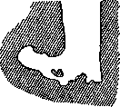
Figure 4. Bone stump after amputation with free bone plate (after 9 months).
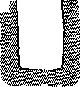
mascial plasty. Similar to free bone plasty, free fascial plasty (Ritter) was also introduced into the technique of amputation. This method in recent years has undergone experimental and clinical verification (Lukyanov), and it turned out: 1) due to its rapid fusion with the bone saw, the transplanted fascial flap protects the bone and its contents from ascending infection from the soft tissues of the stump; 2) the process of formation of the bone plate closing the bone marrow cavity in A. with fascial plasty is significantly accelerated, which in shortens the period of stump formation; 3) in terms of technical ease of execution, this method differs little from the methods without periosteum and sub-periosteum (see Figure 5).
Quite apart from all the methods of A. described above stands the method of Hirsch. This is, strictly speaking, not a method of A., but a method of functional education of the amputation stump, consisting of training it, after which the stump becomes resistant to all kinds of irritants (mechanical, thermal, painful). Hirsch's method should not be opposed to other methods, but should be an essential addition to any other method of amputation. The desire of surgeons to restore the dynamic properties of an amputated upper limb led to its kinematization. The beginning of kinematization was laid by the operation of Vanghetti (Vanghetti, 1898) on the forearm; he arranged on the anterior surface of the forearm stump a skin-muscle loop, to which the mechanism of an artificial hand was later attached. The idea of kinematization of the stump received its further development in the proposals of Paier (Paug), Vreden, Sauerbruch, Albrecht and others. When describing the technique of A., one cannot pass over in silence the toilet of the operative wound in relation to muscles and nerves. The importance of these tissues, especially muscular tissue, for the subsequent function of the amputation stump has been recognized since ancient times. All proposals aimed at combating the results of inevitable muscle retraction can be reduced to numerous variations of the excision and trimming of muscle tissue (Alanson, Dupuytren, Deso, Pirogov) of one size or another, capable of more than covering the bone saw cut, in order to prevent retraction to the extent that usually leads to the formation of a conical stump. To achieve this purpose, the old authors worked out rules indicating where, at what level, and of what size the excess muscle tissue should be taken. - In connection with the teaching that the life of muscle tissue lies in its activity, constant contractions, which are sharply and long disrupted in A., until new points of attachment are formed for the severed muscles, Kocher made the following conclusion: 'Prevention of muscle atrophy and at the same time of a conical stump can be achieved by arranging new points of attachment for the muscles at the moment of the operation itself, and, if they do not find a place on the bone, then it is best to seek them on the antagonists, sewing the muscles in front of the bone stump.' - As for the severed nerves, the following is done: some authors (Mosetig-Moorhof) sever the strongly stretched nerve 1-2 cm above the plane of section of the soft tissues and bone, whereby the severed nerve, having contracted, sinks into the surrounding soft tissues above the future operative scar. Other authors (Ritter) cut a wedge from the severed nerve, with the base turned downward, after which the split ends of the nerve are sutured together. Bardenheuer proposed to sew the end of the nerve in a bent loop to the freshened surface of the same nerve trunk. Cushing sutures the severed nerves end to end, while Kriiger, before severing the nerves, applies crushing to them. Evaluation of different methods of A. According to the method of sectioning soft tissues, A. are distinguished as circular, flap, and oval. From the point of view of modern requirements imposed on the amputation stump, the question of the form of section of soft tissues has lost its acuteness. All authors agree that on different limbs and at different levels of their amputation, it is necessary to resort to different types of section of soft tissues, guided only by the consideration that the future operative scar should not be located on the working surface of the stump (correspondingly, the skin incision). Since such a working surface for the lower limb will be the inferior one, in case of its direct support on the bottom of the prosthesis, and the anterior one, bearing the weight of the prosthesis when it is brought forward, it is desirable to locate the operative scar on the posterior surface of the stump. This possibility is provided by the flap method of tissue section with a large anterior flap. Here the flap method is preferable to all others. In contrast to this, the working surface for the stump of the upper limb is mainly the lateral surfaces and least of all the inferior surface, which is why it will be correct to locate the operative scar in amputation of the upper limb on the inferior surface of the stump, i.e., here the circular method will be more convenient. Some authors generally consider that the normal method of A. should be the flap method in its various (depending on the circumstances) forms, as giving the greatest possibility of application to each individual case. As for the circular and oval methods, they, in their opinion, should be used only for special indications. - According to the processing of the bone sawn through in A., the methods of section are divided into subperiosteal, non-periosteal, and plastic (with bone or fascial, on a pedicle or free plastic). In the evaluation of different methods of A., from this point of view, the greatest disagreement is observed among surgeons. But still, the greatest unanimity among them is noticeable in the denial of the advantages of the subperiosteal method of Walter and Brunninghausen. More favorable are the reviews of authors regarding the non-periosteal treatment of the stump according to Bunge, although this method is by no means flawless. Hofstatter, in his 39 cases of A. according to Bunge, found only in two cases no sequestra or proliferations, although the latter did not spread to the supporting surface of the stump. As for the bone-plastic methods, while giving an undeniable advantage in their final results, they are not generally accessible, as they are somewhat complex (especially bone-plastic A. of the Pirogov and Bier type) and require perfect asepsis, which, under the conditions of the disease requiring removal of the limb, cannot always be guaranteed without losing at the same time in the length of the stump lever - a fact of extraordinary importance for subsequent prosthetics. Amputation stump. Turning to the consideration of the question of the modern requirements that an amputation stump must satisfy, the latter can be formulated, according to the opinion of most authors, as follows: 1) the stump must be painless both in a state of stress and in a free state; 2) the stump must have a resistant bone stump, smooth, without bone proliferations; 3) the stump must be covered with good, not prone to irritation soft tissues; 4) the stump must be of sufficient length so that a prosthesis can be well adapted to it. - In contrast to this there are: 1) conical stumps in their geometric form, which are either the result of a technically unsatisfactory operation or the result of subsequent muscle retraction, both primary and secondary, or atrophy and scarring of soft tissues, and 2) conical stumps in their function, when the skin cover of the stump, of whatever form, is tense, sensitive, swollen or ulcerated. However, it should be noted that authors of recent times have rather sharply diverged in their views on the necessity of striving to obtain a 'resistant' stump for direct loading of it. If Bier, Bunge and other, mainly German, surgeons taught and teach that for a stump to have good functional qualities, certain anatomical conditions are necessary, without which a resistant stump is unthinkable, then a whole series of surgeons, predominantly French, denied not only the importance of this fact but also the very necessity of end support of the stump. Gauzele even considers the teaching of the resistant stump as a phenomenon with which it is necessary to fight. According to these authors, the most important thing to keep in mind in A. is the length of the stump, i.e., the bone lever, which, increasing in arithmetic progression, entails an increase in the function of the stump in geometric progression (Martin). - Despite the advantages of modern methods of wound treatment and the perfection of amputation technique, the long-term results of A. still leave much to be desired. According to Janssen, of the hundreds of amputees he examined, only 17% could be fitted with a prosthesis. In Kelliker's cases, 81% of the stumps were unserviceable. The Russian material does not differ from the one given: by Kolesnichenko, 87.8% of defective stumps of the lower limbs; by Stepanov, 93% for the leg and 80% for the thigh; by Lukyanov, 84.6%. This phenomenon depends on the unfavorable conditions in which surgeons are forced to resort to A. Here are meant traumatic causes, especially in wartime, where it is impossible to take into account the resistance of individual damaged tissues, the shock to which they were subjected, the possible degree of their quantitative and qualitative infection, and the individual characteristics of each patient. On the other hand, one cannot but note that 'there is no other operation to which surgeons would relate so undeservedly externally and superficially' (Bogoraz), considering it technically simple, a 'student' operation.'
And yet the significance of amputation is far from being studied and evaluated from the standpoint of determining the nature and degree of trauma inflicted on the organism as a whole and on the amputated limb in particular, where the operation changes the conditions of life for numerous tissues of different functions and different vital importance. In addition to the remote results directly affecting the life and function of the amputated limb, after A. a whole series of secondary changes develops, depending, mainly, on the change in the statics of the amputated limb. Thus, due to the disruption of muscular equilibrium on the side of the amputated limb, deformities of the chest, curvatures of the spine, as well as deformities of the shoulder or pelvic girdle develop, depending on whether the upper or lower limb was subjected to amputation. The degree of this deformity depends on the level of amputation, as well as on the time of prosthetics: the higher the limb is amputated, the more pronounced the deformity, and vice versa. Changes on the part of the shoulder or pelvic girdle consist in the fact that on the side of amputation, the corresponding half of the girdle stands higher than the other half - the healthy one. In the spine, a lateral, double, S-shaped curvature usually occurs: first in its upper part in the direction opposite to the amputation, and then in the lower part, compensatorily, on the side of the amputation. In this case, the later the amputated person began wearing a prosthesis and the more he used crutches, the more pronounced this curvature of the spine is. As a later complication after unilateral amputation of the lower limb, flattening of the arch of the foot on the other side occurs. The timing of amputation. The question of the timing of amputation in the presence of traumatic indications has currently lost its acuteness. The former desire to perform so-called primary amputation (within the first 24 hours after injury) among surgeons armed with modern methods of wound treatment has disappeared. In this respect, modern surgeons have much more freedom in deciding this issue compared to their predecessors. Nevertheless, cases requiring immediate amputation are not uncommon. These are the cases about which Chassaignac said: 'Before asking how he will walk, one must know if he will walk at all.' In such cases, there can be no question of choosing the method of amputation; only the method of necessity is applicable, the characteristic feature of which should be the speed and simplicity of surgical manipulations. Here the operation of amputation of the 'en saucisson' type (Pauchet) is appropriate in order to subsequently perform one or another of its corrections (re-amputation). Choice of amputation site. The question of the site of amputation is acquiring increasing importance. In the pre-antiseptic era, statistics on amputation showed that the higher the amputation was performed, the greater the danger of losing the patient. Depending on this, a rather uniform requirement was developed, according to which the surgeon spared every extra centimeter of the amputated limb. This rule of the old surgeons has retained its force to the present time with only the qualification that it was given a more modern content: the surgeon sought to preserve the greatest possible length of the amputated limb, keeping in mind that thereby he obtains a longer, and therefore more powerful, lever for controlling the artificial limb; further, he believed that the longer the lever, the closer the amputated person is to the ground, the more stable and confident he will be when walking (in amputation of the lower limb). However, this basic requirement, especially as a result of the experience of the world war, underwent a significant revision. This rule proved not to be valid for all cases of amputation. In connection with this, a proposal appeared to divide the limbs, from the point of view of subsequent prosthetics, into areas of greater and lesser value (Radike, zur Verth). Zur Verth distinguishes in each limb: a) valuable, b) unimportant, and c) hindering prosthetics. What is valuable must be preserved at all costs. Thus, on the thigh, the most valuable part is the thigh up to the border of its middle and lower thirds. The lower segment of the thigh in the area of the condyles (about 6 cm) and the upper segment of the tibia of the same length are places that hinder prosthetics. If it is impossible to have a stump of the tibia of less than 10 cm, it is better to perform the Gritti operation. The most valuable part of the tibia is considered to be its length up to the middle of its extent. Downward from the middle, the tibia can be divided into three zones with gradually decreasing value downward. The last 6 cm, as on the thigh, hinder prosthetics. The thigh should not be amputated higher than 8 cm from the joint edge; the shoulder - not shorter than 15 cm from the acromion, and the forearm - not below the border between its middle and lower third. On the other hand, when removing fingers of the hands, it is necessary to cherish every millimeter. When choosing the site of amputation, it is sometimes necessary to take into account the age of the patient, his profession, social status, and even cosmetic requirements, since modern prosthetics can to a large extent satisfy both functional and cosmetic requirements (Albrecht). The operation and subsequent construction of artificial limbs for heavy physical labor and light functional use can be different. For example, partial amputations posterior to the Scholaresque line for a person who makes maximum cosmetic demands should be replaced by bone-plastic amputation in the lower third of the tibia with subsequent prosthetics with a light prosthesis on the tibia, with support on the end of the stump, etc.
Related articles
Mentioned in
Cite this page
“Amputation.” Soviet Medical Encyclopedia. English translation of Bolshaya Meditsinskaya Entsiklopediya, 1st ed. (Moscow, 1928–1936), ed. N. A. Semashko. https://sovietmedicalencyclopedia.pages.dev/article/amputation/