Hysterectomy
Historical document, translated for reference. It reflects medical knowledge of the 1920s–30s and is not medical advice.
Summary
Hysterectomy is the surgical removal of the uterus for various conditions such as fibromyoma, uterine cancer, inflammation of the uterus and appendages, uterine prolapse, and metropathy. The operation can be performed abdominally or vaginally, with various techniques described for complete or partial removal.
Encyclopedia article (1928–1936)
HYSTERECTOMY, removal of the uterus for various diseases, such as: fibromyoma, cancer of the uterus, inflammation of the uterus, ovaries and tubes, prolapse of the uterus, metropathy, etc. (for details see respective entries). H. is performed by abdominal or vaginal route and consists of removal of the entire uterus or only its body (supravaginal amputation). When indicated, the ovaries and tubes are removed along with the uterus. There are several methods of complete H. per laparotomiam. According to Doyen, after opening the abdominal cavity, the uterus is pulled toward the symphysis, and a longitudinal incision is made between the uterosacral ligaments in the area of the cervix. After opening the vagina, the cervix is grasped with Muse forceps and pulled forward; then the vaginal vaults are cut with curved scissors. After cutting the anterior vault, the bladder is pushed away bluntly (see Figure 1). The uterine arteries are grasped with Kocher forceps and ligated; the vesicouterine fold of peritoneum is opened; the uterus remains attached only to the broad ligaments, which are cut along their edge. The peritoneum of the bladder is sutured to the anterior wall of the vagina, and the posterior peritoneum to the peritoneum of the Douglas space. Node sutures or continuous suture are used for peritonization (see Figure 2). Doyen's method is applicable only when the uterus is quite mobile (free access to the posterior vault). In cases where these conditions are not present, the anterior vaginal vault is opened to perform H. After cutting the broad ligaments, the peritoneum is incised at its junction with the bladder, which is pushed downward bluntly (see Figure 3). When the vagina is exposed, its anterior wall is grasped with strong forceps and cut with scissors. The cervix of the uterus, grasped with Muse forceps, is pulled forward; then the vaginal vaults, uterosacral ligaments, and the peritoneum of the posterior Douglas space are cut with scissors. Some authors preliminarily strip the visceral leaf of the peritoneum above the attachment of the uterosacral ligaments. The uterine arteries can be grasped and ligated when cutting the broad ligaments and separating the bladder. Others prefer to do this after separating the vaginal vaults from the uterus. Hemostasis is not always easily achieved, and it is often necessary to ligate individual branches of art. uterinae. This should be done carefully to avoid injuring the ureter. Kelly, in cases of an immobile uterus, prefers, when cutting the broad ligament from the uterus, to descend to the lateral vault and begin removal of the uterus from there (see Figure 4). When the uterus is immobile and the appendages are surrounded by adhesions, Faure proposes to cut the entire uterus along the median line (hysterectomie totale par hemisection). After pushing the bladder downward, the uterus is cut along the median line with strong scissors, starting from its fundus down to the vagina, which is also opened. Grasping one half of the uterus, it is pulled out and separated from the vagina (see Figure 5); the art. uterina, which tenses at this moment, is grasped with Kocher forceps and ligated, then the broad ligament with the appendage is cut across. After removing the other half of the uterus in this way, the operation is completed in the usual manner. In cancer of the uterus, when the uterus and its appendages as well as the surrounding cellular tissue are removed, H. is performed by the so-called extended method (see Cancer, Uterus). Supravaginal amputation of the uterus per laparotomiam consists of cutting the broad ligaments down to the area of the internal os, which is done while following the edge of the uterus. The peritoneum of the anterior Douglas is incised; ligation of the ascending branch of the uterine artery. The cervix of the uterus is cut out wedge-shaped, with the apex of the wedge directed toward the lumen of the cervical canal, which is smeared with Tinctura Jodi; 2-3 node sutures of catgut are placed at the incision site, the peritoneum of the bladder is attached to the edge of the posterior surface of the cervical stump. After placing sutures on the vessels of the broad ligament, peritonization is performed. The performance of supravaginal amputation is facilitated by removal of the uterine appendages (see Figure 6). Initially, around 1863, extraperitoneal care of the stump was used, with the cervical stump being ligated with iron wire. Subsequently, a rubber tourniquet was placed on the stump, and with the help of two cross-inserted needles, it was kept outside the abdominal cavity, which was closed with sutures in the rest of its extent. After 4-5 weeks, necrotic tissue was rejected. Intraperitoneal care of the stump began after Schroeder's proposal to preliminarily incise the cervix of the uterus along the median line.
Figure 1. Hysterectomy according to Doyen: the cervix of the uterus has been cut from the vaginal vaults, the bladder has been separated from the uterus; only the broad ligament and the vesicouterine fold of peritoneum remain to be cut.
Figure 2. Suturing of the vaginal walls and peritoneum after hysterectomy.
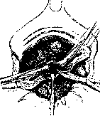
Figure 3. The uterus is pulled upward, the broad ligaments are cut, the vesicouterine fold of peritoneum is incised, the bladder is pushed downward, the uterine arteries are cut and ligated.
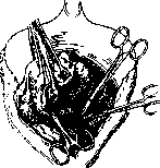
Figure 4. Hysterectomy according to Kelly's method: after cutting the broad ligament, the vagina is opened through its lateral vault.
The apex of the wedge should be directed toward the lumen of the cervical canal, which is smeared with Tinctura Jodi; 2-3 node sutures of catgut are placed at the incision site, the peritoneum of the bladder is attached to the edge of the posterior surface of the cervical stump. After placing sutures on the vessels of the broad ligament, peritonization is performed. The performance of supravaginal amputation is facilitated by removal of the uterine appendages (see Figure 6). Initially, around 1863, extraperitoneal care of the stump was used, with the cervical stump being ligated with iron wire. Subsequently, a rubber tourniquet was placed on the stump, and with the help of two cross-inserted needles, it was kept outside the abdominal cavity, which was closed with sutures in the rest of its extent. After 4-5 weeks, necrotic tissue was rejected. Intraperitoneal care of the stump began after Schroeder's proposal to preliminarily incise the cervix of the uterus along the median line.
Faure's method (hysterectomy by amputation of the uterus body par hemisection). On the uterus body, a rubber tourniquet is applied, which is removed after ligation of the vessels. The proposal to cut out separate flaps from the anterior and posterior surfaces of the uterus for better closure of the cervical stump in the future was abandoned. In cases of a well-mobile uterus, Faure performs supravaginal amputation in the following way: palpating through the posterior vault the external os and retreating 2-3 cm above, the cervix of the uterus is cut from front to back with scissors. After complete removal, by pulling the uterus forward, the uterine arteries are found, which are grasped with Kocher forceps. The peritoneum of the posterior surface of the broad ligament is incised on the right,
Figure 5. Hysterectomy with
Figure 6. Supravaginal amputation of the uterus with appendages. (According to Kelly.)
Figure 7. Suturing of the wound after removal of the uterus per vaginam according to Doyen's method through the posterior vault.
the fingers of the left hand enter the space formed, lift the appendages, and cut the broad ligament, if necessary with the appendages. Grasping the uterus with Muse forceps and pulling the left broad ligament, it is cut across; usual suturing of the wound surface. In inflammatory changes of the pelvic peritoneum, Faure, after opening the peritoneum of the anterior surface of the broad ligament and incising the vesicouterine fold of peritoneum, prefers to cut the cervix in the area of the internal os from front to back. Then, grasping the body of the uterus at the incision site with Muse forceps, the body is pulled upward. The uterine arteries are ligated, after which, by inserting fingers into the area of incision of the broad ligaments, the posterior leaf is protruded, which facilitates the separation of existing adhesions and fusions. In cases of significantly limited mobility of the uterus, Faure recommends making an incision of the uterus along the median line to the internal os (hemisection uterine), from here to remove each half of the uterus separately, and after ligation of the uterine artery, by strongly pulling the uterine stump, to cut the attachments of the broad ligaments, adhesions, and remove the appendages. Against supravaginal H., the possibility of development of malignant new formation in the remaining cervix is usually cited, but the possibility of this is small. Advantages of supravaginal H.: simplicity of technique, better asepsis, good hemostasis, speed of operation. Vaginal H. was developed by Doyen, Pean, Segond, Landau, and D. Ottom. After exposing the cervix of the uterus with specula, the latter is grasped with strong forceps and pulled downward and forward. The posterior lip and posterior vault are sagittally incised, which is separated from the uterus by lateral incisions. Grasping the edges of the incision and pulling the uterus, the incision is continued on the anterior surface of the uterus, the vesicouterine fold is incised, and the bladder is separated bluntly, after which the anterior vault is cut. Clamps are applied to the broad ligament, lig. ovarii propr., and round ligament, which are replaced by ligatures (see Figure 7). Elastic clamps, according to Pean, can be left and removed after 2-3 days. Another modification of Doyen: circular incision of the cervix of the uterus; the bladder is pushed upward, after which the peritoneum is opened; the uterus is cut along the median line, strong clamps are applied to the edges of the incision and the uterus is pulled downward, gradually reaching the fundus of the uterus, after which it is easy to evert the uterus, bring it down to the vulva and
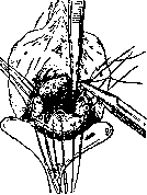
Figure 8. The uterus is prolapsed through the anterior vault; the peritoneum of the bladder is sutured to the vaginal wall; the appendages are cut and ligated; the lower part of the parametrium and the posterior attachments of the uterus are cut.
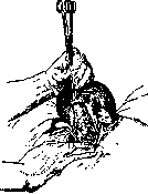
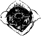
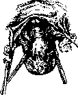
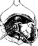
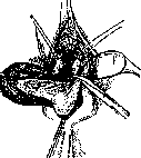
to remove, by cutting the ligaments, peritoneum, vagina, etc. H. by retroverting or anteverting the uterus in cases of significant mobility of the uterus may be performed without preliminary incision of the uterus (see figure 8). When the uterus is immobile or there are adhesions, H. is performed by the "direct descent" method. After a circular incision of the cervix, the bladder is pushed upward, clamps are applied to the paracervical tissue, which is then excised after preliminary ligation (see figure 9). As the uterus is lowered, clamps are applied, the vessels, the superior parts of the tissue, the broad and round ligaments, the proper ligament of the ovary, and the site of origin of the tubes are ligated and cut. After removal of the uterus, the peritoneum is sutured to the vaginal mucosa, to which the stumps of the appendages are also attached, after which usually a gauze tampon is introduced into the abdominal cavity and vagina. In cases of a large uterus (fibromyoma), various methods are used to reduce its size (see Uterus).- Per vaginam hysterectomy may be performed as a partial vaginal or supravaginal amputation according to Rieck: anterior colpotomy, anteversion of the uterus; the appendages and broad ligament are excised to the area of the internal os and ligated, the peritoneum of the bladder is sutured to the uterus below the area of the internal os. The body of the uterus is removed by a wedge-shaped incision at the cervix, and the wound surface is closed with 2-3 nodal sutures of catgut. Sutures on the vagina.
Related articles
Mentioned in
Cite this page
“Hysterectomy.” Soviet Medical Encyclopedia. English translation of Bolshaya Meditsinskaya Entsiklopediya, 1st ed. (Moscow, 1928–1936), ed. N. A. Semashko. https://sovietmedicalencyclopedia.pages.dev/article/hysterectomy/