Douglas Space
Historical document, translated for reference. It reflects medical knowledge of the 1920s–30s and is not medical advice.
Summary
Douglas space is the deepest part of the abdominal cavity, formed by the peritoneum between the rectum and bladder in males or the uterus in females. This article describes its anatomical relationships, variations between sexes, and pathological conditions affecting this space.
Encyclopedia article (1928–1936)
DOUGLAS SPACE (James Douglas, English anatomist, 1675-1742), the deepest part of the abdominal cavity, is formed by the peritoneal recess between the rectum and bladder in males or the uterus in females. The topographic-anatomical relationships and relief of the pelvis are simpler in males. The peritoneum of the anterior abdominal wall, descending into the pelvis, covers the posterosuperior wall of the bladder, forming a distinct transverse fold (plica transversa vesicae) when the bladder is empty. In the lateral parts of the pelvis, folds of peritoneum are visible; they extend from the internal inguinal rings backward and toward the midline and are formed by the vas deferens lying here. Descending from the superior surface of the bladder to its posterior aspect, the peritoneum covers the organs adjacent to the posterior wall of the bladder, namely the seminal vesicles, ampullae of the vas deferens, and the lower parts of the ureters. All these structures are arranged such that their lower ends converge toward the midline, consequently the space between them has the appearance of a triangle with the apex directed downward. The sides of this triangle are formed by the ampullae of the vas deferens, laterally to which are located the seminal vesicles and the terminal portions of the ureters. Covering these structures, the peritoneum of the posterosuperior surface of the bladder descends particularly low in this triangular area and only by 1½ cm does not reach the more deeply situated prostate gland. From this, the deepest part of the peritoneum, it turns backward and upward, covering the anterior surface of the rectum. The resulting peritoneum-lined recess between the bladder and rectum is called the Douglas space. Laterally it is bounded by distinct, semilunar folds of peritoneum extending from front to back, the so-called Douglas folds (plicae Douglassi), which in their base contain muscle-connective tissue bundles of the posterior bladder ligaments. In women, the relationships are more complex, as in the transverse fold of peritoneum between the bladder and rectum lie the uterus, tubes, and ovaries. When the bladder is empty in women as well, the peritoneum covering the bladder forms transverse folds. After covering the posterosuperior wall of the bladder, the peritoneum in women turns onto the anterior surface of the body of the uterus, forming on the sides the anterior layers of the broad ligaments of the uterus. The resulting space between the bladder and uterus, the vesico-uterine recess (excavatio vesico-uterina), is called by some authors the anterior Douglas space. When the uterus is normally anteverted, this space usually appears as a narrow transverse slit, somewhat widening at its lateral ends and forming depressions lateral to the lateral parts of the bladder. Under the peritoneum of the lateral wall of each recess passes the obliterated umbilical artery, and under its floor-the terminal portion of the ureter. After covering the anterior surface and fundus of the uterus, the peritoneum descends onto its posterior wall, forming laterally from the edges (margo lateralis) of the uterus the posterior layers of the broad ligaments of the uterus. Unlike the anterior wall of the uterus, posteriorly the peritoneum covers not only the entire body of the uterus but also its cervix, leaving only the vaginal portion of the uterus uncovered. Thus, after covering the entire posterior surface of the uterus, the peritoneum descends further, covering from above the posterior fornix of the vagina to a depth of approximately 3 cm. Having reached the deepest level, the peritoneum turns upward and backward, and, heading toward the rectum, covers its anterior and lateral surfaces. Thus, between the posterior surface of the uterus and the posterior layers of the broad ligaments on one side, and the peritoneum covering the posterior wall of the pelvis and mainly the anterior surface of the rectum on the other, a rather considerable space is formed, which is given the name recto-uterine space (excavatio recto-uterina) or posterior Douglas space. This space is usually divided by means of Douglas folds into two floors-upper and lower. These ligaments, as in men, begin in the lateral parts of the sacrum, embrace the rectum laterally, and in the form of semilunar folds extend forward, ending in the lateral parts of the uterine cervix (approximately at the level of the internal os). In women, these folds contain muscle-connective tissue bundles, known under the name of sacro-uterine ligaments (lig. sacro-uterina). In a narrower sense, the proper Douglas space should be considered only the lower, lying below the Douglas folds, part of the recto-uterine pouch, leaving for the upper part the general name recto-uterine space. As already stated, the anterior wall of this space is formed by the posterior layers of the broad ligaments of the uterus and the peritoneum covering the posterior surface of the uterus and the posterior fornix of the vagina. The posterior wall is formed by the peritoneum lining the posterior wall of the pelvis, formed by the sacral bone, and the peritoneum of the rectum. When the rectum is not greatly distended, depressions of the peritoneum are visible laterally from its ampulla-pararectal fossae (fossae pararectales). Under the posterior layer of peritoneum lie laterally to the rectum the hypogastric nerve plexuses, and at the border with the lateral wall-the sacral plexuses, as well as the ureters, which descend into the pelvis approximately at the level of the sacroiliac joints and at this place form slight protrusions of the peritoneum, known under the name of ureteric folds (plicae uretericae). The lateral wall of the described space is formed by the peritoneum of the lateral wall of the pelvis. On this wall, the so-called ovarian fossa (fossa ovarica) can be noted-a depression of the peritoneum in the angle between the hypogastric (a. hypogastrica) and external iliac (a. iliaca externa) arteries. As for the contents of the Douglas space, in the male pelvis this consists only of intestinal loops. The contents of the Douglas space in women are more varied. With the normal position of the organs of the genital sphere, in the lower floor of the Douglas space only intestinal loops can be present, whereas in the upper floor, in addition to loops of the small intestine, the lower portion of the S-Romani, the tubes and ovaries with their associated ligamentous apparatus are normally also present. The depth of the Douglas space varies considerably in individuals. It is particularly different in various age groups; in children it is significantly larger due to the peculiarities of the embryological development of the peritoneum in this area; in embryos (and even for some time after birth) the peritoneum descends so deeply that in males it reaches the prostate gland, and in females it reaches the floor of the pelvis. If the low position of the Douglas space peritoneum persists in adults, it may become a predisposing factor to the development of various pathological conditions, such as perineal hernias, prolapse of the rectum, and in women-special forms of prolapse of the vagina and uterus (enterocele vaginale posterior). Pathology. Various diseases of the Douglas space are essentially lesions of the peritoneum. Usually such diseases arise secondarily, as a result of the spread of the pathological process from one or another organ, the peritoneal cover of which has topographic connection with the Douglas space. In this respect, it should be noted the possibility of transition to the peritoneum of the Douglas space of malignant neoplasms from the rectum, urinary and genital organs (especially in women). Inflammatory processes in the Douglas space are particularly important, which also do not arise independently, but as a result of the spread of inflammation from adjacent organs (pericystitis, periprostatitis, perisigmoiditis, perityphlitis, posterior perimetritis, etc.). Being the deepest part of the abdominal cavity, the Douglas space can serve as a site for the accumulation of inflammatory exudates from more distant organs (e.g., in cholecystitis); depending on the nature of the primary lesion and the severity of the infection, the nature of the pathological products in the Douglas space will vary-from a relatively insignificant exudate to extensive suppurations. The clinical picture and course of the disease in these cases correspond to the primary disease, so that it cannot be a question of a lesion of the Douglas space per se. Treatment should be directed against the underlying disease (treatment of pelveoperitonitis). In the acute stage, strictly conservative treatment is required-complete rest, bed rest, position with the upper part of the body elevated, strict liquid diet, care of the intestines, ice on the abdomen, narcotic agents (morphine, pantopon), if necessary-cardiac agents, saline solution under the skin or into the rectum (drop enemas). When a clearly defined exudate is present in the Douglas space, a trial puncture (puncture) of the posterior fornix is performed. The latter is now rarely used, mainly in cases where the nature of the effusion causes doubt. In the presence and detection by puncture of a purulent exudate in the Douglas space, an opening of the posterior fornix is required. The technique of trial puncture
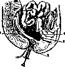
is sterilized
the usual instrumentation for vaginal manipulations (specula, bullet forceps, uterine forceps, and a syringe with a long needle); after appropriate disinfection of the external genitalia and vagina (smearing with iodine tincture), the latter is opened with specula, the posterior lip port, va-ginalis is grasped with bullet forceps and sharply retracted toward the symphysis. The posterior fornix is smeared with iodine tincture, and, retreating 1-2 cm from the vaginal part, an injection is made with a needle along the midline (it can be done through the rectum-see the figure). The piston draws the contents of the Douglas Space. The opening after needle extraction is smeared with iodine.-After healing of the primarily affected organ, persistent changes often remain in the peritoneum of the Douglas Space, which can be considered as lesions of the Douglas Space in the narrow sense. French authors drew special attention to these diseases in women, who also proposed a special term 'douglasitis.' An attempt to classify and systematize douglasitis was made by Condamin. Depending on the severity of the lesion and its topographical spread, he distinguishes three forms of douglasitis. 1. The lesion is localized only in the peritoneum and the adjacent layer of cellular tissue (hardening of these tissues without noticeable shrinkage), without affecting the correct position of the uterus, subjectively manifesting mainly as pains in the lower back. 2. The process of shrinkage and scarring progresses further, involving the posterior and lateral parts of the parametrium. The cervix is pulled backward, creating a sharp anteflexion. To the symptoms are added dysmenorrhea and a number of intestinal disorders (meteorism, dyspepsia, mucous enterocolitis); in many women, dyspareunia, frigidity, and a whole complex of psycho-sympathetic disorders develop, based on reflex influences from such powerful nerve plexuses as the hypogastric and cervical plexuses, directly affected in the process of scarring of the cellular tissue in which they are located. 3. The scarring takes on a sclerotic character and spreads to the retrovesical, retrovaginal, and pararectal cellular tissue. In addition to the described symptoms, the clinical picture shows disorders from the bladder, simulating the picture of cystitis, and from the rectum and vagina in the form of spastic states (tenesmus, vaginismus). To clarify the concept of 'douglasitis' in women, it is necessary to emphasize that its occurrence is not necessarily associated with a primary lesion of the genital organs. Appendicitis, even surgically treated (especially in women of infantile type), apparently plays a very important etiological role here. As for the diagnosis of douglasitis in women, it can be easily made on the basis of subjective complaints and data from bimanual examination. The therapy of douglasitis should consist in the use of resorbent agents, among which hot water, dry air, and mud baths, diathermy, and in women-hot vaginal water and dry air douches, hot enemas can be particularly mentioned. Condamin especially recommends tight glycerin tamponade (colunization) of the vagina and massage followed by the introduction of a pessary. With persistent treatment, a good effect can be obtained even in far-advanced cases.
Related articles
Mentioned in
Cite this page
“Douglas Space.” Soviet Medical Encyclopedia. English translation of Bolshaya Meditsinskaya Entsiklopediya, 1st ed. (Moscow, 1928–1936), ed. N. A. Semashko. https://sovietmedicalencyclopedia.pages.dev/article/douglas-space/