Rectum
Historical document, translated for reference. It reflects medical knowledge of the 1920s–30s and is not medical advice.
Summary
The rectum is the terminal section of the large intestine, extending from the sigmoid colon to the anus. This article details its anatomy, development, fascial relationships, and topography in both male and female pelvis.
Encyclopedia article (1928–1936)
590 II. Methods of examination of the anus and rectum.
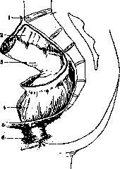
599 I. Anatomy. The rectum (rectum) represents the terminal section of the intestine; it begins at the place where the meso-sigmoid ends on the sacrum and the intestine is fixed to the posterior wall of the pelvis by means of connective tissue. The lower boundary of the rectum is the anal opening. Some authors describe under the name of rectum the segment located below the left sacroiliac joint, and distinguish the intraperitoneal part of the rectum, which has a mesentery (mesorectum), and the extraperitoneal terminal section. In relation to the pelvic bones, the upper boundary of the rectum projects onto the sacrum. Development. Initially in the embryo, the terminal intestine together with the openings of the allantois and Wolffian duct opens into the cloaca. Subsequently, the bladder and sinus urogenitalis develop from the allantois, which in higher animals is completely separated from the posterior anal part of the cloaca. By means of an outpouching of the ectoderm going toward the cloaca, the anal membrane is formed. The latter subsequently ruptures and in this way the anal opening is formed. The rectum in the embryo after the third month lies along the median line and has a complete mesentery. At the fifth month of intrauterine life, after the rotation of the intestine and the formation of the sigmoid flexure, the rectum deviates somewhat to the left. General data, peritoneum, fasciae. The length of the rectum in adults varies on average from 12 to 15 cm, with 10-12 cm falling on the pelvic and 2-3 cm on the perineal sections. The circumference of the pelvic section of the rectum with moderate filling is 8-16 cm, with excessive stretching - 30-40 cm. The circumference of the perineal section is 5-9 cm. The peritoneum covers the rectum on its sides and in front in its upper sections, and only in front in its lower sections. The terminal section of the rectum is completely not covered by peritoneum, which in men passes forward onto the posterior surface of the bladder and upper sections of the seminal vesicles, forming the Douglas space (fig. 1), and on the sides onto the walls of the pelvis, where also bulges of the peritoneum are located, situated on the sides of the rectum and bearing the name fossae retropelvien-nes (Jonnesco). In women, the peritoneum from the anterior wall of the rectum passes onto the uterus, between which a blind peritoneal sac is formed, called excavatio recto-uterina or 'posterior Douglas' (figure 2). The bottom of this sac descends to the level of the posterior fornix of the vagina. It is located at an average distance of 5-8 cm from the sinus. In the lower sections, the anterior wall of the rectum, not covered by peritoneum, adjoins the posterior wall of the vagina and is separated from the latter by a connective tissue layer called septum recto-vaginale. This layer thickens in the lower section and on a longitudinal section has the appearance of a triangle, whose base is directed toward the perineum, and the apex toward the upper section pars analis recti. This triangle is called trigonum recto-vaginale. The part of the rectum not covered by peritoneum is clothed in fascia (fascia recti), which is a process of the pelvic fascia and is particularly clearly expressed and dense behind. Between the fascial covering and the wall of the rectum there is loose connective tissue, thanks to which the rectum can be fairly freely isolated from its fascial sheath. In front of it, f. recti fuses with the fascia covering the seminal vesicles and prostate gland; this fascia is also a process of fascia pelvis and on the sides and below connects with processes of its parietal sheet. The posterior sections of the rectal fascia are dense and have the appearance of a fibrous sheet. In front of that part of the rectum which is not covered by peritoneum lies a ligament called aponeurosis Denonvillier or lig. peritonaeo-perinaeale. It is formed as a result of the fusion of the leaves of the primary Douglas sac. The fascial capsule in which the rectum lies, surrounded by connective tissue, is called capsula Amussati. Three sections of the rectum are distinguished - pars supraampullaris, pars ampullaris and pars analis. The first section is short, has the appearance of a cylindrical tube and represents a continuation of the sigmoid colon; the second is the largest and lies above the pelvic diaphragm in the pelvic cavity (cavum pelvis proprium), the third is located in the perineal area (cavum pelvis subcutaneum). The shape of the rectum is very variable depending on filling, the nature of the contents, etc. Two types of rectum are distinguished - ampullary and cylindrical. In the first case, the intestine is thin and wide, has the appearance of an ampulla and its axis is inclined toward the sacrum, in the second - it is long, narrow and has the appearance of a cylinder. The first type is more often observed in asthenics, the second in hypersthenics with a symphysopetal type of pelvic topography (Moskalenko). The rectum forms a series of curvatures (figure 3) in the sagittal and frontal directions. Two sagittal curvatures are distinguished: one is convex backward and corresponds to the concavity of the sacrum, which is why it bears the name flexura sacralis, the other is convex forward and curves around the apex of the sacrum. This bend is called flexura analis. There are several frontal curvatures; they are not constant and to a large extent depend on the degree of filling of the intestine and the nature of its contents. Usually two frontal curvatures are observed, located on the right side, with the upper one lying at the level of the promontorium, and the lower one somewhat higher than the anus.
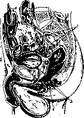
Figure 1. Median section of the male pelvis: 1-intestines; 2-vesicula seminalis; 3-prostata; 4-anus. (After Corning.)
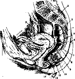
Figure 2. Median section of the female pelvis: 1-peritonaeum parietale; 2-uterus; 3-plica recto-uterina; 4-m. recto-uterinus; 5-fornix vaginae; 6-cavum Douglasi; 7-os coccygis; 8-m. recto-coccygeus; 9-rectum; 10-labium posterius; 11-orificium ext. uteri; 12-labium anterius; 13-anus. (After Spalteholz.)
connective tissue, thanks to which the rectum can be fairly freely isolated from its fascial sheath. In front of it, f. recti fuses with the fascia covering the seminal vesicles and prostate gland; this fascia is also a process of fascia pelvis and on the sides and below connects with processes of its parietal sheet. The posterior sections of the rectal fascia are dense and have the appearance of a fibrous sheet. In front of that part of the rectum which is not covered by peritoneum lies a ligament called aponeurosis Denonvillier or lig. peritonaeo-perinaeale. It is formed as a result of the fusion of the leaves of the primary Douglas sac. The fascial capsule in which the rectum lies, surrounded by connective tissue, is called capsula Amussati. Three sections of the rectum are distinguished - pars supraampullaris, pars ampullaris and pars analis. The first section is short, has the appearance of a cylindrical tube and represents a continuation of the sigmoid colon; the second is the largest and lies above the pelvic diaphragm in the pelvic cavity (cavum pelvis proprium), the third is located in the perineal area (cavum pelvis subcutaneum). The shape of the rectum is very variable depending on filling, the nature of the contents, etc. Two types of rectum are distinguished - ampullary and cylindrical. In the first case, the intestine is thin and wide, has the appearance of an ampulla and its axis is inclined toward the sacrum, in the second - it is long, narrow and has the appearance of a cylinder. The first type is more often observed in asthenics, the second in hypersthenics with a symphysopetal type of pelvic topography (Moskalenko). The rectum forms a series of curvatures (figure 3) in the sagittal and frontal directions. Two sagittal curvatures are distinguished: one is convex backward and corresponds to the concavity of the sacrum, which is why it bears the name flexura sacralis, the other is convex forward and curves around the apex of the sacrum. This bend is called flexura analis. There are several frontal curvatures; they are not constant and to a large extent depend on the degree of filling of the intestine and the nature of its contents. Usually two frontal curvatures are observed, located on the right side, with the upper one lying at the level of the promontorium, and the lower one somewhat higher than the anus.
8 7
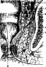
Figure 4. Fossa ischio-rectalis: 1-vas deferens; 2-vesicula seminalis; 3-os ilii; 4-m. levator ani; 5-m. obturator int.; 6-subcutaneous tissue; 7-lower hemorrhoidal vessels; 8-m. sphincter ani ext.; 9-ampulla recti. (After Testut.)
In front of the rectum lie in men the bladder, seminal vesicles (figures 4 and 5), deferent ducts and prostate (separated from it by Denonvillier's aponeurosis), and in women - the uterus and vagina. In the upper sections, as mentioned above, the rectum, covered in front by peritoneum, protrudes into the peritoneal cavity, resp. into the 'Douglas cavity' (posterior in women). Behind the rectum are the sacrum and coccyx (fig. 6), the initial parts of m. piriformis and the anterior roots of the sacral plexus, covered with fascia. The space between the sacrum and the rectum - spatium retrorectale - is filled with loose connective tissue. In it pass the a. and v. sacralis media and the trunks of the sympathetic nerve. This space above passes into the connective tissue located between the leaves of the mesosigmoid, on the sides it is limited by the place where the fascia recti passes onto the pelvic fascia, and below by the pelvic floor. On the sides of the rectum in the pelvic cavity, intestines (left and right), sigmoid colon (left), cecum and vermiform appendix (right) can be located (separated by the peritoneal sac). In addition, under the peritoneum in the fossae, which are formed at the place where the peritoneum passes from the lateral walls of the pelvis onto the rectum, the ureters and branches of the a. and v. hypogastricae pass. Structure. The wall of the rectum is smooth on the outside and has neither the clearly expressed taeniae nor the haustra characteristic of the large intestine; however, noticeable constrictions on the outside, especially the two or three lower ones, resemble haustra, and small thickenings of the longitudinal muscle on the anterior and posterior surfaces of the rectum resemble the taenia libera of the sigmoid colon and taenia mesocolica. These accumulations of longitudinal muscle of the rectum are called taeniae recti. In the region of pars analis, the taeniae interlace with the fibers of m. levator ani. The mucous membrane in the contracted state of the rectum forms a series of folds - longitudinal and transverse. The longitudinal folds are due to a conglomerate of venous plexus; they bear
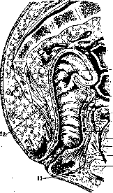
Figure 5. Rectal bed: 1-peritonaeum; 2-rectum; 3-aponeurosis prostato-peritonaealis; 4-bladder; 5-vesicula seminalis; 6-prerectal space; 7-prostata; 8-urethra; 9-fibromuscular part of the perineum; 10-bulbus urethrae; 11-m. sphincter ani; 12-m. levator ani. (After Testut.)
and the trunks of the sympathetic nerve. This space above passes into the connective tissue located between the leaves of the mesosigmoid, on the sides it is limited by the place where the fascia recti passes onto the pelvic fascia, and below by the pelvic floor. On the sides of the rectum in the pelvic cavity, intestines (left and right), sigmoid colon (left), cecum and vermiform appendix (right) can be located (separated by the peritoneal sac). In addition, under the peritoneum in the fossae, which are formed at the place where the peritoneum passes from the lateral walls of the pelvis onto the rectum, the ureters and branches of the a. and v. hypogastricae pass. Structure. The wall of the rectum is smooth on the outside and has neither the clearly expressed taeniae nor the haustra characteristic of the large intestine; however, noticeable constrictions on the outside, especially the two or three lower ones, resemble haustra, and small thickenings of the longitudinal muscle on the anterior and posterior surfaces of the rectum resemble the taenia libera of the sigmoid colon and taenia mesocolica. These accumulations of longitudinal muscle of the rectum are called taeniae recti. In the region of pars analis, the taeniae interlace with the fibers of m. levator ani. The mucous membrane in the contracted state of the rectum forms a series of folds - longitudinal and transverse. The longitudinal folds are due to a conglomerate of venous plexus; they bear
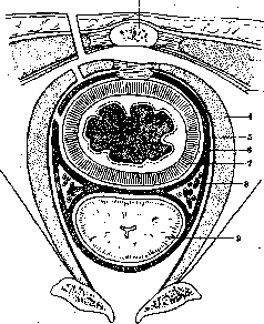
Figure 6. Schematic section at the level of the coccyx and symphysis: 1-coccyx; 2-m. glutaeus max.; 3-lig. sacro-tuberosum; 4-m. levator ani; 5-fascia diaphragmatica pelvis sup.; 6-fascia visceralis recti; 7-rectum; 8-plexus vesico-prostaticus and vas deferens; 9-prostata.
the name plicae longitudinales recti. They are most developed in the upper sections of the rectum and disappear in the lower sections. The transverse folds are formed by the mucous membrane and submucosa and are called plicae transversae recti. They are located at the level of the third sacral vertebra and at the level of the ampulla of the rectum. The latter is called plica transversa terminalis or valva ani. The mucous membrane of the rectum is covered with a single-layered cylindrical epithelium, which in the lower sections, in the region of the anal canal, is replaced by stratified squamous epithelium. The submucosa contains a rich venous plexus, which in the lower sections forms the hemorrhoidal plexus. The muscular coat of the rectum consists of an outer longitudinal and an inner circular layer. The circular layer is thickened in the lower sections and forms the m. sphincter ani internus. The longitudinal layer forms the taeniae recti. The adventitia of the rectum consists of loose connective tissue. Blood supply. The rectum is supplied with blood from the superior hemorrhoidal artery, a branch of the inferior mesenteric artery, the middle hemorrhoidal artery, a branch of the internal iliac artery, and the inferior hemorrhoidal artery, a branch of the internal pudendal artery. The veins correspond to the arteries and form the hemorrhoidal venous plexus, which communicates with the portal system through the superior hemorrhoidal veins and with the systemic system through the middle and inferior hemorrhoidal veins. Lymphatic drainage. The lymph from the rectum flows into the lymph nodes along the superior hemorrhoidal vessels, then into the aortic lymph nodes. In the lower sections, the lymph flows into the internal iliac and sacral lymph nodes. Nerves. The rectum is innervated by the pelvic plexus, formed by branches from the superior hypogastric plexus and the sacral plexus. The sensory nerves are the pelvic splanchnic nerves (nervi splanchnici pelvini).
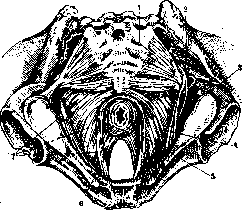
Figure 7. Muscles of the pelvis: 1-m. ischio-coccygeus; 2-m. pyramidalis; 3-m. obturator int.; 4-vessels and nerves; 5-m. sphincter ani ext.; 6-m. levator ani (deep portion); 7-m. levator ani (superficial portion). (After Testut.)
the name columnae rectales (numbering 10-12) and are separated from each other by depressions (sinus rectales). When the intestine is filled, as well as during contractions of the muscular layer, the folds change their shape and direction and can even completely disappear. The most constant are three annular folds of the mucous membrane: the middle one, bearing the name of Kohlrausch's (the largest), is located to the right of the longitudinal axis of the rectum, 6-7 cm above the anal opening. To the left of the mentioned fold there are two more. It corresponds to a depression. Some authors attribute to the annular folds the role of valves (Gaily). In the area of pars analis, the transverse musculature of the rectum, participating in the formation of annular folds, receives reinforcement from the sphincters. There are three of them: 1) m. sphincter ani ext. (fig. 7)-begins from the fibrous plate in the region of the tip of the coccyx,-goes forward and divides into 2 parts, embracing the anus, merging at the anterior edge. the latter and ending in septum perinae-ale, where they interlace with the fibers of the m. transversus perinaei prof, and- m. bulbo-cavernosus in men, and in women-with the m. constrictor cuinip
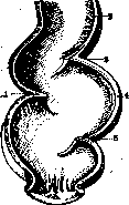
Figure 8.Valvulae recti: 1-middle-Kohlrausch's; 5-valvula inf. (According to Testut.) 2) m. sphincter ani int.-is located at the lower segment of the rectum, slightly above the external sphincter; 3) m. sphincter ani tertius-lies above the internal sphincter; first described by Nélaton (KeShup). Both m. sphincter ani int. and m. sphincter tertius represent only a thickening of the layer of circular fibers of the muscular layer of the rectum, unlike the external sphincter, which is a striated muscle. The walls of the rectum consist of three layers: 1) tunica adventitia and in places tunica serosa, 2) tunica muscularis and 3) tunica mucosa. As has already been noted, the rectum is covered by peritoneum only for half its length (approximately to the level of S iv) on the anterior surface and very insignificantly on the sides; in other places it has a connective tissue covering (adventitia). - Tunica muscularis consists of two layers: the outer longitudinal and the inner circular. The longitudinal musculature forms a thick layer around the rectum, the fibers of which in places interlace with the fibers of the circular muscles, and-in the lower part approach the cП?olKoTstЬИkИГз- m-levatoranLA few valvula sup.; 4--rectum; above the latter behind, the m. recto-coccygeus (sometimes paired) approaches the longitudinal muscular layer of the rectum, originating from the Soc-sh. In front, the longitudinal musculature of the rectum in men connects with a bundle of muscles going to the capsule of the prostate gland and to the posterior edge of the trig, urogenitci-le, and in women - to the outer longitudinal layer of muscles of the uterus (behind) (mm. recto-uterini) and to the posterior wall of the vagina. From the sides and above to the wall of the rectum in men approach mm. recto-vesi-cles-bundles of muscle fibers from the urinary bladder, and in women-muscle fibers lying in the plicae recto - utc-rinae. The circular muscle layer forms around the anal opening a thickening 1-2 cm in height, bearing the name of the internal sphincter, and somewhat higher-6-7 cm above the anus-another thickening (m. sphincter ani tertius), which corresponds to the mentioned Kohlrausch fold.-Tunica mucosa has no villi and is covered with a single-layer cylindrical epithelium with numerous goblet cells. Where the columnae rectales appear, the cylindrical epithelium gradually transitions into a multilayered flat one, in the area of the anal opening pigmented
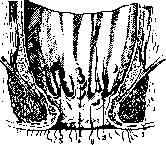
Figure 9. The anal part of the rectum: 1-skin; 2- valvula se-milunaris; 3-edge of the anus; 4- bundle of hemorrhoidal veins; 5- column of Morgagni.
The mucous membrane of the rectum forms a series of semilunar folds, which bear the name plicae transversales recti. There are usually three of them, with the middle one (plica Kohlrausch's) (fig. 8), located 6-7 cm above the anal
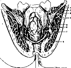
Figure 10. View of the rectum with pelvic vessels: 1-ureter dex.; 2-m.'ilio-psoas; 3-m. glutaeus med.; 4-a. hypogastrica; 5-a. glutaea sup.; 6-a. haemorrhoidalis med.; 7- n. ischiadicus; S-m. glutaeus max. (According to Corning).
opening, is especially clearly expressed (see above). The longitudinal folds present on the mucous membrane of the rectum are very inconstant and disappear when the walls of the intestine are stretched. Only in the anal part they are more constant and smaller; at their base are muscles (forming columnae rectales). The depressions between these folds are called sinus rectales Morgagni (fig. 9). The arteries of the rectum belong to four systems: 1) a. mesenterica inf., 2) a. hypogastrica, 3) a. pudenda int. and 4) a. sacra-lis media. The largest artery of the rectum is a. haemorrhoidalis sup., which is a continuation of the inferior mesenteric artery and goes along the base of mesocoli sigmoidei to the upper part of the rectum, where it divides into two branches-right and left (fig. 10- 12). The artery lies under the fascia on the tunica muscularis recti, where the branches of its right and left branches anastomose with each other, penetrating further into the tunica mucosa. Here they form a dense submucosal plexus, also anastomosing with the branches of other
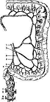
Figure 11. Vessels of the S-romanum and rectum: 1- a. mesenterica sup.; 2-a. co-lica sin.; 3-a. sig-moidea; 4 and 5-a.haemorrhoidalis sup.; 6-a. sigmoidea ima; 7-a. hypogastrica; 8-a. haemorrhoidalis med.; 9-a. pudendainf.; 10-a. haemorrhoidalis inf.
arteries of the rectum. The middle parts of the rectum (mainly the lateral and anterior walls) are supplied by two aa. haemorrhoidales mediae, which are branches of a. hypogastricae. Just like the branches of the previous artery, its branches form two plexuses-one on the muscular coat, the other in the submucous layer. With the help of its branches, the system of aa. haemorrhoidales mediae communicates with the arterial system of the prostate gland, seminal vesicles, and urinary bladder. The lower parts of the rectum are supplied by the inferior hemorrhoidal arteries. The latter are branches of a. pudendae int. and
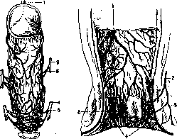
Figure 12.
Figure 13.
Fig. 12. Arteries of the rectum from the front: 1-a. haemorrhoidalis sup.; 2-a. hypogastrica; 3-a. haemorrhoidalis med.; 4-a. pudenda int.; 5-a. haemorrhoidalis inf. Figure 13. Veins of the rectum: 1-v. haemorrhoidalis sup.; 2-v. haemorrhoidalis med.; 3-colum-na rectalis; 4- vv. haemorrhoidales inf.; 5-venous node. branch in the anal area. At the top they anastomose with the plexuses of the aa. haemorrhoidales mediae. The posterior wall of the rectum receives branches from a. sacralis media. These branches, like other arteries of the rectum, participate in the formation of the muscular and submucosal plexuses. The veins (fig. 13) form around the rectum three plexuses-submucosal (plexus haemorrhoidalis int.), subfascial (pi. haemorrhoidalis ext.) and subcutaneous (pi. subcutaneus ani). Individual plexuses anastomose with each other and flow into the upper and middle hemorrhoidal veins, which correspond to the arteries of the same name. V. haemorrhoidalis sup. flows into the inferior mesenteric vein and belongs to the system v. portae. Vv. haemorrhoidales mediae flow into v. hypogastrica and belong to the system of the inferior vena cava. The subcutaneous plexus (pi. subcutaneus ani) is located in the area of the external sphincter of the anus and flows into vv. haemorrhoidales inf. (system v. pudendae int.). It should also be added that from the posterior surface of the rectum small branches depart, anastomosing with the sacral venous plexus. In addition pi. haemorrhoidalis ext. anastomoses with the veins of the bladder, prostate gland and seminal vesicles. There is a connection between the veins of the rectum and the veins of distant areas of the body. In particular, anastomoses with the lumbar, renal, renal and azygos veins are known (Vishnevsky). The lymphatic vessels of the rectum lie perivascularly and, like the veins, form three plexuses: one-the most developed-is located in the submucosal layer, another-in the muscular coat and the third-in the area of the external sphincter under the skin. Lymph from the walls of the rectum flows toward the mesenteric glands and along the middle hemorrhoidal artery to Igl. hypogastricae; only lymph from the skin part of the anus flows into the external lymphatic glands (lgl. in-gui-nales). The lymphatic vessels of the pelvic part of the rectum and the mucous membrane of the anal part flow into Igl. ano-rectales (number 5-7). These glands are located under the fascia in the lower part of p. pelvinae recti. From here the lymph flow is directed to the upper hemorrhoidal lymphatic glands, which are located on the border with S-romanum behind the rectum. The nerves of the rectum belong partly to the sympathetic system, and partly emerge from the sacral plexus. Three plexuses are distinguished: 1) pi. haemorrhoidalis sup., located in the upper part of the rectum and formed by sympathetic branches departing from pi. mesentericus inf.; these branches accompany a.haemorrhoidalis sup.; 2) рг. haemorrhoidalis medius, located on the sides of the rectum; it follows the course of a. haemorrhoidalis mediae and is formed by branches departing from pi. hypogastricus and II, III and IV sacral nerves; 3) pi. haemorrhoidalis inf., located in the anal area and formed mainly by branches of n. pudendis, which accompany the inferior hemorrhoidal artery (innervate the external sphincter of the anus and the skin in its area). The two upper plexuses of the rectum (pi. haemorrhoidalis sup. and pi. haemorrhoidalis medius) anastomose with each other. In addition, branches from the sacral parasympathetic nerve (n. pelvicus) also go to the rectum. Two types of plexuses are distinguished - wide-meshed and fine-meshed. In the latter case, there are fewer nerve ganglia. In addition, in some cases there is a predominance of sympathetic nerve branches, in others - branches from n. pelvicus.
F. Vals'er. Physiology of the rectum-see Defecation. II. Methods of examination of the anus and rectum. The main methods of examination of the anus and rectum are palpation and examination of the anus and cavity of the rectum and analysis of the shape and appearance of fecal masses (see Defecation). The most important is the physical examination of the anus and rectum, without which no patient complaining of disorders of the functions of the anus and rectum should be deprived of it. Objective examination is performed under good lighting and begins with examination of the anus. It is best to examine the patient in the knee-elbow position-the and and the anus are abundantly smeared with vaseline. The finger is inserted into the anus with slow drilling movements, stops for a moment in the area of the sphincter and then, with the patient's deep breathing, is advanced into the rectum. With the finger, the anterior, lateral and posterior walls of the rectum are systematically palpated to the accessible height and the walls of the pelvis. By this examination, the width of the ampulla, special features of its mucous membrane, its mobility, ulcerative processes, tumors, narrowings are established. In tumors, sometimes their size and extent in height and width, their mobility, displaceability can be determined. For completeness of results, it is necessary to examine patients in different positions: in the position on the side, the anterior wall is not sufficiently examined, in the knee-elbow position-the upper parts of the rectum are difficult to reach; in the squatting position with straining, high-located tumors descend and can become accessible to the finger. Bimanual examination in the gynecological position often helps the case. The value of finger examination is great, but it is limited to a height of 9-10 cm. More accurate data are obtained when examining the mucous membrane of the rectum, the lower part of which was previously examined by introducing various mirrors (see Medical mirrors), which are now almost replaced by the proctoscope (see Proctoscopy). Sometimes X-ray examination is used, but in general its role in determining diseases of the rectum is limited. III. Pathology of the rectum. Congenital malformations of the anus and rectum. are observed in the form of complete absence of the anal opening or outlet of the rectum and in the form of narrowing of the latter. Congenital absence of the anus (atresia
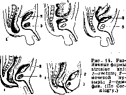
ani) or outlet of the rectum (atresia recti) (fig. 14) occurs as a result of violation of embryonic development. These malformations can be complicated by abnormal communications of the rectum with adjacent hollow organs, for example, with the urinary bladder, with the urethra in men and with the vagina in women, or by the formation of special hollow passages from the rectum, opening with openings or ending blindly on the perineum, on the scrotum, along the penis in men or at the entrance to the vagina in women. All these malformations are divided into 3 groups (Stieda). I. Atresia ani, s. recti, simplex: a) atresia ani simplex-the anal opening is absent, and the blind end of the rectum reaches the external coverings; b) atresia recti simplex-the anal opening exists and leads into a short blind canal, while the blind end of the rectum reaches the latter but does not open into it; c) atresia ani et recti-the anal opening is absent and the blind end of the rectum lies b. or m. high. II. Atresia ani, s. recti, complicata cum communicationibus: a) atresia ani et communicatio" recti cum vagina- communication between the rectum and the vagina in women-
sin; b) atresia ani et communicatio recti cum vesica urinaria-communication between the rectum and the urinary bladder in men; c) atresia ani et communicatio recti cum parte prostatica urethrae-communication between the rectum and the prostatic part of the urethra. III. Atresia ani, s. recti, complicata cum fistulis: a) atresia ani cum fistula perinaeali-a passage extending from the blind end of the rectum opens in the region of the perineal suture; b) atresia ani cum fistula scrotali-a passage extending from the rectum opens on the suture of the scrotum; c) atresia ani cum fistula suburethrali-a passage extending from the rectum opens on the suture of the penis; d) atresia ani cum fistula vestibulari-a passage extending from the rectum opens into the vestibule of the vagina. In 6,000-15,000 newborns there is 1 case of closure of the anus. The described malformations are considered as a result of a delay in embryonic development during the period of division and differentiation of the cloaca (see). According to Frank, part of these malformations represents a pure developmental delay, part of them arises secondarily due to the closure of openings that existed in the embryonic period, and part is formed as a result of a combination of these two processes. Fistula-complicated atresias are particularly controversial. Frank considers these passages and fistulas as remnants of the unobliterated Reichel's groove; Stieda considers them pathological formations. Clinic. If in atresia ani, s. recti the rectum communicates widely with the vagina or with the vestibule of the vagina, then fecal masses are excreted outward and such children can continue to live. When the rectum communicates with the urinary bladder, urine is excreted with meconium. Further entry of intestinal contents into the urinary bladder leads to the development of cystitis, from which children die. When the rectum communicates with the urethra, the intestinal contents are excreted with difficulty and the communication itself may close. With complete atresia of the anus or rectum, severe symptoms of intestinal obstruction develop shortly after birth. Meconium, due to the ingestion of bacteria, undergoes decomposition and the resulting gases strongly stretch the intestine, causing the abdomen to sharply swell and take on a spherical shape. Due to the elevation of the diaphragm, breathing is difficult and cyanosis appears. Vomiting occurs, first expelling the ingested food, and later meconium. Convulsions, hiccups, and body cooling occur; after 4-6 days, sometimes later (up to 13 days), death occurs with symptoms of collapse and peritonitis (in case of intestinal rupture). External fistulous passages (see III group) are often very narrow, sometimes ending blindly, which is why obstruction usually also develops with them. In the literature, however, cases are described in which they were sufficiently patent and children continued to live. Treatment should be undertaken before the development of meteorism and cyanosis. Statistics show that the operation on the first and second day gives less favorable results than the operation performed on the 3rd day. Operations must be preceded by precise examination and study of anatomical relationships: with complete absence of the rectum, the pelvis is small; with a low position of the blind pouch of the rectum, a fluctuation is noted in the region of the anus, and when the child cries, this area bulges and tenses; When meconium is excreted with urine, a catheter is inserted into the urinary bladder; then in vesical communication, urine will contain meconium, while in urethral communication it will be transparent. The operation is simplest in atresia ani simplexi. The membrane closing the anus may consist of a layer of epithelium, and then it can simply be torn with a finger or forceps. If the closing membrane is a connective tissue membrane, it can be incised and the edges cut around with scissors (proctotomy). With a thicker layer, to avoid scarring, it is necessary to create an opening covered with mucous membrane and skin (proctoplastica). In the lithotomy position, an incision is made along the median line from the middle of the perineum to the tip of the coccyx. One penetrates deeply to the blind end of the rectum, through the wall of which meconium is visible. The intestine is pulled out sufficiently to allow it to be brought down to the level of the skin. The wall of the intestine is fixed at the corners of the wound with silk sutures and the blind pouch is opened in the direction of the wound. The edges of the intestinal incision are sutured to the edges of the skin along the entire periphery. With a high position of the blind pouch of the rectum, it is recommended to resect the coccyx (Anders), and with a very high position, also part of the sacrum (according to Kraske), with the peritoneum being opened to facilitate the lowering of the intestine. If it is impossible to reach the rectal pouch from below, laparotomy is performed in the Trendelenburg position. The blind pouch of the rectum is exposed for a length of 7-8 cm and after the pelvic floor is split, it is lowered onto the perineum. In cases of severe condition of the child with distended gas-filled loops of intestine, an anus praeter naturalis is arranged in the left iliac region. After several months, the blind pouch drops so low that it can easily be exposed from below and sutured onto the perineum. When the rectum communicates with the urinary bladder, the urethra, and the vagina, it is necessary not only to create an anus in the normal place but also to eliminate the communication. The opening into the vagina can be left and closed secondarily, but it is better to perform the entire operation in one session. When communicating with the urethra, a probe is inserted into it, and when the blind pouch of the rectum is exposed, it is necessary to find a cord containing the fistulous passage and cut it between two ligatures, after which the pouch becomes mobile. Sometimes it is necessary to open the peritoneum. If the communication with the urethra is wide, it is sutured before lowering the rectum onto the perineum. When the rectum communicates with the urinary bladder, the operation begins with laparotomy, then the rectum is exposed and the communication with the urinary bladder is eliminated, after which the exposed pouch of intestine is lowered onto the perineum. With external fistulas, one can limit oneself to incising the fistulous passage, the mucous membrane of which is removed, and after lowering and securing the blind pouch of the rectum in the normal place, the canal from the fistulous passage is sutured. The long-term results of these operations, with smooth postoperative course, are generally favorable, since the newly created anus is able to retain feces. The outcomes of operations are given in the statistics of Curling, Cripps, and Anders, each covering 100 operated cases. Mortality in Curling (1860) was 47%, in Cripps (1882) 50%, in Anders (1893) 37%. Congenital strictures of the rectum are rare and are considered as not fully developed atresias. These are either membranous or cylindrical strictures over a greater or lesser extent. Membranous strictures usually lie in the region of the ampullary end of the sphincteric part; the strictures here are either circular with a narrow opening in the center or valve-like. The membranes are folds of mucous membrane, at the base of which hypertrophied circular muscle fibers may pass. Membranous strictures are eliminated by endorectal operations, in which the membranes are either incised or crushing instruments are applied to them. With severe cylindrical strictures, resection of the rectum may be required. Functional disorders. Anesthesia and hypesthesia of the anus and rectum are most often based on organic diseases of the spinal cord (tabes, myelitis, etc.). Due to weakening and loss of sensitivity in the lower part of the rectum, on the one hand, involuntary passage of feces may occur, and on the other hand, certain forms of coprostasis. As a result of this stasis, a feeling of pressure on the urinary bladder, heaviness in the body, loss of appetite, palpitations, general malaise, proctitis with the discharge of mucus, etc., occur. Treatment should first be directed toward restoring the feeling of the urge to defecate. Patients should every day at a certain hour visit the toilet and attempt to perform the act of defecation, which accustoms them to the sensation of the urge to defecate. Vibration massage and faradization improve the sensitivity of the rectum. The rectum is freed from fecal masses with the help of small glycerin enemas and liquid paraffin (3-4 tablespoons per day per os). Hyperesthesia and paresthesias of the anus and rectum manifest as a feeling of the urge to defecate with an empty intestine, a feeling of a foreign body in it, a feeling of distressing fullness and incomplete evacuation, sometimes burning, drilling, stabbing, knocking sensations in the rectum. In high degrees of hyperesthesia, pains radiating to the buttocks, perineum, and thighs are observed. Painful spastic conditions occur in the region of the sphincter. The source of hyperesthesia and paresthesia are organic changes in the rectum (proctitis, fissures), changes in neighboring organs (enlargement of the prostate gland, stones in the urethra, gynecological diseases, etc.), functional neuroses (hysteria). (Purely neurogenic hyperesthesias and paresthesias are established by excluding organic diseases.)- Treatment should first be directed toward correct and complete emptying of the rectum, since even with insignificant residues of feces in it, patients experience distressing urges to defecate.
Therefore, after defecation, it is recommended to wash the rectum with lukewarm water or a warm chamomile infusion. Small warm oil enemas, sitz baths, and diathermy with a rectal electrode have a beneficial effect. In case of severe sphincter spasm, manual stretching of it is beneficial. For distressing tabetic crises, epidural injections of 1-3 cm3 of a 10% solution of cocaine-adrenaline in 5-15 cm3 of physiological solution are recommended. Hypotonia and atony of the anus and rectum manifest as weakening of the tone of the sphincters and muscular coat of the ampulla. In hypotonia and atony of the sphincters, there is mainly incontinence of liquid feces, while solid feces are more or less retained. In atony, the anal opening gapes, in hypotonia it is closed, but a finger inserted into the anus either meets no resistance or meets weak resistance. Hypotonia and atony of the sphincters may temporarily occur in mental disorders (fear) and in disorders of consciousness (coma). In diseases of the spinal cord, hypotonia and atony are most often caused by processes in its sacral region (injuries, tumors, myelitis, etc.). Pathological processes in the rectum itself can also cause gaping of the anus (chronic proctitis). Loss of tone in the ampulla occurs in diseases of the spinal cord, in inflammatory peritoneal processes in the pelvis, due to its stretching by stagnant feces. Treatment of severe forms of hypotonia and atony is not successful if the main disease continues to exist. The use of various pessaries, special bandages, and careful cleanliness in the anal region can make the existence of these patients to a certain degree bearable. In atony and hypotonia of the ampulla, care must be taken to eliminate coprostasis and proctitis. Hypertension is observed on the part of the sphincters and on the part of the ampulla. In hypertonia of the sphincters, there is painful contraction of them, associated with the sensation of urge to defecate (tenesmus). Functional spasm of the sphincters is rarely observed, more often it is a symptom in erosions, fissures, inflammatory processes in the pars sphincterica and in the neck of the ampulla, sometimes in diseases of the prostate gland, female genital organs, in low-lying stones of the ureters, etc. Spasm of the ampulla usually occurs in combination with a spastic state of the sigmoid and descending colon. Treatment should be directed at eliminating the underlying disease. Against spasm of the sphincters, heat is applied both externally (warm showers, sitz baths, hot water bottles, etc.) and internally (small warm oil enemas). Sometimes stretching of the sphincters is indicated. In the presence of proctitis, Strauss recommends large sticks from Butyrum Cacao (20.0) with medicinal substances, which at the same time act as oil microclyses. Foreign bodies of the rectum penetrate into it either from the intestine when they are swallowed, or from below through the anus, into which they are intentionally inserted or accidentally fall. Smooth and small swallowed foreign bodies (coins, buttons, etc.) usually pass through the rectum and are excreted outward, while bodies with sharp spines or hooks (pieces of bone, prostheses, nails, etc.) can get stuck in it and cause various injuries. But large objects can also pass through the rectum, and cases are reported of the passage of table forks, spoons, etc. Through the anus, foreign bodies enter the rectum: 1) when falling on a protruding object with the perineum (piece or root of a tree, chair leg, etc.), 2) during masturbation, 3) are forcibly inserted by other persons in a state of intoxication or for the purpose of mischief (bottles, wine glasses), 4) are inserted by patients with the aim of causing bowel movement (stearin candles, bougies), 5) escaping enema nozzles. Foreign bodies that have entered from below are retained in the ampulla and cannot pass due to contraction of the sphincter or upward movement. Injuries to the anus and rectum occur: 1. During perineal and vaginal operations. 2. During childbirth, when the head remains in the pelvic outlet for a long time, necrosis of the wall of the rectum may occur with various consequences (phlegmons, fistulas). 3. From foreign bodies. 4. In children when breaking thermometers inserted into the rectum, as well as fragments of a porcelain vessel on which children are seated. 5. From careless insertion of hard enema nozzles. 6. Cases of injury to the rectum during proctoscopy have been described. 7. Cases of rupture of the rectum or, rather, of the higher parts of the large intestine from the entry of compressed air or gas into it have been observed; multiple ruptures have been described. They occur when, out of mischief or with malicious intent, a nozzle from an apparatus with compressed air or gas is inserted into the anus. But insertion of a nozzle into the anus is not necessary, as the air ejected under high pressure can penetrate through the anus into the rectum even from a distance of 10-20 cm and cause injury. In case of intestinal rupture, air accumulates in the abdominal cavity, in the pelvic cellular tissue, and even under the skin. Sudden pain, severe abdominal distension, subcutaneous emphysema, shock, and peritonitis are the main symptoms. The air is removed by abdominal puncture and shock is eliminated, then laparotomy is performed and the rupture of the intestine is sutured. 8. Spontaneous ruptures of the rectum are observed mainly in persons suffering from its prolapse, but cases of rupture of apparently healthy intestine have been described. In the literature, up to 20 cases of spontaneous rupture of the rectum (Reichle-Tietze) have been described. Ruptures occur during straining of the abdominal press, when lifting heavy objects, or when falling. The anterior wall of the rectum always ruptures, more often transversely, less often longitudinally, at a height of 5-7 cm from the anus (level of the peritoneal sac). The length of the rupture is 2.5-10 cm. Loops of small intestine that protrude through it and then through the anus sometimes form a huge conglomerate. When the rectum prolapses, the mechanism of its rupture is easily understood. It is more difficult to explain the rupture of a healthy intestine. According to Sauerbruch, such a rupture occurs when there is 1) strong filling of the intestine, 2) closure of its entrance and exit, 3) pressure on the abdomen. The intestine ruptures due to strong stretching. According to Bunge, with a sudden increase in intra-abdominal pressure, the anterior wall of the rectum bends and gets into the gap between the edges of thelevator ani, forming a sac. If the pressure inside the sac is higher than outside, it ruptures. This theory does not explain high-located ruptures, which exclude the possibility of the intestinal wall getting into the gap of the levator ani (Heineke). In all cases of rupture of the rectum with prolapse of loops of small intestine, laparotomy in the Trendelenburg position is required, excision of the prolapsed intestinal loops, restoration of intestinal continuity, removal of the prolapsed loops through the anus, suturing the rupture of the rectum, and closure of the abdominal cavity. 9. In fractures of the pelvic bones caused by severe trauma (run over by a vehicle, collapse of 60 ft of earth, compression by car buffers), detachments of the rectum from the anus are observed. The detached intestine must be sutured in its normal place and additional incisions must be made to create favorable conditions for the outflow of discharge. 10. Injuries to the rectum deserve special attention when falling on the perineum or anus on various long protruding objects (metal and wooden parts of gratings, parts of fences, supports for trees, stumps and roots of trees, rakes, forks, hand sticks, bayonets, cow horns, etc.). Objects penetrate into the rectum either through the anus or through adjacent parts of the body: perineum, scrotum, buttocks, vagina. Wounds often have the form of channels, but can be lacerated. They are usually contaminated, contain fragments of objects, pieces of clothing, feces, etc. Depending on the course of the injuring object or when it deviates from the cavity of the rectum, the urethra, urinary bladder, ureters, abdominal cavity, and internal organs may be damaged. The injuring objects often slip back out, but sometimes remain in the body. Madelung collected (1925) from the literature 276 such injuries to the anus and rectum. The anus served as the path for the injuring object in 166 cases. 11. Gunshot wounds of the anus and rectum are rarely observed in peacetime, but ; during war) quite often. The rectum is surrounded by pelvic bones and borders many organs, therefore gunshot wounds of it are often combined with comminution of pelvic bones, injury to the urinary bladder and urethra, injury to loops of intestine, etc. Tissues are injured not only by bullets and projectiles, but also by bone fragments, as a result of which wounds often turn out to be severely crushed and lacerated. Large blood vessels, important nerves, and muscles are often damaged, and the wounds themselves are filled with bone fragments, muscle fragments, blood clots, feces, urine. All this quickly becomes infected, decomposes, and many of these wounded die despite timely surgical assistance. The clinical picture and diagnosis of gunshot wounds of the anus and rectum differ little from the clinical picture and recognition of injuries from falling on sharp objects (see below).
It should be noted that with narrow gunshot wounds, it is more difficult to establish damage to the rectum than with large, wide, and lacerated wounds. Sometimes one can speak generally of a wound to the lower abdomen, and only during laparotomy is damage to the rectum discovered. Clinical picture. Uncomplicated injuries rarely lead to shock and collapse. Immediately following the injury, patients feel acute, cutting, and burning pain in the anus and rectum. Sometimes nausea, vomiting, urge to defecate, and tenesmus appear. When the penetrating object passes through the anus, bleeding is rare; it is more often observed with injuries through the perineum and buttocks. The sphincter is usually relaxed, but even with its damage, there may not be prolonged incontinence of feces and gases. In the perirectal cellular tissue, phlegmons and abscesses can develop. Foreign bodies remaining in the wound channels (parts of penetrating objects, pieces of clothing, etc.) delay healing. When complicated by injury to the bladder, shock and collapse are observed more often. The pain is localized in the area of the bladder, pubis, navel, and above. Sometimes there are frequent urges to urinate, sometimes urination is absent and a catheter inserted into the bladder reveals only a small amount of bloody urine, or feces and gases are excreted with the urine. When the intraperitoneal part of the bladder is injured, urine is excreted into the abdominal cavity and peritonitis develops, and when the extraperitoneal part is injured, urine infiltrates the pelvic cellular tissue, and prevesical phlegmons and abscesses may arise. Urine is excreted from the anus and from the wound channels. In some cases, the penetrating object that has entered the abdominal cavity leaves the intestine undamaged, but in the vast majority of cases, peritonitis nevertheless develops. Out of 103 cases complicated by damage to the peritoneum, peritonitis occurred in 84 cases (Madelung). Out of 103 cases, death occurred in 60 cases, and recovery in 43 (Madelung). Diagnosis. In each case, it is necessary to determine the direction and depth of the wound channel, establish which organs are damaged, whether foreign bodies remain in the wound channel or in the organs, etc. In the case of urine flowing from the rectum or from the wound channel, the injury to the bladder becomes obvious, but it remains to be determined whether the penetrating object passed through the bladder into the abdominal cavity. By examining the rectum with a probe, one can determine the location of the injury, the size of the wound, the prolapse of intestines through the wound, and injury to the bladder. A metal catheter is inserted into the bladder, with which one can establish injury to the urethra. Cystoscopy is not indicated at first, as filling the bladder may lead to the rupture of adhesions formed in the area of its wound. In difficult cases, to determine the question of damage to internal organs, laparotomy is required. Treatment. Any accidental injuries during operations, ruptures during childbirth, injuries in children (see above), etc., should be sutured immediately with careful hemostasis. For the other listed injuries and wounds, it is first necessary to take measures to prevent the spread of infection. When the injury is below thelevator ani muscle of the anus, it is often necessary to limit oneself to stopping the bleeding, cleaning the wound, and loose tamponade. For narrow gunshot wounds to the extraperitoneal part of the rectum, not complicated by injury to other organs, a wait-and-see attitude is sometimes possible. If there is bleeding from the rectum, it must be stopped without delay. To access the bleeding vessels, in some cases it is sufficient to stretch the sphincter, in others it is necessary to incise it from behind, or to expand the wound channels. For wide, lacerated wounds and wounds with foreign bodies stuck in them, the opening of niches, removal of foreign bodies, and then generally accepted surgical measures are applied. To access higher injuries to the extraperitoneal part of the rectum, posterior approaches to exposing the rectum with excision of the coccyx, one or with part of the sacrum, are recommended. When the intraperitoneal part of the rectum is injured, laparotomy is required. In cases of extensive ruptures of the rectum and crushing of surrounding tissues, it is advisable to divert fecal matter from the wound by the formation of an artificial posterior ileal anus. Treatment of rectal wounds complicated by injury to the bladder presents a difficult task. In some of these cases, a wait-and-see attitude is permissible. The task is technically facilitated when the intraperitoneal parts of the bladder and rectum are injured, which are sutured during laparotomy. When the extraperitoneal parts are injured, when the pelvic cellular tissue is infiltrated with both urine and feces, the situation is more complicated. In addition to exposing the rectum and suturing the site of its injury, it is necessary in these cases to open the prevesical space, place drains in it, open the bladder, remove any foreign bodies from it, suture the site of injury from the inside, and establish a siphon for urine drainage. In this case, the formation of an artificial posterior anus may also be required. If, despite all measures taken, rectovesical fistulas are established, they are surgically eliminated only after their healing is complete: the essence of the operations is the separation of the adherent edges of the rectum and bladder and the separate suturing of the openings in each organ. Several approaches are used to suture these fistulas: 1) the transvesical route (Kielleuthner), 2) the perineal route, in which the rectum and bladder are separated from the perineal side, 3) the parasacral route (Bisping), 4) through an incision in the posterior wall of the rectum—the so-called posterior method with excision of the coccyx (Arndt).
Fissures of the anus (fissura ani) represent an open, narrow slit about 1 cm long, lying in a longitudinal direction in the region of the anal opening or in the pars sphincterica. They are usually located in the posterior quadrant of the anus, while in the ampulla they are extremely rare. Fissures are divided into external and internal. The former are visible to the eye, especially if the folds of the anus are spread apart, while the latter can only be detected by finger examination or during endoscopy. They often develop on the basis of hemorrhoids (Quenu, Hartmann), but also from tears of the mucous membrane that occur during the passage of hard feces; fissures are known to occur on the basis of gonorrhea, tuberculosis, and syphilis of the rectum. In women, fissures occur more often than in men, and they are also observed in children, especially in the first years of life. Clinical picture. The main symptom is pain both during defecation and immediately afterward. The pain is burning and cutting, depending on the irritation of the nerve endings exposed in the base of the fissure. The pain can radiate to the genital organs, the bladder, the sacrum, and the thighs. Urine retention may occur. In severe cases, the pain can persist for whole days and nights, depriving patients of sleep. Out of fear of pain during defecation, patients try to delay stool, which becomes hardened and, upon passage, stretches and tears the fissure, preventing its healing. Fissures of the anus can serve as a site for infection entry, with subsequent development of para- and periproctitis. Diagnosis. When a fissure is suspected, the patient is examined in the knee-elbow position or in the lithotomy position, visually and by finger. If there is pain and spasm of the sphincter, it is recommended to introduce a novocaine solution. The fissure is palpated with the finger as a roughness or hardness lying deeper than the surface of the mucous membrane. This area can also be examined with a mirror or proctoscope. Treatment. It is recommended to prescribe food that does not produce much residue and to delay stool for 3-4 days by taking tincture of opium, after which castor oil is taken internally and 50-100 cm3 of warm oil is introduced into the rectum using a well-greased with vaseline soft catheter, in an attempt to painlessly empty the bowels. In cases of very painful defecation, cotton swabs soaked in a 10% solution of cocaine are placed in the anus some time before it. Defecation is recommended to be performed over a night pot with hot water; after defecation, thorough toilet should be performed and suppositories with opium or belladonna should be introduced. Locally, fissures are coated with a 5-10% solution of argyrol, and to relieve pain, they are first coated with a 5-10% solution of cocaine. For fissures of the pars sphincterica, Strauss recommends inserting large sticks from Butyrum Cacao (20.0), containing ichthyol, anesthesin, and belladonna. If therapeutic treatment is unsuccessful, surgical treatment is applied: 1) bloodless stretching of the sphincter with fingers (Recamier), after which the resulting paralysis of the sphincter allows the fissure to heal; 2) incision of the fissure; 3) excision of the fissure with suturing of the wound. Inflammation of the rectum—see Proctitis. Specific inflammatory processes of the anus and rectum. I. Syphilis of the anus and rectum. Infection occurs: 1) during sexual intercourse, especially during unnatural intercourse, 2) by transfer of infection with contaminated fingers and everyday objects (sponges, etc.). In women, primary infection is more common than in men. The site of localization of the primary scleroma is the skin of the anus, the mucous membrane of the pars sphincterica, less often the ampulla. Under the influence of the existing irritation here, the primary infiltrate quickly transforms into a deep ulcerated surface of a pale-gray color. The lymph glands in the groin and along the posterior wall of the rectum swell. In the secondary period, the anus is the site of widespread condylomas. They are also observed in the rectum, where they undergo disintegration and lead to the formation of ulcers. Sometimes they cover the skin of the perineum, scrotum, and labia in women. Infiltration of the sphincters may occur, accompanied by tenesmus. In the tertiary stage, hyperplastic growths (papillary and verrucous), ulcerations, and infiltrates are observed. Occasionally, single or multiple gummatous nodes are found in the anus or ampulla; more commonly, diffuse lesions are observed with changes in the blood vessels in the form of endarteritis and endophlebitis. The submucosal layer is most severely affected by the process, but often the muscular wall and perirectal tissue are also affected. As a result of the disintegration of gummatous nodules, small and large ulcers form, and secondary infection leads to the development of lymphangitis and abscesses in the intestinal wall and surrounding tissue. As a result of these processes, dense scars form, leading to strictures of the rectum and transforming it into a dense scarred tube. Specific treatment in the primary and secondary stages gives favorable results; in the tertiary stage, with scar strictures, it is useless and surgical treatment is indicated (see below). II. Soft chancre (ulcus molle) of the anus and rectum is generally rare. Most often, the soft chancre is localized in the anus and develops as a result of drainage of secretions from the vagina or the introduction of infection with fingers. Under the influence of secondary infection, ulcus molle can take the form of a corrosive ulcer with severe inflammation of the tissue. III. Gonorrhea of the rectum. Examination of the rectum in patients with gonorrhea reveals its involvement in 10% (Mucha), 38% (Baer), 20% (Flugel). In women, rectal gonorrhea is observed more often than in men, which is explained by the proximity of their genital organs to the anus. Rectal gonorrhea occurs in acute and chronic forms. The acute form is characterized by signs of irritation (itching, burning, pain in the sacrum, tenesmus) and more or less abundant discharge of a yellowish, viscous, foul-smelling secretion. Sometimes excoriations and ulcerations, fissures in the anus and in the pars sphincterica, eczema, and condylomas are found on the mucous membrane. The chronic form in most cases presents a picture of chronic proctitis with minor signs of irritation. In severe cases, ulcers are observed that penetrate deeply and lead to the development of perirectal abscesses, fistulas, and strictures. Bacteriological examination is important for establishing the diagnosis. For treatment, it is recommended to irrigate the rectum with a solution of argyrol (1:4,000-1:000), potassium permanganate (1:5,000); the rectum should be examined weekly with mirrors and a proctoscope, and if there are separate foci, they should be coated with a 2-5% solution of argyrol. After the symptoms disappear, treatment is stopped, but the patient remains under observation, as relapses are possible. In case of stricture formation, treatment is surgical. IV. Actinomycosis of the anus and rectum—a not infrequent disease. According to Grill's data, out of 107 cases of intestinal actinomycosis, 12 cases involved the rectum. In 1910, Melchior was able to collect only 18 cases of rectal actinomycosis. Primary and secondary actinomycosis of the rectum are distinguished. The mechanism of entry of the ray fungus into the rectum is not entirely clear. It can be introduced into the intestine with swallowed grains, but it can also enter from below after defecation when using hay and straw (Nove-Josserand). When the lower part of the rectum is affected, limited, often multiple infiltrates and abscesses appear at the anus, which upon opening leave fistulas, sometimes in large numbers. The edges of the fistulas are undermined, the surrounding skin has a leaden color, and granulations protrude from their openings. The process gradually spreads to the tissue posteriorly, less often anteriorly, for example, to the cavernous bodies (Poncet). In the upper part of the rectum, actinomycosis, by infiltrating the intestinal wall, can spread to the peritoneum and loops of intestine, forming abscesses between them; in the retroperitoneal area, it spreads to the pelvic tissue, the bladder, the anterior abdominal wall, where infiltrates in the form of tumors form. The infiltrates and abscesses that form open up and give fistulas from which pus, and sometimes feces, are discharged. When it spreads to the bladder, rectovesical fistulas may form. From the tissue, infiltrates can spread to the walls of the pelvis and open in the lumbar and gluteal regions. With severe infiltration of the intestinal wall and compression of the intestine by surrounding infiltrates, stenoses form. The clinical symptoms correspond to the anatomical changes: irritation of the peritoneum, elevated temperature, discharge of mucus, pus, and urine from the rectum, etc. In accessible cases, when the pelvic tissue has not yet been involved in the process, it is necessary to resort to the excision of all affected tissues. Nonspecific ulcers of the rectum. A traumatic ulcer (ulcus traumaticum) and a simple ulcer (ulcus simplex) are distinguished. A traumatic ulcer can arise on the basis of damage to the rectal mucous membrane, for example by a foreign body or as a bedsore from hard fecal masses (ulcus stercoreale). With a simple ulcer, even with the mucous membrane sutured to the skin, healing is prolonged.

Figure 1. Partial excision of the wall-
of the rectum with an ulcer (1)
When conservative treatment is not beneficial, it becomes necessary to resort to surgical intervention—resection (Figure 15) or amputation of the rectum. Fistulae of the anus and rectum (fistulae ani et recti) are classified by their genesis as: 1) congenital (see above), 2) traumatic (see above), 3) symptomatic (e.g., rupture into the rectum of dermoid cysts), 4) inflammatory. The latter arise as a result of abscesses from the cellular tissue surrounding the rectum and anus opening into the lumen of the rectum, or through the outer coverings in the area of the anus or near it (Fig. 16). Various diseases of the rectum and 2 neighboring organs lead to the formation of abscesses: 1) injuries to the mucous membrane of the rectum, 2) ulcers, 3) inflammatory processes, 4) post-typhoid suppurations 1 in the t area of the anus. Figure 16. Location of abscess: nnnYnTIP пр 1-mucosa recti; 2-muscularis rec- uFUAU«ci aJ -°Ub ti; 3-m. levator ani; 4-os ischii; 5-sphincter ani ext.; 6-sphincter ani int.; 7-subcutaneous abscess; 8-submucosal abscess.
Further suppurations occur in Cooper's and Bartholin's glands. The suppurations are caused by bacillus coli, staphylococci, streptococci, and especially the tubercle bacillus. Statistics show that persons suffering from tuberculosis of the lungs particularly frequently develop fistulae of the anus. Of 132 cases, Melchior notes tuberculosis of the lungs in 81 cases. Tuberculous abscesses run as cold and limited. The tuberculous nature of the fistulae is established by inoculating animals and histological examination of the removed specimens (Quenu, Hartmann), but these studies are few in number. The remaining fistulae develop from acute abscesses which sometimes take on a phlegmonous character. Complete and incomplete fistulae are distinguished; the latter are divided into external and internal (Fig. 17). In complete fistulae, the fistulous tract opens with one end into the lumen of the intestine and the other into the area of the anus or near it. Incomplete fistulae open with only one end either into the lumen of the intestine (internal fistulae) or into the area of the anus or near it (external fistulae). External fistulae are most common, then complete, and internal are least common of all. In Melchior's 173 cases of external fistulae, there were 116 (67%), complete 52 (30%), internal 5 (3%). The external openings of fistulae are usually located on the sides of the anus. Their shape is round, and often they are filled with protrusions above the surface.
Figure 17. Location of fistulae: 1-fistula incompleta int^rna submucosa; 2-fistula completa sub-cutaneo-submucosa; 3-listula completa ischio-rectalis; 4-fistula incompleta exteina pelvi-rec-talis.
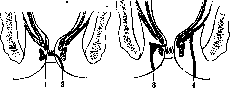
with granulations, sometimes the granulations are sparse and the openings themselves are barely noticeable. The openings of tuberculous fistulas are distinguished by pale pink, jagged, and undermined edges, are relatively wide, and discharge liquid pus. The internal openings are usually narrow and barely allow the head of a thin button probe to pass through. External openings are often multiple, while the internal opening is usually single, rarely two, but not more than three (in tuberculous fistulas). Internal openings most often lie in the sphincteric part, less frequently higher, and very rarely more than 5 cm from the anal opening. -Fistula tracts have various directions and according to them fistulas are divided into types: 1) subcutaneous-submucous fistulas, the tract of which lies under the skin or under the mucous membrane below or inside the external sphincter; in complete fistulas, the internal opening lies in the sphincteric part; 2) ischiorectal fistulas, the tract of which goes from the external opening into the ischiorectal space, often ending in an expanded blind end, and in complete fistulas opens into the lumen of the rectum between the external and internal sphincters or above the latter; 3) rectovesical fistulas, the tract of which goes upward above the levator ani muscle, either ending blindly in a purulent cavity or opening into the lumen of the rectum in the area of the internal sphincter or above it. Fistula tracts are sometimes tortuous and branching, in some cases, after going around the rectum, they open into the skin of the anus on the opposite side. Clinic. The course of fistulas of the anus and rectum depends on their nature, on the length, direction, and topographic relationships of the fistula tracts. In external fistulas, itching is observed in the area of the anus and discharge of secretions that stain underwear, irritate the skin, cause eczema, and sometimes papillomatous growths. In complete fistulas, gases and feces may pass through them. In internal fistulas, feces may accumulate in the fistula tracts and cause pain. If the external opening is closed, secretions accumulate in the fistula tract, forming an abscess-like condition- pain, tenesmus, and fever appear. As soon as the opening reopens, all symptoms disappear. - Diagnosis. Fistulas are often easy to recognize, but sometimes difficulties arise, especially when the fistula opening is small, retracted, and marked only by slight redness or bulging. It is more difficult to determine if there is a complete fistula, as the internal opening is not always felt upon examination with a finger. For greater clarity, examination with a probe is performed, the movements of which are controlled by a finger introduced into the rectum; if the fistula tract is filled with a vaseline-bismuth mixture, an X-ray can be taken of the fistula tract. A colored liquid can also be injected through the external opening: in a complete fistula, it appears in the lumen of the intestine. Treatment. Established fistulas do not heal; this is prevented by a number of factors: 1) constant disturbance of rest due to contraction of the sphincter and levator ani muscle and during defecation, 2) constant infection of the fistula tract with intestinal contents, 3) development of dense scar tissue around the fistula tract, 4) epithelialization of the fistula tract over a greater or lesser extent, 5) narrowness of the fistula openings, due to which secretions are retained in the fistula tracts, 6) presence of fecaliths or foreign bodies in the fistula tracts. Tuberculous fistulas in general are little inclined to heal. Therapeutic treatment of fistulas is pointless and only surgical treatment can be successful. Two methods of operations are used: 1) incision of the fistula tract, 2) excision of it. In incision, a button probe is introduced into the fistula tract from the rectal side under the control of a finger until it penetrates into the cavity of the intestine, from where its end is brought out through the anus. The layer of tissue lying on the probe is incised. If the fistula tract is tortuous, the probe is inserted as far as possible and the fistula tract is incised up to the button of the probe; then the probe is further advanced into the fistula tract and the latter is incised, and so on, until the internal opening is found or the blind end of the fistula tract is determined. In internal fistulas, the opening is located, a probe is inserted into it, and the fistula tract is incised along its entire length. The granulations lining the fistula tract are curetted, and in tuberculous fistulas, the base of the tract is also cauterized with a thermocautery. With this method, it is often necessary to incise the external sphincter, sometimes also the internal one, and in rectovesical fistulas also the levator ani muscle. In terms of healing, the method gives good results even in tuberculous fistulas (Melchior), but incision of the sphincters is not without danger. Therefore, excision of the fistula tract makes more sense, in which without significant damage to the sphincter, along with the tract, all scar tissue is removed and the wound is sutured without leaving dead spaces (subcutaneous catgut sutures or figure-of-eight silk drainage sutures). According to Golk's data in 86 cases of fistula excision, primary healing was achieved in 77 cases (89.5%). The success of the operation depends on careful preparation of patients before the operation and careful care after the operation. - Strictures of the rectum more often occur in the ampulla and at the level of the rectum's encirclement by the levator ani muscle, less frequently in the sphincteric part. Perret examined strictures in 60 corpses and found that 4 times the stricture began in the sphincteric part, 32 times below 6 cm from the anal opening, 3 times at a height of 6 cm, 7 times between 6 and 9 cm, 5 times above 9 cm, 6 times at the transition of the rectum into the sigmoid. In 4 cases, there were multiple strictures. In Finkelstein's 5 cases, the stricture extended to the entire rectum with complete destruction of the sphincters, in 27 cases it began at a height of 2-6 cm and in 6 cases above 6 cm. In Vize's 211 cases, the stricture was tubular, in 36 cases annular, in 11 cases linear. - Pathological anatomy. Strictures of the rectum represent the final stage of various inflammatory processes that involve not only the mucous membrane but also the wall of the intestine and the surrounding cellular tissue, as a result of which the rectum is transformed over a greater or lesser extent into a tube with a narrow lumen and with dense callous walls. The degree of narrowing varies and often only the tip of a finger can barely enter it. The mucous membrane is destroyed and replaced by scar tissue. In the remnants of the mucous membrane, metaplasia of cylindrical epithelium into bridge-like (Hartmann, Napalkov) is sometimes observed. Above the stricture, the intestine is dilated, its mucous membrane is inflamed and often ulcerated, the muscular layer is hypertrophied. Abscesses forming in the perirectal cellular tissue give rise to fistulas in the area of the anus, perineum, buttocks. In 100 cases of strictures, Hartmann observed fistulas in 32 cases. Below the stricture, the mucous membrane rarely ulcerates, but in the area of the anus, under the influence of irritation by flowing secretions, eczema and fissures often develop. The altered rectum fuses with the surrounding sclerosed cellular tissue into a common mass and adheres to the walls of the pelvis. The cellular tissue of the ischiorectal fossae becomes markedly sclerosed. Etiology. The frequency of strictures varies from 0.6% to 1.9% of all patients in general (Schede, Konig). Women get sick more often than men (5.6:1). Many factors play a role in the development of rectal strictures: 1) trauma: after hemorrhoid operations according to Whitehead, after extirpation of the rectum, uterus, after childbirth with prolonged engagement of the fetal head in the pelvic outlet, after treatment of cancer of the cervix uteri with radium, after gunshot and other injuries to the rectum; 2) dysentery (Kummell, Konig). Dysenteric strictures are localized in the upper part of the rectum and sigmoid (Kummell); 3) inflammatory processes, especially gonorrhea and syphilis (the most common cause of strictures); less often the cause was tuberculosis. Strictures based on actinomycosis of the inguinal lymphogranuloma are rarely observed. Pronounced annular strictures of the rectum occur in parametritis, but differ from those listed above in that the mucous membrane of the intestine is not involved in the process and remains movable. In the strict sense of the word, these are not strictures. Soft chancre of the rectum gives rise to strictures, but they are not common. Finally, a tumor of the rectum, especially cancer, can be the cause of strictures. Clinic. In most cases, people aged 20-40 years suffer from rectal strictures, less often in older age. Narrowings develop extremely slowly. Patients come to surgeons usually in the stage of severe stenosis with the duration of the process from 1 to 15 years. When the stricture involves a large extent of the rectum, including the sphincter, there is constant discharge of mucus, pus, and foul-smelling masses; frequent urges to defecate are accompanied by pain, and eczema and papillomatous growths appear around the anus. Patients are striking in their suffering appearance, malnutrition, they lose their appetite, cannot sleep. When the stricture does not involve the sphincter and lies above it, occupying a limited section of the intestine, symptoms of chronic obstruction are observed, sometimes exacerbating. Then, above the narrowing, fecal masses can accumulate in large quantities and be palpated through the abdominal wall as bumpy masses.
When these masses soften and liquefy, constipation is replaced by diarrhea, and the fecal masses that are excreted have a caustic effect on the lower parts of the intestine below the strictures, causing the development of fissures and eczema in the anal region. During defecation, there may be tenesmus and pain. Diagnosis. When the stricture is located low, it can be recognized by simple examination of the rectum with a finger, but this only determines the position and shape of the lower end of the stricture. Proctoscopy allows examination of this end and detection of changes below it. Sometimes the proctoscope can be passed through the stricture and thus determine its length and the condition of the intestine above it. An idea of the degree and length of the stricture is given by X-ray examination, with the contrast substance being administered per os and introduced from below by enema. With the help of a proctoscope, secretions can be obtained for bacteriological examination and a biopsy can be performed, but in fully formed strictures, histological examination of them with respect to etiological factors does not give positive results. For gonorrheal strictures, some authors consider the detection of plasma cells on histological sections (Exner, Gorash) to be characteristic. In the papillae of the mucous membrane, gonococci are sometimes found (Napalkov). In syphilitic strictures, obliterating endarteritis and periarterial infiltration are found. The Wassermann reaction does not always have decisive value. Histological detection of changes characteristic of tuberculosis decides the question of the tuberculous nature of the stricture.--The prognosis for severely expressed strictures of the rectum is unfavorable. Left to their own devices, patients can lead a wretched existence for a long time until they die from perforation of the intestine into the abdominal cavity, from exhaustion, amyloid degeneration of internal organs, from pelvic phlegmon, erysipelas, or from an outbreak of tuberculosis of the lungs. Treatment. Therapeutic treatment of strictures of the rectum can only be applied in the initial stages of development. Specific treatment for syphilitic strictures can give results only in fresh cases. Recently, many authors (Barbilian, Bensaude, Marchand and others) persistently recommend the treatment of strictures of the rectum by diathermy and consider it the method of choice. Diathermy makes the scar tissue soft, elastic, pliable—the symptoms of obstruction disappear. Bougienage is also used. Both hard and soft bougies are used. The introduction of bougies is performed with the help of a proctoscope, which is then removed, and the bougie is left in place for 15-30 minutes. Probing must be preceded by thorough cleansing of the intestine. At first, bougienage is repeated every 2-3 days, later less frequently. It continues for months, even years, since a return of the strictures to their previous condition is observed as a rule. Among the operative methods used are: 1) internal rectotomy, 2) external rectotomy, 3) rectoplasty, 4) anastomosis between the rectum and sigmoid colon, 5) resection or amputation of the rectum, 6) colostomy. Internal rectotomy is permissible for ring-shaped strictures of the lower part of the rectum; in other cases it is useless and dangerous. In external rectotomy, through an incision from the anus to the middle of the sacrum, the coccyx is excised and the posterior surface of the rectum is exposed. The posterior wall of the rectum is incised lengthwise throughout the entire length of the stricture, and the sphincter is not incised. A thick drainage tube is inserted into the rectum up to the sigmoid colon, and the intestine is sutured over it without catching the mucous membrane with the suture. In other cases, the incised intestine is left open and healing is conducted under a tampon. This operation does not give good results, and during the healing of the wound, it is necessary to resort to the expansion of the intestine with bougies. Better results are obtained by a modification of the method, which consists in that the edges of the incised intestine are sutured to the edges of the skin, and after their union, the opening in the intestine is closed plastically with a skin flap turned with the epidermis inward, over which another skin flap is applied from the other side (Polya, Napalkov). Rectoplasty—longitudinal incision of the stricture from within and suturing of the wound in the transverse direction—is applicable only for low-located ring-shaped strictures.--Anastomosis between the rectum and sigmoid colon is possible when the stricture is located above Douglas' pouch. For a lower location of the stricture, another method is applicable, in which the sigmoid colon is divided, both ends are sutured, the perineum is divided transversely, and the peritoneum is opened; the sutured end of the sigmoid colon is brought down and passed through an incision in the anterior wall of the rectum into its cavity, after which it is fixed with sutures from the side of the wound and the cavity of the rectum (Ali Krogius, Rotter). If the stricture extends upward to the descending colon, the transverse colon can be drawn down (Kimmell).--Circular resection or amputation with or without preservation of the sphincter is performed by methods from below or by a combined route (see below—cancer of the rectum).--Due to the extent of adhesions and sclerosis of the pelvic cellular tissue, these operations may be technically difficult to perform and their results are not always satisfactory. Radice, in reporting on 135 such operations by various authors, indicates that 14 died (10.3%), of 58 traced patients 9 recovered, in 21 cases a partial success was obtained, in 28 cases a relapse occurred. Recently, many are advocating for a palliative operation—the creation of an artificial anus in the left iliac region. The condition of patients after this operation improves and they continue to live without suffering. An artificial anus is also created: 1) for the purpose of bougienage by the method without end (ohne Ende), 2) for washing the affected part of the intestine with disinfectant and astringent solutions.--Diverticula of the rectum are extremely rarely observed; in the literature only 4 cases are known (Morgagni, Terrier, Neumann, Fantozzi). Benign tumors of the anus and rectum. Some benign tumors of the anus and rectum are extremely rare and reports of them are isolated. 1) Myomas and fibromyomas have been described 18 times (Dukhina). They originate from the muscular layer of the intestine, at first grow into the submucous or subserous membranes, but later penetrate through them. Three forms of these tumors are distinguished: a) polyp-like tumors, hanging into the cavity of the intestine; they are most common and are observed in young age; b) tumors of the anterior wall of the rectum, penetrating into the abdominal cavity; c) tumors of the posterior wall, penetrating into the sacral fossa. As they grow, myomas and fibromyomas may give symptoms of cancerous tumors: pain in the sacrum, mucus and blood in the stools, repeated bleeding, emaciation, sometimes obstruction. Treatment is only surgical. 2) Lipomas of the rectum are very rare. They originate from the submucous or subserous membranes, are situated on a thin stalk. They grow either into the cavity of the intestine or into the abdominal cavity. They may intussuscept into the anus or twist on their stalk: then they become necrotic and are spontaneously discharged (Vogeler). In their thickness, foci of calcification and bone tissue are sometimes found. 3) Lymphangiomas of the rectum—a great rarity. 4) Cavernous angiomas of the rectum have been described about 10 times. They are located mainly in the ampulla. They originate from the submucous layer, but later they may involve the entire wall of the intestine and spread to the perirectal cellular tissue. Clinically they manifest with severe bleeding, which can lead to the death of the patient. Among the operative procedures, the most rational is combined extirpation of the rectum with preservation of the external sphincter. 5) Dermoids of the rectum have been described about 10 times. They are more often situated in the perirectal cellular tissue and secondarily penetrate into the cavity of the rectum. In three cases there was a pathognomonic symptom—the exit of hair from the anus. Cases of perforation of the rectum by ovarian dermoid have been observed. 6) Condylomata acuminata of the anus are observed relatively often and develop mainly under the influence of irritation from the secretions of fractures. They are located on the skin of the anus and represent small warty growths, which sometimes reach considerable size, close the anal opening, and spread to the skin of the perineum.--Treatment consists in the removal of the growths with scissors or a sharp spoon. Recurrences usually do not occur.--Polyps of the rectum—see Intestine—new formations of the intestine. Villous tumor, or papillary adenoma of the rectum (tumor villosus). The tumor originates from the mucous membrane of the rectum, has a broad base or a broad stalk, is divided into lobules covered with villi of varying lengths. The stroma of the tumor originates from the submucous tissue, which thickens in this place. The muscular layer of the mucous membrane becomes delicate, in places disappears altogether, and the tumor lies on the muscular coat of the intestine, not extending beyond its limits. A villus represents a connective tissue thread covered with a single row of cylindrical epithelium. Tumors are inclined to malignant degeneration in 45%, but they penetrate inward slowly and for a long time remain accessible to surgical intervention.--Clinical picture. Villous tumors for many years may not manifest clinically. The general condition of the patients remains satisfactory for a long time; in rare cases, repeated bleeding leads to anemia.
In general, the bleeding is insignificant, often in the form of drops of blood in the stool. Inconstipation is frequently replaced by diarrhea. There is no pain, but there may be sensations of pressure and a foreign body in the rectum. A characteristic symptom is the discharge of mucus resembling egg white, sometimes in large quantities. Accumulating in the rectum, it causes frequent urges to defecate (up to 20-30 times a day), but these urges are not accompanied by tenesmus or burning in the anus. The tumor may prolapse from the anus, become irreducible, and require emergency assistance. Pieces of the tumor are passed in the stool, which indicates its transition to a malignant state. Diagnosis. In 80% of cases, villous tumors are localized in the rectum at a height of 6-12 cm from the anal opening and are accessible to examination by finger. Sometimes the contours of the tumor escape the finger, which sinks into it as if into a gelatinous mass. During proctoscopy, a collection of mucus is found on the surface of the tumor, which at times is so abundant that it fills the proctoscope, while in other cases it is possible to see the mucus oozing from the tumor like from a sponge. In case of malignant degeneration, the surface of the tumor is less villous, flattens less easily under pressure from the proctoscope, and begins to bleed, and the secretion of mucus is less abundant. Biopsy is significant only when there are positive findings in terms of malignancy and should be performed from many places. Treatment. Treatment with radium and X-rays leads only to superficial sloughing of the tumor and relapse occurs quickly. With repeated sessions of electrocautery, the disappearance of the tumor can be achieved, but a relapse soon follows. The only radical method of treatment is surgery: 1) excision of the tumor through natural passages, 2) resection, and 3) amputation of the rectum. Removal of the tumor through the anus is applicable in many cases, especially with benign tumors and those on a stalk. In elderly and weakened patients, this method finds broader indications and not only for benign but also for malignant tumors, if they are small in size and mobile. For the operation to be performed, it is important that the position of the tumor allows it to be brought down by stretching the sphincter. The excision of the tumor itself must be performed within healthy tissues, otherwise a relapse may occur. In cases inaccessible to this operation, resection of the intestine over a certain length or amputation with preservation of the sphincter or a combined method (see below - rectal cancer) is used.
Cancer of the rectum (cancer recti). In terms of frequency of cancerous involvement, the rectum occupies 5th place among other organs (Birch-Hirschfeld) or even 3rd (Borst). Out of 12,730 autopsied cancer patients at the Basel Pathological-Anatomical Institute, there were 1,078 (8.5%), and of them, patients with rectal cancer were 57 (5.4%) of all cancer patients (Krasting). According to Ball, rectal cancer constitutes 4% of all cancerous lesions of the body and 80% of cancerous diseases of the intestine. Other figures are also given (see Intestine, tumors of the intestine). - Etiology of rectal cancer - see Cancer. It is noted that people who suffer from hemorrhoids, constipation, chronic catarrh of the large intestine for decades more frequently develop rectal cancer. Cases of development of rectal cancer during its prolapse, further on the basis of tuberculous ulcers, syphilitic scars, trauma have been observed. Cancer of the ampulla often originates from the anterior wall; the reason for this is seen in the stronger irritation of it by fecal masses. Cancer quite often develops from rectal polyps, especially in so-called polyposis, and it is possible for several cancers, including in the rectum, to appear simultaneously (see above). Pathological anatomy. The ampulla of the rectum is most often affected by cancer, then the upper part of it, and finally the perineal part. In Funke's 238 cases, the ampulla was affected 162 times, the upper part of the rectum - 64 times, the perineal part - 12 times. Cases are observed where cancer affects the entire length of the rectum (12-15 cm). The external form of cancerous tumors is varied. Tumors of the anus appear as either isolated nodules or nodular or ulcerated tumors involving part or the entire circumference of the anus. Cancer of the ampulla most often appears as either a limited or extensive ulcer with an uneven and infiltrated base, with dense and raised edges. The ulcers occupy in some cases one or another wall, in others the entire circumference of the intestine. In the upper part of the rectum, cancerous neoplasms quickly take on a ring-like form and lead to narrowing of the intestinal lumen. Representing a serious obstacle to the passage of fecal matter, they lead to retention of stool and to dilation of the proximal part of the intestine. In the longitudinal direction, cancerous tumors can occupy various extents - from 1-2 cm to the entire length of the rectum. Histologically, two types of cancer are observed in the rectum - squamous cell and cylindrical cell. The first is found in the anus, the second in the rest of the rectum. Cases of squamous cell cancer in the pelvic part of the rectum (Bohm) on the basis of epithelial covering buds carried in during the embryonic period or on the basis of metaplasia of cylindrical epithelium into flat under the influence of inflammatory processes have been observed. Squamous cell cancers of the rectum are much less common than cylindrical cell cancers. Out of 842 cases, there are 809 cylindrical cell and 33 squamous cell cancers (Braitsyev). Cancers of the rectum have a tendency to mucous metaplasia. Mucus-producing cells die, and their place is taken by connective tissue, as a result of which scar-forming ring-like cancers, characteristic of the upper part of the rectum, are formed. Special mention should be made of mucous diffuse cancers, which predominantly affect young people, possess great energy of growth, are distinguished by high malignancy and destructiveness. This type constitutes approximately 8.5% of all rectal cancers. - Growth into the lumen of the intestine in the form of polyps is characteristic only of papillary and microcystic adenocarcinomas, all other cancers grow deep into the intestinal wall. Penetrating the muscular and mucous membranes, cancerous growths go into the submucosal layer, from where on the one hand they penetrate and destroy the mucous membrane in places, on the other - they penetrate the muscular coat of the rectum, go beyond its limits, pierce the surrounding cellular tissue and spread to neighboring organs. Cancer of the anus penetrates the sphincter and turns this part into a dense, non-contractile tube. Posteriorly it can involve the skin, laterally the cellular tissue of the ischiorectal fossae, anteriorly it spreads to the perineum, the urethra and the prostate gland in men, to the vulva and vagina in women. In case of ulceration of the tumor, gates are opened for infection, suppuration and fistulas develop. Cancers of the ampulla between the levator ani of the anus and the peritoneal recess penetrate the proper fascia of the rectum, posteriorly involve the cellular tissue and the sacrum, laterally can spread to the walls of the pelvis, anteriorly penetrate Denonvilliers' aponeurosis (Denonvilliers), spread to the prostate gland, the seminal ducts and the seminal vesicles, to the bladder and ureters in men, to the vagina and uterus in women. Cancer of the upper part of the rectum posteriorly involves the cellular tissue and the skeleton, anteriorly can penetrate the peritoneum and spread to the bladder in men and to the uterus in women. With ulceration of the tumor, inflammatory adhesions with neighboring organs are formed. But before cancerous growths have time to go beyond the rectum by continuous growth, transfers of cancerous elements occur along lymphatic pathways to regional lymph glands, and then along blood vessels to distant organs. The study of the lymphatic system of the rectum (Braitsyev, Villemin, Huard et Montagne and others) leads to the conclusion that in regard to the spread of rectal cancer along lymphatic pathways, the position of the tumor along the length of the intestine is of great importance. - For cancers of the perineal part, involvement of the inguinal glands, sacral and sacral regions, retrorectal along the branches of the superior rectal artery and subperitoneal glands should be expected. For cancers of the ampulla below Douglas' fold, the subperitoneal glands and glands along the superior and inferior rectal arteries are affected, while for cancers above Douglas' fold, only glands along the superior rectal artery are affected. Due to the presence of anastomoses between the lymphatic pathways of the ampulla and perineal part in cancers whose lower border is not higher than 5 cm from the anal opening, the entire lower part of the rectum should be considered affected and transfers can be expected in all regional glands (Braitsyev). After the regional glands are involved, the second stage for transfers is the lymphatic glands along the inferior mesenteric artery up to the glands along the aorta. Rectal cancerous tumors can penetrate blood vessels, especially veins. Metastases are most often observed in the liver, then in the lungs, in the iliac bones, in the spine, in the shoulder blades, in the femurs, in the spleen, in the kidneys, in the skin, and in the brain. Transfers to lymphatic glands and to distant organs are given by all rectal cancers possessing a clearly expressed infiltrating growth. Only mucous diffuse cancers represent
620 some peculiarity. Rapidly capturing surrounding rectal tissues and most severely affecting the lymphatic pathways, they rarely give rise to metastases to distant organs. The degree of malignancy of rectal cancers is determined by two factors: the speed and extent of growth; the speed and frequency of recurrences after operations. The most malignant are solid cancers, and among them—diffuse mucous ones. A close relationship is observed between the anatomical form and the frequency of recurrences. In 125 cases studied histologically and clinically to the end by Petersen, the following data are available: in 82 cases of simple adenocarcinomas—84% recurrence; in 8 cases of papillary and small-cystic cancers—100% recoveries; in 39 cases of all other forms—100% recurrences. Recurrences are most common in the cellular tissue and in the regional glands: in Petersen's 38 cases—83%, in Zinner's 141 cases—72%. All recurrences are direct recurrences, i.e., those arising from remnants of the neoplasm not removed during the operation. Indirect recurrences, i.e., new diseases of rectal cancer, must be extremely rare. Part of them apparently falls on cancers that existed simultaneously with the operated ones and remained unnoticed during the operation, part—on those newly arising from rectal polyps. Clinical picture. The clinical course of rectal cancer is not always the same. In many cases, the onset of the disease remains hidden and is not accompanied by any symptoms. In other cases, there are small discharges of mucus or an admixture of blood in the stools, but this often attracts little attention from patients. The duration of this latent period is on average 15 months. As the tumor grows and ulcerates, symptoms begin to appear. In cancers of the anus, the first symptoms are pain, itching, burning, and discomfort. With the infiltration of the sphincters, conditions for incontinence are created—gases, feces, blood, mucus, and pus are constantly discharged, which stains the linen and makes the condition of patients difficult for themselves and for others. With the development of ulcerations, phlegmons and fistulas often arise. In cancers of the ampulla during the period of decay and ulceration, bleeding, tenesmus, and frequent urges to defecate (every 1/2-1 hour) appear, with only blood and mucus being discharged. When the process spreads to the surrounding cellular tissue and nerves, pains in the sacrum with radiation to the anus, perineum, penis, vagina, and lower limbs appear. When the cancer spreads forward to the urethra and prostate gland, difficulty in urination up to complete retention of urine may occur, sometimes destruction of the urethra with the formation of a urinary fistula; when it spreads to the bladder, a rectovesical fistula may form, resulting in septic cystitis and pyelonephritis. In case of compression or infiltration of one ureter, hydronephrosis occurs; of two—complete anuria. In highly located cancers, an early symptom is constipation. With appropriate treatment, these patients feel quite well for a long time, but often symptoms of obstruction suddenly arise, and their stools may be absent for 10-20-30 days (Quenu, Kraske, etc.)—Feces are retained above the tumor, gases continue to pass. Sometimes symptoms of true ileus develop. In case of perforation of the intestinal wall, death from peritonitis occurs. Regardless of the position of the tumor, in case of septic decomposition of the products of its decay, chills and temperature increases appear. Metastases to the liver often remain asymptomatic for a long time, sometimes are discovered only at autopsy; metastases to the brain give a picture of local diseases or a picture of psychosis (Kraske); metastases to bones can cause pathological fractures. Ultimately, a sharp decline in strength, cachexia, and death become the inevitable end for all patients left to the natural course of the disease. The average duration of life from the onset of symptoms is 12-19 months, and from the onset of the disease—27-34 months. Recognition of rectal cancer in the initial period is a difficult matter. The disease remains hidden for a long time, the first symptoms are often indefinite, attract little attention from both patients and doctors. Doctors themselves often treat these patients for hemorrhoids, catarrh, without examining the rectum with a finger. The dispensary method of examining patients can help in detecting the initial forms of rectal cancer, provided that mandatory examination of the rectum is carried out. Examination of the rectum with a finger is the simplest and at the same time most perfect method. Once the tumor is accessible to examination by finger, diagnosis is not difficult. By palpating the tumor with a finger, one can get an idea of its form and size, its density and ulceration, its mobility and relation to surrounding tissues and organs. If the tumor is located above the Douglas fold, it may be inaccessible to the finger, it may not be palpated even through the abdominal walls. In these cases, it is necessary to make the patient strain while squatting, then the tumor descends and may become accessible to the examining finger. In highly located ampullary cancers, the ampulla is empty and distended with gases (Hochenegg). It is important to know the height of the upper border of the tumor for choosing the method of operation, but this is not always possible, as one cannot pass freely through the narrowed place either with a finger or with a proctoscope, and to use force is dangerous. Experience shows that the spread of the tumor for 10-12 cm along the length of the intestine is a rarity. In all cases where the finger does not reach the tumor, where it is necessary to check with the eye what was found by finger, where it is necessary to confirm the presence or absence of rectal disease, proctoscopy (see) is of invaluable service. Rectal cancer must be differentiated from syphilis of it, from sarcoma, from polyps, from tuberculous and gonorrheal ulcers. Syphilitic lesions in most cases have the character of strictures, extend over a considerable length, the process lasts for many years (10 or more years). To a certain extent, the Wassermann reaction can help. Tuberculous and gonorrheal ulcers are rare and have their own peculiarities (see above). Sarcomas are rare, biopsy is needed to distinguish them. Polyps are characterized by their form; the question of their benignity or malignancy is also decided by biopsy. Petersen and Kollmers recommend applying biopsy in all cases of rectal cancer for the purpose of determining the histological structure, which has importance in relation to establishing indications for operation and the choice of the method of operation. Substituting the dissected peritoneum to the lying part of the rectum. Methods of operation. In case of doubt in the histological research, whether there is an adenoma or cancer, the neoplasm should be considered as cancer if it has grown through the muscular mucous membrane. Treatment. Neither radium nor X-rays give anything except some alleviation, radical surgery is applicable not often, since most patients turn to surgeons already in such a condition when only palliative operations are applicable. Radical surgery is a complex act, consisting of the operation of access to the intestine, the operation on the intestine itself, and measures for restoring function. For access to the rectum, many operative methods have been proposed, the development of which has its history. On the rectum itself, three operations are performed: 1) extirpation, when the entire rectum is removed together with the sphincters, 2) amputation, when a part of the rectum is removed with the sphincter apparatus, 3) circular resection over a length, when after excision of the affected part of the rectum, its upper and lower ends are connected with sutures (fig. 18 and 19). Lisfrank first performed amputation of the rectum affected with cancer (1826), Dieffenbach first introduced the principle of resection of the rectum for those cases when the sphincter is not affected by cancer, Hueter first performed circular resection of the rectum (1867), Simon proposed to remove movable cancerous tumors located at a height of 4-5 cm through the anus, stretching the sphincter or dissecting it posteriorly along the median line. For access to the rectum, Denonvillier introduced a posterior incision from the anus to the tip of the coccyx. To this incision, Verneuil added the excision of the coccyx. The last method was widely used by Kocher (Fig. 19. Connection of the intestine with a circular suture). In 1885, Kraske proposed to excise in highly located rectal cancers, besides the coccyx, also the left half of the sacrum down to the lower edge of the third sacral foramen. Bardenheuer crossed the sacrum transversely at the level of the third sacral foramina, and Rose—even at the level of the second. In 1889, Heineke proposed temporary resection of the coccyx and sacrum, sawing them lengthwise and crossing the sacrum transversely at the level of the fourth or third sacral foramina. Rehn
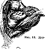
S\
Substituting the dissected peritoneum to the lying part of the rectum. Methods of operation. In case of doubt in the histological research, whether there is an adenoma or cancer, the neoplasm should be considered as cancer if it has grown through the muscular mucous membrane. Treatment. Neither radium nor X-rays give anything except some alleviation, radical surgery is applicable not often, since most patients turn to surgeons already in such a condition when only palliative operations are applicable. Radical surgery is a complex act, consisting of the operation of access to the intestine, the operation on the intestine itself, and measures for restoring function. For access to the rectum, many operative methods have been proposed, the development of which has its history. On the rectum itself, three operations are performed: 1) extirpation, when the entire rectum is removed together with the sphincters, 2) amputation, when a part of the rectum is removed with the sphincter apparatus, 3) circular resection over a length, when after excision of the affected part of the rectum, its upper and lower ends are connected with sutures (fig. 18 and 19). Lisfrank first performed amputation of the rectum affected with cancer (1826), Dieffenbach first introduced the principle of resection of the rectum for those cases when the sphincter is not affected by cancer, Hueter first performed circular resection of the rectum (1867), Simon proposed to remove movable cancerous tumors located at a height of 4-5 cm through the anus, stretching the sphincter or dissecting it posteriorly along the median line. For access to the rectum, Denonvillier introduced a posterior incision from the anus to the tip of the coccyx. To this incision, Verneuil added the excision of the coccyx. The last method was widely used by Kocher (Fig. 19. Connection of the intestine with a circular suture). In 1885, Kraske proposed to excise in highly located rectal cancers, besides the coccyx, also the left half of the sacrum down to the lower edge of the third sacral foramen. Bardenheuer crossed the sacrum transversely at the level of the third sacral foramina, and Rose—even at the level of the second. In 1889, Heineke proposed temporary resection of the coccyx and sacrum, sawing them lengthwise and crossing the sacrum transversely at the level of the fourth or third sacral foramina. Rehn
Substituting the dissected peritoneum to the lying part of the rectum. Methods of operation. In case of doubt in the histological research, whether there is an adenoma or cancer, the neoplasm should be considered as cancer if it has grown through the muscular mucous membrane. Treatment. Neither radium nor X-rays give anything except some alleviation, radical surgery is applicable not often, since most patients turn to surgeons already in such a condition when only palliative operations are applicable. Radical surgery is a complex act, consisting of the operation of access to the intestine, the operation on the intestine itself, and measures for restoring function. For access to the rectum, many operative methods have been proposed, the development of which has its history. On the rectum itself, three operations are performed: 1) extirpation, when the entire rectum is removed together with the sphincters, 2) amputation, when a part of the rectum is removed with the sphincter apparatus, 3) circular resection over a length, when after excision of the affected part of the rectum, its upper and lower ends are connected with sutures (fig. 18 and 19). Lisfrank first performed amputation of the rectum affected with cancer (1826), Dieffenbach first introduced the principle of resection of the rectum for those cases when the sphincter is not affected by cancer, Hueter first performed circular resection of the rectum (1867), Simon proposed to remove movable cancerous tumors located at a height of 4-5 cm through the anus, stretching the sphincter or dissecting it posteriorly along the median line. For access to the rectum, Denonvillier introduced a posterior incision from the anus to the tip of the coccyx. To this incision, Verneuil added the excision of the coccyx. The last method was widely used by Kocher (Fig. 19. Connection of the intestine with a circular suture). In 1885, Kraske proposed to excise in highly located rectal cancers, besides the coccyx, also the left half of the sacrum down to the lower edge of the third sacral foramen. Bardenheuer crossed the sacrum transversely at the level of the third sacral foramina, and Rose—even at the level of the second. In 1889, Heineke proposed temporary resection of the coccyx and sacrum, sawing them lengthwise and crossing the sacrum transversely at the level of the fourth or third sacral foramina. Rehn
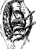
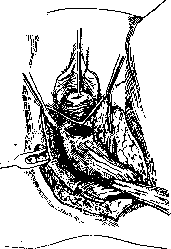
proposed in 1895 an approach to the rectum in women through the vagina, which is dissected longitudinally along the posterior wall with extension of the incision onto the perineum to the anterior periphery of the anus. This operation was performed even earlier by Czerny and Vyalyaminov (in 1887). As a result of these and other proposals, 5 routes were created, following which the rectum can be exposed from below: 1) perineal, 2) vaginal (Figure 20), 3) posterior with coccygectomy, 4) parasacral, 5) posterior with coccygectomy and partial resection of the sacrum. The latter method provides the widest exposure, but even after this method was introduced into practice, cases were encountered in which the removal of cancerous tumors of the rectum from below proved difficult or impossible due to the narrowness of the pelvis and the high position of the tumor or high involvement of the lymph glands. The desire to perform the operation even in these cases led to the application of the combined method, which represents a combination of laparotomy with one of the methods from below. The idea of this method was expressed by Volkmann in 1877, and implemented in practice by Konig in 1882. The first recovery after this operation was achieved by Jeannel, who operated on a woman by the abdomino-perineal method (1894). Figure 20. Vaginal amputation of the rectum. The posterior wall of the vagina has been dissected (on clamps) and the levator ani muscle has been exposed. The rectum has been freed.
operations: 1) combined
1) combined amputation of the rectum (or extirpation) with the lower end of the intestine brought down to the perineum, 2) combined amputation with the creation of an ileal artificial anus, 3) combined resection. Special operations include those for high-located tumors, where circular resection is performed only by laparotomy (Schloffer), where after resection of the intestine from the abdominal cavity, an ileal artificial anus is created from the upper end, and the lower end is sutured and left in place (Hartmann), and finally, when the rectum is mobilized through the sacral route, the affected portion is brought out through the wound, fixed, and resected in a second stage after 2-3 days (Küttner-Vorlagerungs-method). Between circular resection and amputation of the rectum stands the operation of Hochenegg, the so-called method of passage (Durchziehungs-methode), which consists in bringing down the upper end of the intestine to the perineum and passing it through the preserved anal section deprived of mucous membrane and suturing it to the skin. In this operation, the difficulties of circular suture after resection on a segment are avoided, and at the same time the sphincter apparatus is preserved; whereas to restore the closure of the intestine brought down to the perineum or sutured into the upper angle of the sacral wound after amputation of the rectum is a "difficult" task. Gersuny proposed to turn the brought-down end 180° around the longitudinal axis before suturing, but this does not provide secure fixation and threatens to disrupt the blood circulation in the intestine. Willems developed on cadavers, and Witzel performed on living persons, a method of creating an artificial sphincter by passing the brought-down end of the intestine through the thickness of the divided left gluteal muscle along the course of its fibers, but this method also does not give satisfactory results. Therefore, in combined amputation of the rectum, many surgeons abandon bringing down the upper end and establish an ileal artificial anus, which, especially according to the methods of Kappis, Lambret, and others, is more advantageous than an anus on the perineum or in the sacral region without the sphincter apparatus (see Anus praeternaturalis). Indications for radical operation and choice of operative method. High degree of cachexia, spread of the neoplasm to neighboring organs, and metastases to the liver and other organs constitute absolute contraindications to radical operation. Spread of cancer to the posterior wall of the vagina is not a contraindication. To improve immediate and long-term results, it is necessary to narrow indications as much as possible and extend to the utmost limits the extent of removal of the intestine and surrounding tissues together with lymphatic pathways. For this purpose, cancerous tumors that have not yet gone beyond the proper fascia of the rectum are most suitable. When establishing indications for operation, it is important to know the microscopic structure of the tumor (biopsy). For less malignant forms of cancer, indications should be broadened, while for more malignant forms, on the contrary, they should be narrowed. Of the methods of access to the rectum, the perineal method cannot satisfy modern requirements for rectal cancer operations, as it is technically suitable only for low-located tumors, and in these cases, a high and wide amputation with removal of pelvic cellular tissue and regional lymphatic glands should be performed, which is unattainable by this method. Almost the same can be said of the vaginal method. The posterior method is more advantageous, especially with excision of the coccyx and part of the sacrum (Kraske). It allows the rectum to be freed together with its fascia, provides sufficient access to Denonvilliers' fascia in men and to the posterior wall of the vagina in women, permits removal of pelvic cellular tissue and lymphatic glands, opening of the peritoneum, and excision of 20 or more centimeters of intestine and bringing down its upper end to the perineum. But at the same time, this method has disadvantages: it is difficult to excise high-located cancers, especially in a narrow pelvis and with a short sigmoid mesentery; it does not allow removal of lymphatic glands along the inferior mesenteric artery; it is difficult to perform ligation of the inferior mesenteric artery (see Intestine--anatomy) above the "critical point," as a result of which the superior rectal artery has to be ligated, which may lead to gangrene of the brought-down intestine. In defense of the sacral method, Goetze appears, who crosses the sacrum at the level of the third sacral foramina and performs early opening of the peritoneum. Nevertheless, the combined method is being given more and more place. The laparotomy performed in it first of all allows establishing the operability of the case, makes it convenient to ligate the inferior mesenteric artery, excise the sigmoid mesentery including affected lymphatic glands while preserving the marginal arterial arcade, allows freeing the rectum from above downward to the pelvic floor together with pelvic cellular tissue and lymphatic glands, after which the intestine is removed by one or another method from below. In combined amputation, it is better to combine laparotomy with the perineal method than with the sacral method, as damage to bone tissue complicates the operation and increases susceptibility to infection; whereas in resection, it is more convenient to attach the coccygeal or sacral method to laparotomy. However, from the point of view of cancer spread along lymphatic pathways, circular resection of the rectum is permissible only for cancers of its upper part (above Douglas' pouch). At present, voices are being raised against circular resection of the rectum in general (Kirschner, Schmieden, Fischer, and others), as it is technically difficult, complications after it are frequent, the circular suture due to infection and marginal necrosis holds in a limited number, fistulas often develop, many of which remain permanently, and healed ones may lead to stricture of the intestine. In favor of resection is the preservation of the sphincter apparatus, although even with this, patients sometimes suffer from incontinence. In Küttner's cases after resection, 50% had control of feces, in Kirschner's-28.5%. For cancers of the lower part of the rectum (below Douglas' pouch), a high and wide amputation should be performed with removal of cellular tissue and lymphatic glands of the pelvis, with excision of the fatty tissue of the ischiorectal fossae and the parts of the levator ani adjacent to the rectum. Preservation of the external sphincter is possible only in cases where the lower border of the tumor is not lower than 5 cm from the anal opening. But even when it is anatomically possible to preserve the sphincter apparatus, many authors abandon bringing down the upper end of the intestine to the perineum and establish an ileal artificial anus (Quenu, Hartmann, Kirschner, Schmieden, and others), as even after ligation of the inferior mesenteric artery above the "critical point," such bringing down is not without danger of gangrene of the intestine, as its blood circulation may be disrupted due to kinking and stretching of the vessels. Mortality in the creation of an ileal artificial anus is lower than in bringing down the intestine. In Russian authors, out of 25 cases of the first operation, 28% died, out of 23 cases of the second-39% (Brajtsev). In bringing down the intestine to the perineum, combined amputation of the rectum is performed in one session, but when creating an ileal artificial anus, the question of a two-stage operation may arise. In deciding this question, the condition of the patient's strength and the presence or absence of fecal poisoning and adhesions are of essential importance. In case of weakness of the patient and presence of the other factors, first an artificial anus is created with division of the intestine, and after 3-4 weeks, when the patient's strength is restored, removal of the rectum is performed, the operation again beginning with laparotomy. By this time, many adhesions have disappeared, and the operation of removing the rectum is considerably facilitated. Preparation of patients for operation. 7-8 days before the operation, patients should be given nourishing food that leaves little residue, and the rectum should be irrigated with weak antiseptic solutions. In the last two days, laxatives are given. In the evening before the operation, the intestine is irrigated and the patient receives 10 drops of tincture of opium. In case of stricture of the intestine and accumulation of fecal masses above it, a rubber tube can be passed through the stricture and regular washing of these masses carried out for 1-2 weeks. This improves the general condition of patients and reduces inflammatory changes around the tumor. In case of severe exhaustion and fecal poisoning, a preliminary artificial anus is applied with elimination of the possibility of fecal matter entering the lower part of the intestine. In these cases, the radical operation is performed after the patient's strength is restored. Care should also be taken to strengthen the vascular system. Technique of operations. Operations on the rectum for cancerous neoplasms are performed under general anesthesia or under spinal anesthesia. The latter is more widely used as it prevents shock. Many operations have been proposed, but the following methods are most commonly used. I. Transanal method-removal of the tumor through the anus.
The operation is permissible only for benign, growing into the lumen of the intestine adenocarcinomas. In the lithotomy position, the sphincter is stretched, the mucous membrane is grasped with Luer forceps above the tumor and brought down, the tumor is surrounded by an incision of the mucous membrane at a distance of 2-3 cm and removed. The wound of the mucous membrane is sutured. II. Perineal method. The patient in the lithotomy position. A metal catheter is inserted into the urinary bladder. The anal
Figure 21. Incisions for a
Figure 22. Incisions for amputation of the rectum:
mputation of the rectum: a - according to Volcker; Ь - according to
a - according to Hoohen^gg'y; Ь - according to Zuckerkandl-Wolfler'y.
Kociier'y.
anal opening is closed with a subcutaneous purse-string suture of thick silk, which is tightly tightened. At a distance of 2-3 cm the anal opening is surrounded by an elliptical incision and its median edges are sutured, which achieves hermetic closure of the rectum. From this incision in front on the midline of the perineum an incision of 4-5 cm is made, behind - to the tip of the coccyx (figs. 21 and 22). Behind the edges and tip of the coccyx are exposed and the
ano-coccygeal ligament is severed. On the sides the fat of the ischio-rectal fossa is incised, the levator ani muscle is exposed, which is incised behind the perineum (figs. 23-25). The center of the perineum is incised and the dissecting space between the rectum and the prostate gland is opened. On the lower part of the posterior surface of the prostate gland a transverse incision is made, separating the rectum from m. transversus perinei superficialis and m. bulbo-cavernosus (above), and Denon-villiers' fascia is separated from the prostate gland. The lateral plates of the aponeurosis and the anterior parts of the levator ani are incised, and the intestine is freed from the pelvic floor. The middle rectal vessels are ligated and the rectum is dissected upward outside its own fascia. After separation of the intestine from the seminal vesicles and urinary bladder, the intestine is detached from behind the sacrum, after which the peritoneum is opened and the superior rectal vessels are ligated. The intestine is brought down sufficiently so that it can be severed 5-6 cm above the tumor. The peritoneum is carefully sutured to the intestine, the levator ani and the ano-coccygeal ligament are also sutured to it. The perineal muscles are sutured, the brought-down intestine is incised transversely, and the edges of its incision are sutured to the edges of the skin. Drains are inserted into the ischio-rectal fossae and the skin wound is sutured. A rubber tube wrapped in gauze and vaselined is inserted into the intestine.In women, excision of the rectum through the perineum is facilitated by the simplicity of the anatomical relationships in front. Together with the intestine, it is recommended to remove the posterior wall of the vagina - here the perineal method is in a way combined with the vaginal. In other respects the operation does not differ. At present the perineal method has almost lost its independent significance and
Figure 24. Liberation of the rectum
from m. levator ani and lig. ano-coccygum. serves mainly as a component of the combined abdomino-perineal method. III. Sacral method. The patient on the right side, legs bent and brought to the abdomen, the back is bent so that the pelvis protrudes and slightly protrudes beyond the edge of the table. Some prefer the position on the left side (Hochenegg) or on the abdomen (Depage, Voelcker, Goetze). The anus, if amputation is planned, is sutured. A midline incision 12-15 cm long from the level of the sacro-iliac joints to the periphery of the anus or an arcuate incision from the left sacro-iliac joint to the tip of the coccyx, convex to the right (figs. 26-29). The coccyx and sacrum are exposed and muscles and ligaments are separated from them on the left. The coccyx is excised and the left edge of the sacrum is beveled to the III sacral foramen. The pelvic fascia is incised, and after separation of the fat the proper fascia of the rectum is exposed. The intestine is bluntly dissected together with the fascia and fat. Around the intestine a plate is formed and the intestine is extracted in a bent form into the wound. After separation of the intestine from the seminal vesicles and urinary bladder, the peritoneum is exposed and opened. The intestine is dissected from behind, and the superior rectal vessels are ligated as high as possible. After this the intestine is brought out through the wound for a considerable length. The peritoneum is sutured to the intestine. In circular resection the affected part is excised within the limits of healthy tissues (10 cm on each side from the tumor), and the ends of the intestine are sutured. Kuttner leaves the brought-out intestine outside the wound for 2-3 days and then resects in the second stage. In amputation the lower part of the intestine is dissected from the perineal side, as described above (see art. 626). After excision of the affected rectum the brought-down intestine is sutured either in place of the anus or in the upper angle of the sacral wound, or is passed through the thickness of the left gluteal muscle. After the operation by the sacral method a large wound cavity remains behind, which is filled with loose packing and somewhat narrowed by sutures. IV. Combined method. The operation begins with laparotomy in the Trendelenburg position. The lower part of the sigmoid intestine is pulled upward and vertical incisions of the peritoneum are made on both sides of the mesentery of the intestine, which are connected below by a transverse incision on the posterior surface of the bladder above the bottom of Douglas' pouch. The lateral edges of the peritoneum are separated from the underlying fat, which is shifted to the middle and separated from the posterior abdominal wall together with mesorectum, containing lymphatic vessels and glands. The rectum is separated from the sacrum together with the
Figure 27. Dorsal amputation of the rectum. The midline incision exposes the coccyx and it is resected. A flap is cut from the aponeurotic plate, which hangs on the posterior surface of the rectum.
fat as far as the coccyx. Between the urinary bladder and the rectum one penetrates deeply to the seminal vesicles and the upper part of Denonvilliers' fascia, at the level of which the dissection of the intestine ceases. The lateral fibrous strands containing the middle rectal blood vessels are ligated between two ligatures, and the intestine is freed from above. Above the promontory or above the 'critical point' the inferior mesenteric vessels are ligated and severed. At the same level the sigmoid
Figure 28. Dorsal amputation of the rectum. After separation of fascia propria recti the pelvic part of the rectum is dissected under control of the finger. m. levator ani is severed.
intestine is severed. Both ends are invaginated with the help of purse-string sutures and sutured to complete hermeticity. The lower end is shifted into the small pelvis and the peritoneum over it is carefully sutured. The upper end is sutured in the upper angle of the abdominal wound or in a separate wound in the left iliac region and serves as a permanent artificial anus. The abdominal wound is sutured. The operation is completed below by the perineal method
Figure 29. Dorsal amputation of the rectum. Incision of the peritoneum and liberation of the rectum.
(see above - perineal method). If a resection is decided upon, the sacral or coccygeal method is added to the laparotomy. In women it is recommended to remove from above in
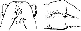
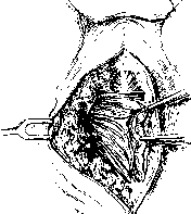
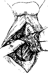
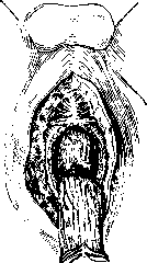
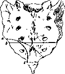
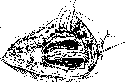
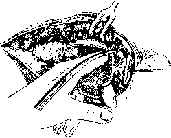
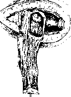
- = with the R. to the uterus, and from below to the posterior vaginal wall. Some surgeons (Kirschner, Colovov, Solovyov) perform preliminary bilateral ligation of the hypogastric artery through the abdominal wound, while others (Braitsky, Schmieden, Fischer) consider such ligation unnecessary, since with the correct course of the operation it is not needed to prevent blood loss, but at the same time it devascularizes the pelvis and reduces tissue viability. V. The abdominal-transanal or intravaginal method is used for high-lying rectal cancers. Its essence is that the mobilized intestine from above is invaginated through the dilated anus. To facilitate invagination, a thick probe with a button on its end is passed through the anus, and the intestine above the tumor is tied with a thick thread below the button. By pulling on the probe, the tumor is brought out through the anus, the intestine is resected, and its ends are sutured (fig. 30-31) (Maunsell, Trendelenburg, Moszkowicz). Postoperative care and complications. Operated patients require intensive attention. In addition to general measures, measures must be taken to prevent and eliminate complications, which are particularly common in these patients. Among the complications, the following should be noted: 1. Wound infection, which is particularly common with the sacral method. After operations by this method, up to 60% of all deaths are due to infection. 2. Shock. It is more often observed after operations by the combined method. The danger of shock has significantly decreased since spinal anesthesia began to be used instead of inhalation anesthesia. It is recommended to prepare patients for surgery by prolonged bed rest (up to two weeks) and the use of cardiac tonics.

Figure 30. Connection of the intestine through the anus.

Figure 31. Connection of the intestine by the invagination method. 631
In case of blood loss during surgery - apply blood transfusion. 3. Sequential bleeding. Rarely observed. 4. Complications from the urinary tract: a) retention of urine - it is necessary to evacuate urine with a catheter with all precautions in terms of asepsis; b) urinary fistulas due to injury to the bladder or urethra - catheter a demeure; c) with the combined method, injury to the ureter is possible - it is necessary to suture or transplant the ureter into the bladder or extirpate the kidney; d) in rare cases, urinary incontinence is observed for several weeks (Mummery).5. Necroses of the intestine brought down to the perineum. To prevent this, it is necessary to observe all rules regarding the ligation of blood vessels during the operation, avoiding tension of the brought-down intestine. 6. Bronchitis and pneumonia. 7. Thromboses and embolisms. 8. Hemiplegias. In elderly people, they can occur on the basis of embolism from formed thrombi. 9. Acute obstruction. It can be caused: a) strangulation of loops of the small intestine in the separated pelvic floor wound, b) kinking of loops of the small intestine due to adhesion to the suture line of the pelvic floor or abdominal wall, c) strangulation of loops of the small intestine in the area of the artificial posterior ileal passage. 10. Delayed wound healing. It is caused by a decrease in the patient's general strength, the presence of fistulas due to retention of ligatures, the formation of loops and pockets in the wound. Mortality after radical operation for cancer of the R. k. Until 1910, the immediate mortality after radical operation for cancer of the rectum in individual authors varied with the perineal methods from 6.22% to 32%, and with the combined method - from 14.3% to 50%. Recently, according to the compiled statistics of Gaudier on 2,400 cases, mortality with the perineal method was 20%, with the coccygeal - 16%, with the sacral - 17.4%. Heze determines mortality with the sacral method at 10%, with the combined at 20%. On 441 cases of Russian authors, Braytsev gives a general mortality of 19.7%, with the perineal method - 6.85%, with the vaginal - 28%, with the coccygeal - 29%, with the sacral - 26.92% and with the combined - 33.3%. In this regard, in addition to the complications mentioned, the experience of the surgeon, clarity in determining the indications for the operation and the ability to correctly assess the patient's strength in relation to the severity of the operation are important. -Distant results. According to followed-up observations, over 50% of operated patients have recurrences, and in the first 2 years after the operation. On 1,263 cases collected from the literature by Braytsev, recurrence occurred in 721 cases (57%). The critical period of 3 years is survived without recurrence by 35-54%, but this does not eliminate the possibility of recurrence occurring at a later period. There are no figures for good long-term results after combined operations yet, but according to reports from individual authors they are higher. In Poshet (Paichet), the combined method gave up to 70% of long-term cures, in Hartmann out of 37 surviving patients, 14 lived without recurrence for 10 and 16 years. Treatment of inoperable cancers of the R. k. Patients with cancer of the R. k. often turn to the surgeon in such an advanced state that a radical operation cannot be performed. To alleviate their condition, it is necessary to resort to palliative operations, and first of all to the creation of an artificial posterior ileal passage (see Anus praeternaturalis). The second operation in these patients is posterior linear rectotomy (rectotomia linearis posterior). It is indicated: 1) in patients in whom, with an established artificial posterior ileal passage, decomposition of the tumor, pain, and tenesmus continue to exist, which cannot be eliminated by other measures (irrigation from below or above), and abscesses develop; the intestine is incised above or below the tumor (rectotomia superior, rectotomia inferior); in patients who refuse the establishment of an artificial posterior ileal passage to eliminate obstruction, upper rectotomy is applicable; 2) to expose the tumor for local treatment with radium or X-rays; 3) in recurrences, especially if they are located on the anterior wall and the posterior wall is free. As for the treatment of inoperable cancers of the R. k. with radiant energy, X-ray therapy is of little use, while the use of radium gives some relief in terms of reducing pain, bleeding, and ichorous discharges. - Along with primary cancers, secondary cancers of the R. k. are also encountered in connection with metastasis to the Douglas space of cancer of the stomach, gallbladder, etc. These metastases can grow through the walls of the R. k. into its mucosa and simulate primary cancer of the intestine. Sarcoma of the R. k. According to the calculation of Exner (Exner) on 100 cases of cancer of the R. k., 1 case of sarcoma is encountered. Three types of sarcomas are observed: 1) simple, 2) pigmented, 3) lymphosarcoma. Simple sarcoma (round-celled, spindle-celled, and others) is less common than pigmented, and lymphosarcoma is even rarer. Key on 58 cases of sarcoma of the R. k. collected from the literature notes pigmented sarcoma in 65%. Bensaude, Cain, and Horowitz in 1929 report 3 of their own cases and 12 cases from the literature, a total of 15 cases of lymphosarcoma of the R. k. Simple sarcoma has a stalk in 30%, pigmented in 70%, lymphosarcoma grows diffusely. Pigmented sarcoma is located closer to the anus, simple is higher. The size of the tumor in both varies from a pea to a bulky mass the size of a child's head. Sarcomas originate from the submucosal layer, remain mobile for a long time and do not involve the mucous membrane. Over time, they become immobile and ulcerate. Clinically, they manifest with bloody stools, bleeding, tenesmus, frequent urges, and discharge of serum, with rapid onset of emaciation. Both simple and pigmented sarcomas give metastases. With pigmented sarcomas, metastases occur early and occur through lymphatic and blood vessels (to the inguinal glands, to the glands behind the R. k., to the liver, lungs); Metastases to lymphatic glands of simple sarcoma are not described, but through blood vessels they occur and first of all to the liver. Lymphosarcomas arise from adenoid tissue and closed follicles and take on a diffuse character, involving the entire thickness of the wall of the R. k. Usually, lymphosarcoma begins directly above the sphincter, which it involves and extends upward sometimes to the sigmoid colon. The surface of the mucous membrane has a papillary appearance and often ulcerates. The perirectal cellular tissue is rarely involved in the process, but involvement of the lymphatic glands is the rule. The lumen of the intestine is narrowed, but not enough to cause obstruction; often the lumen is even aneurysmally dilated. In some cases, lymphosarcoma represents a limited tumor that protrudes into the lumen of the intestine and subsequently ulcerates. The course of lymphosarcomas of the R. k. is treacherous. They often involve the entire organ without causing any disturbances. Bleeding is rare, tenesmus is absent. Characteristic are progressive emaciation, pallor, and feverish condition of the patients. For the diagnosis of sarcomas in general, biopsy is of decisive importance. -Treatment. With sarcomas of the R. k., removal through the anus is permissible only for very limited tumors. The operation of choice is amputation of the R. k. The results of surgical treatment are not encouraging, since recurrence is the rule. According to observations of Bensaude, Cain, and Horowitz, lymphosarcomas are very sensitive to X-rays, and in one case after X-ray therapy, they observed a long-term cure.
Related articles
Cite this page
“Rectum.” Soviet Medical Encyclopedia. English translation of Bolshaya Meditsinskaya Entsiklopediya, 1st ed. (Moscow, 1928–1936), ed. N. A. Semashko. https://sovietmedicalencyclopedia.pages.dev/article/rectum/