Mesentery
Historical document, translated for reference. It reflects medical knowledge of the 1920s–30s and is not medical advice.
Summary
This article defines the mesentery as a peritoneal fold connecting the intestine to the posterior abdominal wall, serving as a conduit for vessels and nerves. It details the various types of mesenteries in the adult, including those of the small intestine, colon, and pelvic organs, while noting their embryonic development and potential anomalies.
Encyclopedia article (1928–1936)
MESENTERY, mesenterium (for the large intestine—mesocolon), is a type of peritoneal ligament (see). By mesentery, we specifically mean those ligaments that extend from the posterior abdominal wall to various parts of the intestinal tube; it is a duplication of the peritoneum, representing the transition from the parietal layer of the latter into the visceral layer covering the organ. Between its two laminae, in loose connective tissue, lie lymphatic glands, and blood vessels, lymphatic vessels, and nerves pass through. The mesentery, therefore, acts as a conduit for vessels and nerves and, furthermore, has a certain mechanical significance—the intestine is, as it were, suspended by it and, at the same time, can move within certain limits. In an adult, the following mesenteries are distinguished: 1) mesentery of the small intestine, mesenterium in the narrow sense of the word; 2) mesentery of the vermiform appendix, mesenteriolum processus vermicularis; 3) mesentery of the transverse colon, mesocolon coli transversi; 4) mesentery of the S-shaped curvature (sigmoid), mesocolon flexurae sigmoideae; and 5) mesentery of the rectum, mesocolon recti. The mesentery of the small intestine extends along the small intestine throughout its entire length, with the exception of the duodenum—from the duodenojejunal flexure to the end of the ileum (therefore, the jejunum and ileum, taken together, are combined into one concept—intestinum tenue mesenteriale). The mesentery has two surfaces—right and left—and two edges; one of them is connected to the intestine, whereby the peritoneal laminae forming the mesentery transition into the visceral layer covering the intestine. The other edge is attached to the spine, where the laminae of the mesentery diverge and continue into the parietal peritoneum, peritonaeum parietale; this is the root of the mesentery, radix mesenterii, the line of attachment of which to the posterior abdominal wall runs almost vertically, more precisely—from the left side of the body of the II lumbar vertebra to the right sacroiliac joint. The edge of the mesentery connected to the intestine is many times longer than the other edge; therefore, the shape of the mesentery is compared to a trapezoid or a ruffle (hence the name of the organ). Indeed, if one were to cut the intestine from the mesentery along the line where they connect to each other, the resulting free edge of the mesentery would present a wavy line, and the mesentery itself would be a plate folded into numerous pleats, the depth of which increases toward the free edge. The mesentery of the small intestine contains branches of the superior mesenteric artery and vein, lacteals, and lymph nodes (glandulae mesenteriales); the latter are gathered here in such numbers (more than 150) as nowhere else in the human body, and lie in several rows, with the smallest glands located closer to the intestine, and the larger and more densely arranged ones—at the root of the mesentery. The mesentery of the vermiform appendix, mesenteriolum processus vermicularis, due to variations in the shape, size, and position of the latter, is characterized by great inconsistency. It runs along the upper edge of the appendix (not quite reaching its distal end) and reaches the taenia mesocolica of the cecum. In the free edge of the mesentery lies the appendicular artery (a branch of the ileocolic artery). Sometimes there is a mesentery of insignificant size for the cecum as well—mesocoecum, connecting it to the posterior abdominal wall. The mesentery of the transverse colon, mesocolon coli transversi seu mesocolon transversum, runs
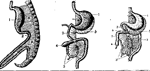
Diagram of the development of the gastrointestinal tract and mesentery in a human embryo (according to O. Hertwig): 1—stomach; 2—duodenum; 3—jejuno-ileum; 4—cecum; 5—ascending colon; 6—transverse colon; 7—descending colon; a—dorsal mesentery; b—greater omentum developing from the mesentery (for a detailed description, see article 143).
in the transverse direction, partitioning the abdominal cavity into upper and lower sections. It attaches to the posterior abdominal wall, along the anterior surface of the head of the pancreas, and then along the lower edge of the body of the latter. It fuses with the two posterior laminae of the greater omentum (see Peritoneum). The mesentery of the sigmoid colon, mesosigmoideum, originates from a line that runs from the middle of the left iliac fossa to the sacral promontory. Its edge attached to the intestine is significantly longer. The lower end of the mesentery of the sigmoid colon, narrowing, transitions into the mesentery of the rectum, mesorectum; the latter is developed only at the beginning of the rectum and varies very greatly. In addition to the intestinal mesenteries listed here, similar formations exist in women in the pelvic region. The cavity of the latter is divided by a frontally placed septum into two spaces—anterior and posterior; this septum consists of two laminae of the peritoneum and, in its middle, unpaired part, encloses the uterus, and in the lateral parts (specifically, in the region of their upper edge)—the oviducts (fallopian tubes). Thus, on both sides of the uterus, between the latter and the lateral wall of the pelvis, there is a duplication of the peritoneum—the broad ligament of the uterus, lig. latum uteri, in the form of a quadrilateral, the lateral edge of which is attached to the lateral wall of the pelvis, the lower one—to its floor (in both cases, the two laminae of the ligament diverge, continuing into the parietal peritoneum), the medial one—to the lateral side of the uterus, and only the upper one is free and encloses the oviduct. Inside the broad ligament of the uterus are located the vascular and nerve pathways serving the uterus. This results in a complete similarity to the mesentery, with the only difference being that here there is a paired formation, inside which, in addition to blood vessels, two special ligaments also pass—the round ligament of the uterus and the proper ligament of the ovary; the ovary is attached to the posterior surface, and the oviduct lies in the upper edge. Since the broad ligament of the uterus is most closely related to three organs—the uterus, the oviduct, and the ovary—a corresponding number of sections are distinguished in it: the uppermost part, long and narrow, located between the ovary and its proper ligament (below) and the oviduct (above), is called the mesosalpinx; the part of the posterior lamina of the broad ligament where it transitions to the ovary is described as the mesentery of the latter, mesovarium; finally, the remaining (larger) part of the broad ligament is assigned the name mesometrium, therefore, this is the mesentery of the uterus proper. In conclusion, mention should be made here of the common mesentery, mesenterium commune, which is observed as a rare anomaly. In this condition, the intestine, starting from the duodenum and ending at the upper section of the rectum, has a common mesentery that attaches to the spine along the midline; in this case, the differentiation of the mesentery has stopped at an embryonic stage of development (regarding the comparative anatomy and embryology of the mesentery, see Peritoneum; there also is the literature).
V. Tankov. Diseases of the mesentery. Pathological processes in the mesentery sometimes arise more or less in isolation in one or another constituent part of the mesentery (e.g., embolism of an artery, inflammation of the lymph nodes); sometimes the mesentery suffers as a whole (e.g., contusions, ruptures of it). Depending on the starting point of the disease, one can provide the following scheme for the pathology of the mesentery: diseases of the mesentery as a whole—congenital and acquired; changes in blood vessels; changes in lymph nodes; changes in nerves; tumors. 1. Diseases of the mesentery as a whole. As developmental defects, the absence of a part of the mesentery is observed (sometimes in its upper section, sometimes in the lower); then the beginning of the jejunum or the end of the ileum are attached immovably to the posterior abdominal wall (like the duodenum). The reverse picture is presented by excessive width of the mesentery; it is observed in both the small and large intestines, giving mobility to a normally fixed section of the intestine (for example, the ascending or descending colon) or imparting excessive mobility to certain sections of the small or large intestine, predisposing to volvulus. Apparently, holes in the mesentery are also congenital; they are most often (2/3 of cases) encountered near the entry of the ileum into the cecum, but have also been noted in other places of the mesentery of the large and small intestines, in the mesoappendix, and in the epiploic appendages of the colon. Some authors explain the origin of such holes by atavism; others by irregularities in the growth of the mesentery, which does not keep pace with the growth of the intestine; and others by atrophy of the mesentery in places poorly supplied with blood vessels (Treves' field). There are, however, undoubtedly acquired holes in the mesentery—on the basis of abdominal injuries or injuries left unsutured after one or another surgical operation on the mesentery. Whatever the origin of the mesenteric hole, it can cause internal strangulation (see); this is its interest for the clinician; many dozens of such cases have been described, with some ending in death despite operative assistance. During abdominal contusions, as well as during the strangulation of hernias, ruptures of the mesentery and its avulsion from the intestine have been observed; this is a very serious complication, accompanied by bleeding from torn vessels and threatening necrosis of the corresponding section of the intestine; immediate surgical assistance is required, which is limited to stopping the bleeding if the damaged vessel is compensated by collaterals, but more often consists of resection of the section of the intestine deprived of blood circulation. Contusions of the mesentery sometimes do not lead to ruptures but damage the blood vessels, causing the formation of hematomas or thrombi; the consequence of this will also be a disruption of the proper nutrition of the corresponding section of the intestine, with subsequent necrosis. To the number of diseases of the mesentery as a whole also belongs 'retractile mesosigmoiditis' ('retractile mesosigmoiditis' of French authors); it is observed in the mesentery of the pelvic segment of the sigmoid colon and consists of the following: the mesentery becomes dense, thickened, whitish in color (due to the appearance of scars); the vessels turn out to be compressed by proliferating connective tissue; the elasticity of the mesentery is lost, it shrinks, bringing together the corresponding intestinal loops, which in sharply expressed cases lie side by side like the barrels of a shotgun. The cause of the disease is likely a slowly progressing inflammation. Its consequences are constipation, sometimes accompanied by bouts of pain and symptoms of obstruction; often on this basis, a volvulus forms. In the case of scarring of the mesentery in the anterior-posterior direction, an approach of individual sections of the sigmoid colon to the root of its mesentery is observed. Often, cicatricial changes of the mesentery of the sigmoid colon also spread to the root of the mesentery of the ileum; in the latter, stellate scars are often observed independently in its lower part. Special attention has been paid recently to inflammation of the mesoappendix, with which unexpectedly arising peritonitis after appendectomies is associated (Jauré). 2. Isolated and independent diseases of the blood vessels of the mesentery fall into diseases of the arteries and veins; one distinguishes their occlusion by emboli, thrombi, and proliferation of the inner lining; then, as a very rare form, one can point to diffuse dilation of the mesenteric vessels. Emboli occur on the basis of heart diseases (endocarditis), more rarely of the aorta; thromboses usually join atheromatous ulcers; proliferation of the inner lining of the vessels most often occurs on the basis of syphilis, sometimes its nature remains unexplained. Of the arteries, the superior mesenteric artery is more often occluded; in far-advanced cases, the consequence of this will be necrosis of the mesenteric part of the small intestine and the right half of the colon; in cases of occlusion of branches of the superior mesenteric artery or other mesenteric arteries, necrosis will seize the corresponding sections of the intestine. Usually, occlusion of the arteries entails a hemorrhagic infarct of the intestine, more rarely its necrosis of an ischemic character. Occlusion of the veins of the mesentery is tolerated somewhat more easily; necrosis on the basis of blood stasis sets in more slowly and is more limited. On the other hand, the possible continuation of a thrombus in the direction of the portal vein is already a serious complication, e.g., in mesenteric phlebitis of the appendix. The clinical picture of acute occlusion of the mesenteric vessels resembles an attack of internal strangulation; patients feel a suddenly onset, most acute pain, the face becomes pale, sweat breaks out, the pulse is weak and frequent; the abdomen turns out to be tense, often it is possible to palpate a tumor; stool is either delayed or diarrhea is observed (often with blood). The natural outcome of this disease is death from general poisoning due to necrosis of the intestine; death occurs earlier than general purulent inflammation of the peritoneum manages to develop; only in rare, more slowly progressing cases is peritonitis the final link of the disease. However, there are exceptions to this general rule; there are cases of spontaneous recovery: such is, for example, the case of Karcher. His patient recovered after an attack of mesenteric vessel occlusion and then (after 2 months) died from another disease; at autopsy, an embolus was indeed discovered in the superior mesenteric artery. Slowly developing closure of the vessels of the mesentery gives a less stormy picture and more often gives time for the development of collateral circulation ('formes incompletes' of French authors), manifesting itself in periodically occurring attacks of intestinal obstruction. One cannot, of course, count on such exceptionally happy outcomes; one should always resort to urgent surgical intervention according to the general rules of abdominal surgery, which gives good results in the first hours of the disease, when there are no signs of general intestinal poisoning; during the operation, one has to either resect the intestine within the limits of healthy tissues with a subsequent fecal fistula or anastomosis, or one sometimes has to limit oneself to bringing the necrotic section of the intestine to the outside; completely hopeless cases end only in an exploratory laparotomy. Dilations of the vessels of the mesentery, sometimes reaching the thickness of a little finger, are described as accidental findings during laparotomies; they have not yet been studied and, apparently, do not cause painful phenomena. Besides the indicated diseases of the vessels with a purely anatomical basis, there exist, apparently, also spastic states of the mesenteric vessels, similar to vascular spasms observed in other areas (dyspragia intermittens angiosclerotica intestinalis). By analogy with angina pectoris, Wieting calls them 'angina intestinalis' and describes their clinical manifestation in the form of attacks of acute pain with signs of intestinal obstruction. 3. Diseases of the lymphatic pathways of the mesentery manifest themselves, mainly, by the involvement of the nodes: mesenteric lymphadenitis is observed in infectious diseases of the intestine, in tuberculosis; sometimes involvement of the mesenteric nodes is observed in lymphogranulomatosis; these nodes are the first focus where metastases of malignant tumors of the intestine stop. The breakdown of inflamed mesenteric nodes is sometimes the cause of general purulent peritonitis with an intact intestinal wall (for example, in typhoid fever). Tuberculous involvement of them, known by the name tabes mesaraica, is most often observed in early childhood, but is also encountered in adults; with a confirmed diagnosis, this form is subject to treatment according to the general rules of conservative treatment of glandular tuberculosis, since operations have given a rather large percentage of mortality (15% according to Lecène et Leriche) and cannot claim to be radical; only in exceptional cases can tuberculosis of the mesenteric nodes require an operation: in cases of compression of the intestine, formation of adhesions, etc. In tuberculosis, more rarely after typhoid fever, petrification of the mesenteric nodes and the formation of sometimes large stony conglomerates are observed. 4.
Diseases of the nerves of the mesentery do not yet constitute an independent chapter; one can only cite individual clinical observations indicating their suffering; thus, for example, it seems that the nerves of the mesentery sometimes play a role in the pathogenesis of intestinal spasms, spastic obstruction, and "rigidity"; in these cases, there is excessive activity of the parasympathetic system, perhaps sometimes on the basis of suffering of the nerves of the mesentery; mechanical irritation of these nerves during surgical operations or during intestinal strangulation sometimes causes general phenomena: shock, respiratory arrest. Postoperative paralysis of the intestine, sometimes leading to death on the 10th-15th day after intestinal operations, is explained by some authors as damage to the mesenteric nerves. 5. The most developed and practically important section is tumors of the mesentery. Cysts and solid tumors are distinguished; among the former, dermoids, echinococcus, enterocystomas, and cysts with bloody, serous, or chylous contents are encountered here; they are more often solitary, very rarely multiple. These tumors are benign, grow slowly, are very mobile, and do not cause any pathological phenomena until they reach a very large size. The origin of cystic tumors with bloody contents can be explained by the encapsulation of a hematoma of the mesentery; serous and chylous cysts are probably of inflammatory origin; an admixture of blood in them may be due to secondary hemorrhages. Treatment is possible only surgically—enucleation of the cyst, which is often successful by blunt dissection after incision of the serous covering and without damage to the vessels. Solid tumors of the mesentery are encountered less frequently; they can be benign (lipomas, fibromas, fibrochondromas, angiomas) and malignant; among the latter, one should distinguish primary tumors—sarcomas, lymphomas—and metastatic ones—transfers of various malignant tumors from other organs. Benign tumors sometimes proceed without symptoms—such is the case of Brooklouse, when the patient did not suspect the existence of a 13-pound tumor of the mesentery in himself; but often tumors manifest themselves with vague pains, gastric phenomena, diarrhea, or constipation. Malignant tumors retain the usual signs here—rapid growth, infiltration of neighboring tissues, metastasis, and causing ascites. Diagnosis is difficult, as it is extremely difficult to accurately establish the relationship of the tumor specifically to the mesentery; the most characteristic feature for tumors at the beginning of their existence is significant mobility. Treatment is possible only operatively; however, technically these operations are very difficult, and the prognosis is significantly worse than for cystic tumors, since even benign solid tumors are often closely connected with vessels, the intestinal wall, and the ureter.
Related articles
Mentioned in
Cite this page
“Mesentery.” Soviet Medical Encyclopedia. English translation of Bolshaya Meditsinskaya Entsiklopediya, 1st ed. (Moscow, 1928–1936), ed. N. A. Semashko. https://sovietmedicalencyclopedia.pages.dev/article/mesentery/