Gastroenterostomy
Historical document, translated for reference. It reflects medical knowledge of the 1920s–30s and is not medical advice.
Summary
This 1930s encyclopedia article details the history, surgical technique, and potential complications of gastroenterostomy, a procedure connecting the stomach to the jejunum bypassing the pylorus. It covers the evolution of the operation from Wölflers method to improved techniques and discusses the pathology of the vicious circle syndrome.
Encyclopedia article (1928–1936)
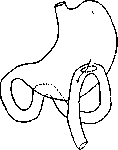
GASTROENTEROSTOMY (from Greek gaster—stomach, enteron—intestine, and stoma—mouth), the name of an operation that consists of connecting the stomach with the jejunum via an anastomosis, bypassing the pylorus. The first gastroenterostomy was performed by Wölfler on September 28, 1881, in Billroth's clinic quite by accident: while operating on stomach cancer with the intention of excising the tumor, Wölfler opened the abdominal cavity, became convinced that resection was impossible, and decided to close the wound. At that time, Nicoladoni, who was present at the operation, suggested connecting the stomach to the intestine with a new opening for the passage of food masses, which was executed. The method by which this first gastroenterostomy was performed bears Wölfler's name and has been preserved in its general features to the present day. Through subsequent modifications concerning details of great practical importance, the technique of Wölfler's method was brought to the highest possible perfection, but nevertheless, it was gradually superseded by other methods that yielded significantly better results (Hacker, Roux, Petersen). The history of the improvement of gastroenterostomy followed two lines: the improvement of the technique of intestinal suturing in general (specifically in the placement of anastomoses) and the combat against severe complications that often occurred after the operation in direct dependence on inadequate operative methodology and technique. Wölfler's method (gastroenterostomia anterior antecolica) as the primary method consists of the following: the abdominal cavity is opened by an incision along the midline from the xiphoid process to the navel. After examination establishes the necessity of a gastroenterostomy, the stomach is withdrawn from the abdominal cavity and the intestinal loop required for the anastomosis is located; for this purpose, the transverse colon is withdrawn, its mesentery (mesocolon) is stretched, the surgeon goes down to the spine with the right hand, and in the left corner of its intersection with the mesocolon, the duodenojejunal flexure is found by touch; 40-50 cm are measured downward along the intestine from here, and this loop is brought outward. It is extremely important to make sure with absolute precision that the intestine has been found correctly—otherwise, the anastomosis will be made in the wrong place. Therefore, if doubts arise, one should be guided not only by palpation but also by inspection: having spread the edges of the wound and pushed the intestinal loops downward, one must make sure that the loop taken to be the duodenojejunal flexure actually emerges from beneath the root of the mesocolon. The selected loop is brought to the anterior wall of the stomach in front of the transverse colon and the greater omentum, giving it an isoperistaltic direction relative to the stomach (i.e., the afferent end to the left, the efferent end to the right), the abdominal cavity is carefully walled off with gauze pads, a separate pad is placed between the stomach and the intestinal loop beneath the site of the future anastomosis, clamps are applied to the stomach and intestinal loop, and the placement of sutures is begun in such a way that the opening in the stomach falls as close as possible to the greater curvature and the direction of the opening runs from upper left to lower right (see Fig. 1). The opening in the intestine must be made longitudinally along its axis on the side directly opposite the site of mesenterial attachment; regarding the size of the opening, debates were held in due time, and some authors proposed making it very large—up to 10 cm, while others were satisfied with 3-4 cm; the majority now make an incision of 5 cm. The first gastroenterostomy operations using the indicated method were performed in such a way that first
Figure 1. The stomach and intestine were opened with incisions and only then did they proceed to placing the sutures. Braun (1891) pointed out a technique that was adopted by everyone and now remains classical: before opening the stomach and intestine, the posterior serous suture is placed; parallel to it, retreating by 1/4 cm, incisions are made through the seromuscular layers of the stomach and intestine, avoiding the opening of the mucous membranes; this is followed by a second row of seromuscular sutures on the posterior lip of the wound, and only now are the mucous membranes opened; sometimes they are also sutured with a separate row of catgut sutures (see Figures 2 and 3) and then the entire operation is completed by placing seromuscular and serous sutures on the anterior edges of the anastomosis (see Figure 4). With this technique, the entire operation is significantly simplified and performed with the least contamination. In the described typical form of the operation, modifications were subsequently introduced concerning
Fig. 2.
individual details; most of them were dictated by the fact that after gastroenterostomy, a perversion in the movement of food masses, bile, and pancreatic juice often occurred as a fatal complication; the so-called circulus vitiosus developed, when bile and pancreatic juice entered not the efferent limb, but the stomach, and the gastric contents entered the afferent loop—i.e., a peculiar form of intestinal obstruction set in. The study of the conditions for the development of the circulus vitiosus and the various interpretations of this complication were the main stimuli for later improvements in the operation technique and caused the very numerous modifications of Wölfler's method and the emergence of other gastroenterostomy methods. The clinical pictures of the circulus vitiosus are quite diverse in their manifestations, time of development, persistence, and outcomes. The most typical cases manifest in the form of persistent vomiting, beginning from the very first days regardless of food intake, consisting of a dark brown liquid and often taking on a fecal odor. Sometimes after gastric lavage, high enemas, and changing the patient's position from recumbent to sitting, a striking turn for the better occurs and even complete cessation of vomiting, but more often these measures lead to nothing. In other cases, analogous phenomena develop after several days and even weeks, but subsequently become equally persistent and lead to a fatal outcome. Sometimes the entire course and development do not bear such a stormy character, increasing gradually, subacutely, or chronically, but in the end also lead to general exhaustion and death if a secondary operation is not undertaken. Initially, all these phenomena tended to be explained by the reflux of bile into the stomach, but it was soon established that this was not the case, but rather that a partial or complete obstruction of the stoma is created (magen ileus, according to Petersen), the essence of which reduces to the fact that the stomach contents and bile with pancreatic juice do not have a free exit into the efferent limb, accumulate in the stomach or in the afferent loop, which at the same time is extremely stretched, including the duodenum. The fact that it was often possible to eliminate the fully expressed picture of the circulus vitiosus by placing an anastomosis between the afferent and efferent limbs of the loop (Braun) finally clarified the essence of this perversion in the movement of gastrointestinal contents. The study of the casuistry of the circulus vitiosus showed that various factors underlie this complication, the principal ones being the following: 1. Incorrect, unfavorable positioning of the stoma itself for emptying the stomach—too far from the greater curvature or close to the cardiac part—a position that is especially dangerous if gastric atonia is present. 2. Artificial (due to poor technique in placing sutures on the stoma) formation of folds or puckers at the efferent limb or axial twisting of the efferent limb, hindering access to it both from the stomach and from the afferent loop
\
\ cause—the formation
I
\ of the so-called spur
/
I
in the stoma. Under
y
the name "spur" (see
rO \
Figure 5) is understood
U^
a threshold that
pis. 5. narrows the lumen of the intestine and at the same time divides the opening of the anastomosis into two parts, such that the opening, when viewing the specimen from the side of the stomach, acquires the appearance of the end of a double-barreled shotgun; with further development, the spur moves towards
Figure 3.
Figure 4.
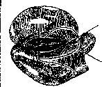
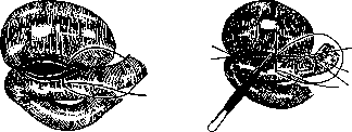
31 side of the efferent limb, sometimes to such an extent that the passage into it is closed as if by a valve; from this moment, the flow of gastric contents into the peripheral section of the intestine ceases completely, everything spills over into the afferent end, which becomes stretched to extreme proportions, compressing the efferent limb at the same time. To this stage, however, things do not always come, because patients die earlier from exhaustion and intoxication. The causes of spur formation are diverse and, apparently, not yet fully elucidated. The former view of many surgeons that a freely permeable pylorus contributes to the development of circulus vitiosus due to the fact that food masses continue to pass not through the anastomosis, but via the normal pathway through the pylorus, and thus stretch the afferent limb and compress the efferent one, does not withstand criticism and has been refuted by subsequent observations. Therefore, the exclusion of the pylorus, proposed by Doyen and Mayo, proves to be superfluous as a preventive measure. Just as untenable proved to be the explanation given at the time by Mikulicz, who attributed the formation of the spur to atonia of the stomach. The true and principal cause of spur formation must be considered to be the primary kinking of the intestinal loop at the anastomosis; initially it may be insignificant, but then, under the influence of the disturbances in the movement of food masses and duodenal juices caused by it, the spur protrudes fatefully sharper and sharper, and the main role in the entire phenomenon begins to belong to it. The immediate causes of the kinking of the intestine are: a) the inclusion in the suture of too wide a strip of the intestinal walls, which leads to their flattening out against the anastomosis in the shape of a patch; b) the position of both limbs of the intestine side by side, closely one next to the other, especially if they grow together in such a position, e.g., in the Billroth-Brenner and Courvoisier method (see below) or with a very small anastomosis; c) any tension of the intestinal mesentery forces both limbs to come together and form an acute kink; for example, if the afferent loop is taken very short, close to the duodenojejunal flexure, or the anastomosis in the stomach is made far upwards, or the stomach, forcibly pulled out of the abdominal cavity, upon being replaced draws the intestinal loop upwards along with it, which can also, in addition, change its position in another sense: the efferent limb can rise above the afferent one or rotate 180°, so that it stands in relation to the afferent one in the sagittal plane, with one limb crossing over the other. Excessive overfilling of the stomach, e.g., during its atonia or postoperative paresis, also contributes to such twisting of the intestinal loop.
)
\ to the spine in lordosis.
/
| As you
/
V U/ ) ) mentioned above) is entero
y f-^J anastomo-sis according to Braun
/ A- ynu between the afferent
W and efferent limbs
pиc. 6. (see figure 6); in many cases of already fully pronounced circulus vitiosus, this technique during secondary intervention made it possible to eliminate the phenomena of obstruction and save patients doomed to death. The majority of surgeons at present, if circumstances compel the use of Wölfler's method, always combine it with Braun's enteroanastomosis. Among other techniques, the following should be mentioned: 1. Suturing the efferent, and especially the afferent, limb to the walls of the stomach far beyond the boundaries of the anastomosis itself ("suspension of the loop" according to Kappeler), in order to avoid kinking of the intestine (see figure 7). 2. So-called "valvular anastomoses"; their essence boils down to certain modifications of the suture in order to create a semblance of a valve at the anastomosis, directing the stomach contents and intestinal juices into the efferent limb: such are the methods of Chaput,
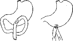
Figure 7.
Figure 8.
Sonnenburg, Faure (Chaput, Sonnenburg, Faure), Zykov, and especially Kocher. Gastroenterostomy according to Kocher is performed as follows: the intestinal loop is sutured to the wall of the stomach not in the frontal plane, but in the sagittal one, the afferent limb is placed posteriorly, the efferent anteriorly; in addition, the incision in the intestine is made semilunar, with a convexity towards the periphery (see figure 8). Of all methods of valvular anastomosis, only Kocher's method had practical significance, mainly due to the high authority of the author; at present, valvular anastomoses, even in this form, have been abandoned by everyone. The most rational method was proposed by Roux. It consists in the fact that the intestinal loop is transected and the peripheral end is implanted into the stomach, the central one into the peripheral: thus, a Y-shaped anastomosis figure is obtained (see figure 9). Roux's method completely guarantees against the development of circulus vitiosus and at one time had fairly wide application, but then had to yield its place to later proposals that turned out to be simpler and just as effective. The biggest and most decisive shift in the improvement of the methodology of gastroenterostomy occurred from the moment when the anastomosis began to be made not on the anterior, but on the posterior wall of the stomach and behind the transverse colon (gastroenterostomia posterior retrocolica). This modification, first proposed by Hacker, quickly began to displace anterior gastroenterostomies. Even before Hacker's proposal, some surgeons tried
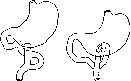
Figure 9.
Figure 10.
to eliminate the disadvantageous aspects of antecolic gastroenterostomy (gastroenterostomia anterior antecolica) by passing the intestinal loop behind the transverse colon through the mesocolon and the gastrocolic ligament and suturing it to the anterior (Brenner) or posterior wall of the stomach (Courvoisier). Both of these proposals were not successful, as they were associated with the danger of incarceration of the intestine in the ring of the mesenteries. With Hacker's method, all those complications are eliminated which are associated with the abnormal position of the intestinal loop in any antecolic (anterior) gastroenterostomy; the operation by this method is performed as follows: the stomach and transverse colon are withdrawn from the abdominal cavity; the mesentery of the latter is stretched and the duodenojejunal flexure is found; 25–30 cm from the duodenojejunal fold are measured out and clamps are placed on the intestine. Then an opening is made in an avascular place in the mesocolon of the transverse colon, and the assistant protrudes the posterior wall of the stomach into this slit. The edges of the slit in the mesocolon are fixed with several interrupted sutures to the posterior wall of the stomach, closer to its greater curvature, in order to prevent subsequent incarceration of the intestine in the opening of the mesocolon, on the one hand, and the sinking of the intestines into the omental bursa, on the other (see figure 11). Further on, the formation of the anastomosis itself proceeds in the same way as with Wölfler's method. Posterior gastroenterostomy immediately gave a significant jump in the outcomes of the operation in terms of improving immediate and functional results, however, circulus vitiosus was also observed with this method. A further and most essential improvement in the methodology of posterior gastroenterostomy was introduced by Petersen from Czerny's clinic;
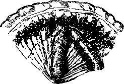
Figure 11.
its essence boils down to making a gastroenterostomy "without a loop." Petersen called his method physiological, since with it the mutual relationship of the organs (the stomach and the beginning of the jejunum) remains normal; the opening in the intestine is made at the duodenojejunal fold itself or, with marked dilation and lowering of the stomach, no further than 10 cm from it; in this case, the intestine retains its natural position and direction not from left to right, but from top to bottom, and, thus, we are no longer talking about a right and left limb, but rather about an upper and lower one (see figures 10 and 12; gastroenterostomia verticalis). Under such conditions, there is no ground for the formation of a kink and spur. Clinical experience soon confirmed the correctness of Petersen's reasoning, and since then this method has occupied a dominant position, serving almost as a faithful guarantee against circulus vitiosus. Those few cases where it nevertheless developed, upon closer inspection, turned out to be either cases operated on with a departure from Petersen's method
fig 12 Various modifications of the posterior G. (Petersen's method) concerned only minor additions, which moreover are not universally accepted. Besides the circulus vitiosus, a number of other complications after G. pointed to the necessity of 1) improving the technique of the gastro-intestinal anastomosis (suture) and 2) clarifying the detailed particulars of the operation as a whole. After all methods of G., intestinal obstruction was consistently observed due to internal incarcerations or volvulus of the intestines. The cause of these is the circumstance that after the operation artificially created slits remain between organs, as, for example, after anterior G.-a slit under the intestine sutured to the stomach; sometimes the slit in the bursa omentalis remains unclosed. Even after posterior G. according to Petersen, 'without a loop,' a narrow slit remains between the mesentery of the small intestine and the transverse colon, and cases are observed where individual intestinal loops slip into these slits or even a large part of the intestine goes in and becomes incarcerated there. This complication is of such grave significance that it should always be kept in mind and prevented by closing all slits with sutures. As for the technique of intestinal suture, we will mention here only certain particulars directly related to G. In the early years, when the operation still gave a high percentage of mortality from insufficiency of the sutures, numerous 'safe' methods were tested; these include the so-called 'two-stage G.s,' the essence of which is that communication between the stomach and intestine is established spontaneously after several days; during the operation, however, a constricting elastic ligature is placed between the stomach and intestine, or the walls of the stomach and intestine are turned into a slough (by cauterization or with chemically cauterizing substances) and a protective suture is placed around them. All these methods have only historical interest and have finally disappeared from the scene. On the other hand, numerous attempts were made to replace the gastro-intestinal suture with the help of 'prostheses' consisting of various kinds of plates on which the edges of the future anastomosis were covered. Of these proposals, the Murphy button proved the most durable, but it has now been abandoned by everyone, finding application only in exceptional cases. Surgery has entirely returned to the starting point-the silk or catgut suture, which, however, has many modifications, each with its own positive and negative sides. The question of the most perfect suture for G. remains unresolved to this day; the striving for improvement is dictated, mainly, by two sequentially observed complications-narrowing of the anastomosis and the development of secondary peptic ulcers in the anastomosis. Both these complications currently attract the most attention from surgeons. The first-gradual narrowing of the anastomosis can reach the degree of its complete overgrowth; usually this is the result of gross technical errors, but to a certain degree narrowing is inevitable in all cases due to scarring of the edges of the anastomosis. The scarring narrowing will be the more pronounced, the worse the process of wound healing was, the more pronounced was the infiltration along the edges of the anastomosis. Meanwhile, in the healing of stomach wounds, the inflammatory reaction is inevitable, since the wounds are not aseptic and irritation is maintained by the presence of sutures; of the silk sutures, only a part is absorbed, another part is cut through into the intestine, and at autopsies it often happened to find a silk thread of a continuous suture hanging in the form of a loop at the edges of the anastomosis; in other cases, after the threads are cut through, deep fistulous tracts remain for a long time. All this indicates that in the technique of creating an anastomosis, the last word has not yet been said. It is necessary to note the following most essential points in applying a gastro-intestinal suture: 1) the edges of the anastomosis should be covered with mucous membrane, since only the stomach mucosa protects them from the corrosive action of gastric juice; 2) for applying sutures in general, it is more advantageous to use thin catgut, or 3) according to Bir's proposal, to apply a knotted silk suture in one row. At one time Kelling expressed the view that the anastomosis narrows if the pylorus remains patent and the anastomosis does not function; this view is completely erroneous. The second complication that forced a re-examination of certain technical questions of G.-these are secondary peptic ulcers developing in the anastomosis itself or in the nearest sections of the jejunum. The frequency of development of such ulcers to this day remains controversial: while some authors consider them a great rarity, in others the percentage of ulcers reaches 1.5-4, and according to some even 9%. In the Mayo Clinic, out of 7,000 cases of G., a secondary ulcer was observed in 168 cases (1.4%). According to Russian statistics, it was observed in 0.7-2% of cases. In reality, the frequency of secondary ulcers should be evaluated with a higher figure, since in many cases the ulcer proceeds latently and cannot be diagnosed on the basis of clinical symptoms. Even ulcers that have perforated into the transverse colon sometimes remain unrecognized until the end. The symptoms of perforation are-rapidly developing exhaustion, sharp intensification of pains, foul eructation with a clear fecal odor, vomiting with fecal odor in the absence of obstruction symptoms, diarrhea of undigested food. Sometimes a very good diagnostic procedure is the introduction per clismam of a liquid colored with some dye (for example, methylene blue), followed by extraction of the gastric contents with a probe. The appearance of colored liquid in the stomach speaks for the presence of a fistula. According to statistics of various authors (Brams, Meyer), this complication occurs, approximately, in 20% of peptic ulcers of the jejunum. Considering the unclear clinical manifestation, some authors evaluate the frequency of secondary ulcers with extremely high figures-up to 34% (Lewson) and even 40%. If these last figures at least approximately reflect reality, the situation could be called catastrophic; in any case, the question deserves the most attentive study. The immediate cause of secondary peptic ulcers is obviously the same, not yet more precisely clarified factors that underlie the occurrence of round ulcers of the stomach itself, since both these forms represent phenomena of the same order; without dwelling on them in more detail, one can only note the conditions that are created by the G. operation itself. After every G., the edges of the anastomosis and the nearest sections of the jejunum are subjected to the digestive action of gastric juice, and this danger must be measured not only by the height of the acidity figures but also by the general peptic power of the gastric juice. The mucous membrane of the jejunum is physiologically unadapted to resist this digestive action, meanwhile the gastric contents enter the intestine not neutralized by bile and pancreatic juice, since the conditions of their secretion after G. are sharply disrupted due to the loss of physiological reflexes. The circumstance that after exclusion of the pylorus according to Eiselsberg-Duayen ulcers develop especially frequently (in 20% according to Haberer, 27% according to Clairmont) clearly shows that this moment is extremely important. A second factor contributing to the development of ulcers is the trauma inflicted on the walls of the stomach and intestine during the operation itself: the edges of the anastomosis, not protected by mucous membrane, the presence of sutures, hematomas at the site of application of clamps, and other factors. It should be considered sufficiently proven that it is dangerous to supplement G. with exclusion of the pylorus; similarly, one should not operate according to the Roux method, and even the entero-anastomosis according to Brown is disadvantageous, since (in both methods) the neutralization of the food bolus occurs only below the intestinal anastomosis. Bearing in mind the severe consequences and persistence of secondary peptic ulcers, a corresponding regimen and treatment should be prescribed for patients after the operation for a prolonged time for preventive purposes. After the operation of creating a gastro-intestinal anastomosis, significant changes can be naturally expected both in the motor and in the secretory activity of the stomach. Similarly, the chemistry of digestion of food and its assimilation should also change. A huge number of clinical and experimental works are devoted to the study of these questions, but the conclusions of various authors are far from uniform and partly contradictory. This finds its explanation in the fact that the changes in the chemical and, especially, motor activity of the stomach that occur after G. depend also on how perfectly the operation itself was performed.
Meanwhile, the quality of the operation often suffers as a result of imperfect operative methods or from successive complications (such as: insufficient size of the anastomosis from the very beginning, its inconvenient position) or successive narrowing of the anastomosis, from the development of new ulcers or adhesions, formation of various other mechanical obstacles to the passage of gastric contents into the efferent loop, and so on; it is not always possible to take all these factors into account when evaluating the functional results of G. clinically; the diversity of these factors, due to the heterogeneous selection of cases studied, explains the inconsistent and partly contradictory data, both presented by different authors and obtained by the same authors in different cases. The methods for studying the motor activity of the stomach are either pumping out with a probe or roentgenoscopic examinations; the latter are particularly indicative, but it must be borne in mind that they do not always coincide with the former. On the screen, one can be convinced that only in exceptional cases does the stomach contents 'fall through' the anastomosis into the intestinal loop; usually, however, the time for emptying the stomach is only slightly shorter than normal. Obviously, from the very beginning there is some apparatus regulating the passage of food masses into the intestine. The assumptions made by some authors that a new sphincter develops in the anastomosis over time are already refuted by the fact that the regulating apparatus begins to function from the first days; microscopic studies of the edges of the anastomosis many months after the operation reveal no anatomical substrate for speaking not only of the development of a new sphincter, but even of local hypertrophy of the muscular layer. In addition, it remains incomprehensible how such a sphincter could function without a special nervous apparatus. The thought naturally suggests itself that the intestinal loop takes on the role of the sphincter, which allows food masses to pass only in the quantity and gradualness as is characteristic of its physiological function. The entire mechanism can be roughly compared to a faucet, which is periodically opened, and the contraction of the walls of the intestinal loop can be equated to the periodic opening of the faucet. With regard to functional results, cases of cancerous and cases of benign diseases of the stomach should be considered separately, and in addition, the result should be evaluated taking into account not only physiological norms, but comparatively, i.e., taking into account the condition in which the functions of the stomach were before the operation. Cases of cancerous diseases. Since in cancer G. is considered indicated only in the presence of stenosis of the pylorus, i.e., with sharply delayed emptying of the stomach, the motor activity after the operation shows marked improvement; in many cases it reaches physiological norms, in others, when pumping out on an empty stomach, food residues can still be obtained; accelerated emptying of the stomach is observed only in rare cases. The change in the chemical nature of gastric digestion and, in particular, the state of acidity in cancer patients is not of great interest, since even before the operation free HCl is absent in most patients, and further changes occur less depending on the operation than on the spread of the new growth.-Cases of benign diseases of the stomach are of the greatest interest, all the more so that here observations can continue for an indefinitely long time. Changes in chemical activity manifest differently: in some cases, a sharp decrease in the quantitative content of free HCl is noted, sometimes reaching its complete absence. Hypersecretion can also cease immediately after G.; conversely, in cases where HCl was absent before the operation, after more or less significant periods, its reappearance was sometimes noted. But in most cases, the amount of free hydrochloric acid does not show significant changes. The motor function is restored to normal only in a minority of cases: as an exception, accelerated emptying of the stomach compared to normal is observed, sometimes even rapid 'falling' of food into the intestine; but in most cases, motor function remains slowed down. Severe disturbances remain in cases complicated by atony of the stomach and adhesions. The area of application of G. First performed for cancer of the pylorus, G. found much greater application in benign forms of stomach diseases. Monastyrsky in 1885 was the first to operate on a patient with stenosis of the pylorus on the basis of a round ulcer, and since then in the casuistry of G. cases of scar strictures predominate, in which the operation is a radical method, whereas in cancer it is only a temporary palliative. Attempts to expand the area of application of G. in cancer to cases where the tumor occupies areas remote from the pylorus and, consequently, is not accompanied by pylorostenosis, proved fruitless and were abandoned; at present, most surgeons prefer in inoperable cancers, if the tumor is located far from the pylorus, to limit themselves to a trial laparotomy, rather than creating a gastrojejunal anastomosis. But in the sphere of benign diseases, G. has gone far beyond the area of pylorostenoses and throughout its history has been tried in almost all kinds and forms of stomach diseases. The area of its application remains insufficiently precisely defined at present, but on the other hand, it has become sufficiently clear that with incorrect indications G. sometimes causes more harm than good. With full definiteness, areas can be indicated where G. should be considered indicated and where it has advantages over other competing operations (resection, pyloroplasty, etc.). These include: 1) scar strictures of the pylorus after burns and healed stomach ulcers; 2) congenital strictures; 3) strictures of the pylorus caused by adhesions after trauma or inflammatory processes in the bile ducts or of other origin; 4) ulcers occupying the antral part of the stomach or duodenal. In stomach ulcers located far from the pylorus, G., according to the observations of some authors, is just as indicated as in pyloric ulcers (Galpern), while others consider it in these cases an operation that gives a satisfactory result in only half of the cases. The basis for G. in these cases is the assumption that the presence of an anastomosis paralyzes the spasm of the pylorus; indeed, roentgenoscopy shows that with the presence of an anastomosis food masses begin to pass more quickly through the pylorus, and soon almost all gastric contents leave by this way, bypassing the anastomosis, which thus, as an exit from the stomach, proves to be superfluous. In other cases, the beneficial effect of the anastomosis is explained by a significant decrease in the acidity of gastric juice, but, as has already been pointed out above, in most cases no marked change in this respect is observed. Thus, in ulcers remote from the pylorus, the effect of G. is problematic, and in each individual case it cannot be predicted in advance. As for cases of functional diseases of the stomach, such as hypersecretion, hyperchlorhydria, atony, neuroses, or cases with an anatomical basis but without signs of stenosis, such as gastoptosis, dilatation of the stomach, then here G. should be applied with great caution, since it is precisely in these cases that the operation itself creates a new peculiar state, described by Pribram (Pfi-bram) under the name 'gastroenterostomy as a disease' (see below). The results of G., both immediate and remote, should be evaluated differently for cancerous and benign cases. - Cancerous cases. The immediate mortality, initially reaching very high figures (up to 75%), gradually decreased as the technique of the operation was developed and at present hardly exceeds, on average, 15-20%. Only a relatively small percentage become victims of technical errors, the majority of patients die from the underlying disease and associated complications (marasmus, diarrhea, bleeding from the tumor, pulmonary complications). The effect of the operation and its duration in cancer patients depends on the stage of the disease, the degree of spread of the new growth and the associated secondary complications; in some patients after the operation all painful symptoms cease, and the general condition improves remarkably quickly, weight sometimes increases by 20 kg, in others the operation gives almost no relief or only some symptoms are eliminated by it; finally, sometimes its effect is too short-lived, and soon the old or other symptoms appear, depending on the further growth of the tumor. Here everything depends on how much the picture of pylorostenosis is complicated by symptoms from other organs; if the entire symptom complex depends exclusively on the obstruction of the pylorus, the operation has a remarkable effect, but where the symptoms of stenosis recede into the background, much cannot be expected a priori from the operation.
In all cases of gastroenterostomy performed for cancer of the pylorus, the result can hardly be considered good in half of them. The average duration of life after G. is 7-8 months, but in individual cases patients lived after the operation for 2 and even 3½ years (Alsberg, case of colloid cancer); at the same time, erroneous calculations are possible, since the tumor, considered cancer at the time of the operation, could in reality have been a callous ulcer, only later undergoing cancerous degeneration. Cases of benign diseases. For this group of patients, the immediate mortality rate, currently varying in the hands of individual surgeons from 12% to 0%, is on average approximately 3%, with the larger part of fatal outcomes belonging to the group of open ulcers, and patients become victims of bleeding, perforation of ulcers, or postoperative pulmonary and other complications. The immediate effect of the operation in most cases is good, often brilliant, however, in a significant percentage of cases it is still not complete or turns out to be only temporary and sometimes short-lived. In this regard, cases must be divided into different categories: in cases of completed ulcerative processes, in cases of stenosis of the pylorus, the effect of the operation is complete from the very beginning, and if sometimes new deteriorations occur, this is mainly due to the various complications mentioned, inherent in gastroenterostomy. In cases of open ulcers, the effect of the operation, besides the complications just mentioned, is sometimes unsatisfactory from the very beginning - either because the ulcer has no tendency to heal, or because new ulcers develop or recurrences appear at the site of the old ulcer; finally, some ulcers subsequently undergo cancerous degeneration. The percentage of consistently occurring deteriorations or 'recurrences' is assessed very differently by various authors, but very authoritative names indicate figures of 37½% (Garre) - 50% (Mayo). Many surgeons draw attention to the fact that gastroenterostomy, performed in the absence of proper indications, itself becomes a disease, and the condition of patients only worsens compared to what it was before the operation: pains become constant, belching, vomiting, accumulation of a large amount of acidic gastric juice in the stomach develop, and general exhaustion sets in. The reason for this, according to Pribram, is that the gastric contents, entering from the anastomosis into the afferent loop, causes reflex secretion of gastric juice, which more often occurs after posterior G. with a short loop, i.e., after the modification which is undoubtedly the best of all methods of G. In a number of cases, after unsuccessful attempts to correct the situation with various additional surgical techniques, which often do not lead to the goal, recovery occurs only after gastroenterostomy is eliminated and the status quo ante is restored. In the Mayo clinic, out of 7,000 cases of gastroenterostomy, 343 required this measure.
Related articles
Mentioned in
Cite this page
“Gastroenterostomy.” Soviet Medical Encyclopedia. English translation of Bolshaya Meditsinskaya Entsiklopediya, 1st ed. (Moscow, 1928–1936), ed. N. A. Semashko. https://sovietmedicalencyclopedia.pages.dev/article/gastroenterostomy/