Billroth Operations on the Stomach
Historical document, translated for reference. It reflects medical knowledge of the 1920s–30s and is not medical advice.
Summary
The Billroth operations on the stomach refer to two methods of gastric resection developed by Theodor Billroth in the 1880s. These surgical techniques revolutionized the treatment of stomach cancer and established the foundation for modern gastric surgery.
Encyclopedia article (1928–1936)
BILLROTH OPERATIONS ON THE STOMACH, two methods of one of the most important operations in practical surgery—resection of the stomach. Experimental surgery began to approach this goal 70 years before Billroth (experiments by Merrem), but despite the participation of such major surgeons of his time as Gussenbauer and Czerny, operations on animals invariably ended in failure. Attempts to directly transfer these experiments to the clinic (Reap, Riediger) were also negative. B. was the first to achieve complete success in his case of pyloric cancer, operated on by him on February 28, 1881; from this date begins the rapid development of stomach surgery. Its flourishing was facilitated by the teachings of Pasteur and Lister about wound infection, about antiseptics and asepsis, which marked the beginning of a new era in surgery. The first resection operation consisted in circular excision of the pyloric part; this technique remains the same for more extensive excisions (ending with removal of the entire organ), essentially remaining to the present time. Modifications introduced later by other authors concerned either minor details or did not receive wide development. Almost simultaneously, Billroth proposed two methods of resection. -1st method of Billroth consists in circular excision of one or another part of the stomach and connecting the segments end-to-end with sutures (see Figure 1). The abdominal cavity is opened along the midline from the xiphoid process to the navel; with extensive resections, low mobility of organs, as well as under all other conditions requiring expansion of access, the incision is increased either by excision of the xiphoid process or by a transverse incision through the rectus abdominis muscle to one side or the other. After examination (see below) has confirmed that resection (removal of the cancerous tumor) is possible, the lesser omentum is first separated up to the boundaries of the planned resection by means of a ligature needle and double ligatures, then in the same way the gastrocolic ligament is separated, with the glands lying along the greater and lesser curvatures being left on the side of the tumor and removed together with it. The last ligatures at both ends must include the main gastric vessels (right and left, upper and lower coronary arteries and veins); isolated ligation of vessels is usually not performed. Under the part separated from the ligaments and adhesions, a wide layer of gauze is placed, and the entire abdominal cavity is carefully isolated with gauze sponges. On both sides of the planned lines of incision, clamps are applied (see Figure 2), and between them the part to be removed is severed. Then they proceed to sewing the end of the duodenum into the stomach incision; since the latter is always significantly larger in diameter than the duodenum, it is first sutured, starting from the lesser curvature,
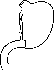
Figure 1. Diagram of Billroth I operation.
so that the remaining unsutured part equals the diameter of the duodenum (see Figures 3 and 4). Other modifications in the sewing method initially proposed by Billroth were abandoned as irrational; but even in the typical method of Billroth, the "joint" of the two
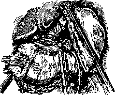
Figure 2. (After Bier-Braun-Kummell).
lines of sutures remains the weakest point, and special attention must be paid to this point; in this case, it must be borne in mind that a too dense and tightly drawn suture itself can lead to necrosis of the edges and rupture of the sutures. The danger of rupture of the sutures is greater the more extensive the resection, the less mobile the organs, Figure 3. Billroth I. The larger part of the stomach incision is sutured; the sewing of the duodenal lumen into the stomach has begun (after Bier-Braun-Kummell). since all this leads to tension in the sutures; in addition, the end of the duodenum is poorly supplied with blood vessels, and its posterior wall, if the resection is performed far from the pylorus, is not covered with a serous membrane.-Taking into account these disadvantages, Billroth as early as 1881 proposed for cases of extensive resections his 2nd method (see Figures 5, 6 and 7), differing from the 1st in that both ends—the stomach and duodenum—are completely closed and the connection between the stomach and intestine is restored by means of anterior or posterior gastroenterostomy. In the technique of the 2nd method of B., it is necessary to note a very critical moment—complete closure of the end of the duodenum (see Figure 6), since its stump most often served as the site of insufficiency of the sutures. The cause of this are the two above-mentioned features of the structure and blood supply of the intestine, and in addition, the technical difficulty of applying a hermetic suture to a difficult
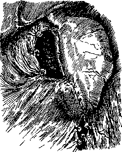
Figure 4. Billroth I. The lumen of the duodenum
is sewn into the stomach; the operation is completed (after Bier-Braun-Kummell). accessible and poorly mobile part. The best method for closing the duodenum—ligation of its end with a ligature or suturing through its entire thickness with a Graser clamp, followed by a second row of purse-string sutures applied 1 cm from the ligation, and burying the stump inward. In the technique of gastric resection, clamps are of great importance. In addition to the usual gastroduodenal clamps, here clamp-excrasers are used, which facilitate the technique of suturing itself. Such are, for example, Graser clamps with a longitudinal slot along the entire length of both branches; after removal of the part to be removed, all layers of the wall are sutured together through this slot with a straight needle, then the clamps are removed and the line of sutures is buried inward with a second row of sutures (see Figure 7). Clamp-excrasers (Duayen, Kocher, Martel) are used with the aim of crushing linearly all layers of the wall: in the groove only the serous membrane remains uncrushed; a continuous suture applied behind the clamp closes the lumen of the organ, and in addition,
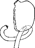
Figure 5. Diagram of Billroth II.
a second row of sutures is applied. The abdominal wound after any resection is usually completely closed; the introduction of tampons is only harmful and sometimes served as a cause of rupture of the sutures. This technique remains the dominant one to this day. As for the comparative merit of the 1st and 2nd methods of Billroth, each has its own negative and positive sides. In addition, the following must be added: with the 1st method, anat.-physiol. relationships remain least disturbed, distortion in the movement of food masses cannot occur, and in a functional sense this method is the most perfect. But in terms of technical feasibility, it cannot be considered universal and always feasible due to the great tension in the sutures; mobilization of the duodenum according to Kocher does not always prove sufficient. In this respect, the 2nd method of Billroth has all advantages and can be Figure 6. performed even with the most extensive resections. Meanwhile, in the removal of stomach cancer, the basic condition is to excise the affected part as far as possible from the boundaries of the neoplasm, and the 2nd method of Billroth makes it possible not to be constrained by the size of the tumor. So-called subtotal and total resections are essentially also performed according to the technique of Billroth.-Total gastrectomy (first performed by Schlatter in 1897) can be called Figure 7. Closing of the stump only when KO-stomach^by^Vrapp- when the section is made along the esophagus, but usually a part of the cardia and dome of the stomach remains. The preservation of this area, however small it may be, greatly facilitates the technique of sequential restoration of the continuity of the gastroduodenal canal

Closing of the duodenal stump (after Bier-Braun-Kittel).

and ensures the strength of the suture. In addition, from the remnants of the cardia, a cavity is formed—something like a new stomach—with a capacity of up to 100 cubic cm. With total resection, the esophagus is either connected to the end of the duodenum or sewn into a loop of the jejunum. - The area of application of resections—above all, stomach cancers. Removal of the tumor is always performed by circular resection, more or less extensive, depending on the size of the neoplasm. In the absence of other methods of treatment for stomach cancer, resection is currently the only possibility to save the patient; therefore, practically the question reduces not to establishing indications for the operation, but to determining the possibility
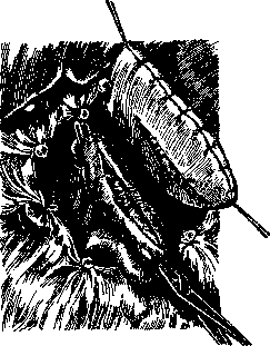
Figure 8. The serous and muscular membranes of the stomach and a loop of jejunum are incised for the purpose of applying an anastomosis (after Bier-Braun-Kummell).
of resection and the general condition of the patient. The size of the tumor is least of all a contraindication to resection. The possibility of the latter is determined by the absence of cachexia, metastases, seeding of the peritoneum (ascites), infiltration of neighboring organs (liver, pancreas, transverse colon), and the degree of damage to the lymphatic apparatus. Resections are also used for gastric ulcers; the attitude of modern surgery to the question of excision of ulcers is not yet fully determined. As for the outcomes of resections, in gastric cancer the immediate mortality remains high, varying among different surgeons from 20 to 50%. Any complications of the operation, so-called 'extended operations', significantly worsen the prognosis of the immediate outcome, without providing any benefit to the patient in other respects. Depending on the stage of the disease and the complexity of local conditions, mortality rates for different groups vary within very wide limits - from 7 to 37% (Finsterer). Total and subtotal resections, the number of which now exceeds a hundred, give a mortality of about 50%. In gastric ulcers
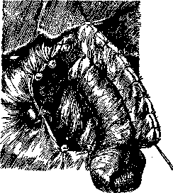
Figure 9. Billroth II. Posterior anastomosis has been applied. Operation completed (according to Bier-Braun-Kilmmell).
the outcomes of resections are significantly better; mortality, on average, is about 6%, but with individual surgeons this figure rises to 12%.-Hemostatic forceps, see Surgical instruments.
Related articles
Cite this page
“Billroth Operations on the Stomach.” Soviet Medical Encyclopedia. English translation of Bolshaya Meditsinskaya Entsiklopediya, 1st ed. (Moscow, 1928–1936), ed. N. A. Semashko. https://sovietmedicalencyclopedia.pages.dev/article/billroth-operations-on-the-stomach/