Peritoneum
Historical document, translated for reference. It reflects medical knowledge of the 1920s–30s and is not medical advice.
Summary
The peritoneum is a serous membrane lining the abdominal cavity and covering abdominal organs. It consists of a thin transparent sheet with connective tissue foundation and is divided into parietal and visceral layers, forming various ligaments and mesenteries.
Encyclopedia article (1928–1936)
Peritoneum, peritonaeum, a serous membrane lining the internal surface of the abdominal wall and covering the organs located in the abdominal cavity (see Abdominal cavity). The peritoneum is a thin transparent sheet, the free (facing the peritoneal cavity) surface of which in the normal state is smooth, shiny, and moistened with serous fluid. -Structure of the peritoneum. The foundation of the peritoneum consists of connective tissue containing elastic fibers and numerous bundles of collagen fibrils; from the surface it is covered with a single layer of flat epithelium (so-called endothelium). Between the epithelial cells are scattered the smallest pores-stomata; through these the peritoneal cavity communicates with the intertissue spaces. With the underlying parts (abdominal walls, viscera) the peritoneum is connected by means of loose connective tissue (so-called subserous membrane, tunica subserosa), which contains blood and lymph vessels, nerves, numerous elastic fibers, and fat cells in varying amounts. Tunica subserosa is not equally developed everywhere (e.g., on the liver it is completely absent); where it is well developed, the peritoneum is mobile. Cavum peritonaei, the peritoneal cavity, coincides with the boundaries of the abdominal cavity: above it is bounded by the diaphragm, in front and on the sides by the abdominal muscles, below it freely passes into the pelvic cavity and is closed by means of the pelvic diaphragm (m. levator ani). The peritoneal cavity in the male is closed, in the female it communicates with the external world through the paired openings of the uterine tubes. In the peritoneal cavity, under normal conditions, there is a minimal amount of transparent serous fluid, liquor peritonaei, which moistens the surface of the peritoneum and fills the slits between organs and walls with a very thin layer. These slits form a whole labyrinth and constantly change their configuration depending on the position of the organs, most of which possess considerable mobility. The displacement of organs in relation to each other and to the abdominal wall occurs easily and without any friction because all contacting surfaces are smooth and moist: both the viscera and the abdominal wall are covered with peritoneum; therefore, in the peritoneum one can distinguish the parietal sheet, lamina parietalis, and the visceral, lamina visceralis; in many places the sheets pass into each other in the form of so-called peritoneal ligaments, ligamenta peritonaei. Therefore, under the term ligament in this case is meant the transition of the peritoneum from one organ to another or from an organ to a wall; at the same time only certain ligaments appear in the form of clear, more or less isolated folds, which freely project into the peritoneal cavity; among these are the mesenteries and omenta. Under the name mesentery, mesenterium, is understood specifically those peritoneal ligaments that go from the posterior abdominal wall to various parts of the intestinal tube (see Mesentery). The omentum, epiploon, differs from other ligaments by containing a significant amount of adipose tissue and a special, very thin structure. Not all organs of the abdominal cavity relate to the peritoneum in the same way: some are covered by it to a greater, others to a lesser extent. On this basis they are distributed into three groups: 1. Organs covered by the peritoneum on all sides, i.e., lying completely inside the peritoneal cavity, intraperitoneally (with the exception of only a narrow strip or line along which the mesentery is attached); example-the small intestine. 2. Organs covered by the peritoneum only on one side, namely in front; on the other sides the serous membrane is absent, the organ lies outside the peritoneal cavity, extraperitoneally; example-the kidney. 3. Organs covered by the peritoneum on three sides; only on one side (usually the posterior) the organ is devoid of a peritoneal covering, and in front and on the sides it is clothed with peritoneum, therefore it is located mesoperitoneally; example-the ascending and descending colon. When studying the abdominal cavity opened from the front, under normal conditions from the entire complex of internal organs only the liver and part of the anterior surface of the stomach are visible above; below-only small segments of the intestine, the rest is covered by the greater omentum, which descends from the greater curvature of the stomach [see Table I (pp. 147-148), Fig. 11. If one takes the omentum by its lower edge and lifts it upward, then together with it the transverse colon is also raised, the mesentery of which-mesocolon transversum-going from right to left across the abdominal cavity, divides it into 2 floors-upper and lower. In the upper part lies the liver; it is covered with peritoneum on most of its surface and is devoid of it only in the region of the posterior edge, in the adjacent part of the upper side (where the liver is fused with the diaphragm) and in the grooves. The serous covering is very firmly fused with the liver. The transition of the peritoneum from the liver, in the form of special ligaments, partly occurs to the diaphragm (ligamentum falciforme hepatis, lig. coronarium hepatis dextrum et sinistrum and lig. triangulare dextrum et sinistrum), partly to neighboring organs (lig. hepato-gastricum, lig. hepato-duodenale and lig. hepato-renale). Of these ligaments, lig. falciforme hepatis, starting immediately above the navel from the anterior abdominal wall, connects it and the lower surface of the diaphragm along the median line with the upper side of the liver; in the free edge of the ligament is included the obliterated vena umbilicalis-lig. teres hepatis. Not reaching the posterior edge of the organ, the two sheets of peritoneum forming lig. falciforme begin to diverge-one to the right, the other to the left, continuing into ligamentum coronarium on each side, connecting the liver and diaphragm in the frontal plane (ending on the left and right in the form of a free triangle-lig. triangulare hepatis). Lig. hepato-gastricum s. omentum minus [see Table II (pp. 147-148), Fig. 4] goes from the porta hepatis to the curvatura minor of the stomach and passes into lig. hepato-duodenale, connecting the porta hepatis with the beginning of the duodenum; in this ligament are included the vena portae, a. hepatica and ductus choledochus. The right free edge of the ligament limits in front a special opening leading from the general peritoneal cavity into the cavity of the lesser omentum (otherwise-into the vestibule of the greater omentum, vestibulum bursae omentalis), so-called foramen epiploicum s. Winslowi. The latter is bounded behind by the transition of the peritoneum from the liver to the right kidney-lig. hepato-renale, below-by means of lig. duodeno-renale and above-by the substance of the liver itself, namely-tuber caudatum of it. -The stomach is covered by the peritoneum on all sides and, besides lig. hepato-gastricum (see above), has the following ligaments: lig. phrenico-gastricum (connects the diaphragm with the cardia of the stomach); lig. gastro-lienale (goes from the fundus of the stomach to the hilus of the spleen; in its origin it represents, together with the following ligament, a modification of the dorsal mesentery of the stomach-see further the outline of the development of the peritoneum); lig. gastro-colicum s. omentum majus, the greater omentum, consists of 4 layers, with the 2 anterior layers, starting from the curvatura major, descend far downward and at the free edge of the omentum pass into the 2 posterior layers; the latter, ascending upward, fuse with the transverse colon, with the mesocolon transversum and then continue into the parietal peritoneum of the posterior abdominal wall. Between the anterior and posterior layers of the omentum is a slit-like space-the omental cavity, bursa omentalis (in adults it often is absent), which communicates with the vestibule of the omentum through an opening that also has the form of a slit-foramen pancreatico-gastricum s. isthmus bursae; the latter is bounded in front by the stomach, behind by the pancreas, on the sides by the transitions of the peritoneum between these organs, especially sharply on the left side, where plica gastro-pancreatica is described. -The spleen is covered by the peritoneum everywhere, except at the hilus; with the diaphragm it is connected by lig. phrenico-lienale. Here it is necessary to mention lig. phrenico-colicum; this ligament is not directly connected with the spleen, but supports it from below, forming the so-called saccus cecus lienis; it is thrown below the spleen from the pars costalis of the diaphragm (in the region of IX-XI ribs) to the flexura colica sinistra. -The duodenum, as well as the pancreas, on most of its length lies outside the peritoneum, crossed in front by the mesentery of the transverse colon; only at the very beginning (at the pylorus) and at the very end (at the transition to the jejunum) it is covered by the peritoneum. Throughout their entire length they have a complete peritoneal covering: jejunum, ileum, processus vermicularis, transverse colon and flexura sigmoidea (description of their mesenteries-see Mesentery). -The cecum is covered by the peritoneum on all sides (more rarely on three), but does not have a mesentery. The ascending and descending colon have only the posterior side devoid of peritoneal covering. The rectum in the upper part is covered by the peritoneum on all sides (it also has a mesentery), in the middle on three sides and in the lower part only in front. The kidneys together with the adrenal glands, ureters, abdominal aorta and inferior vena cava are located in the retroperitoneal cavity and are covered by the peritoneum only from the front. The urinary bladder in the empty state is covered by the peritoneum only from above, when filled it is an organ of the mesoperitoneal type. From the lateral surface of the recti to the urinary bladder, along the wall of the pelvis, on both sides stretches a fold of the peritoneum-plica recto-vesicalis; the space located between the rectum and bladder, below these folds, is called excavatio vesico-rectalis. In the woman this space is divided by the uterus and its broad ligaments into the anterior-excavatio vesico-uterina, and the posterior-excavatio recto-uterina s.
Douglas; the latter is significantly deeper, as the peritoneum, covering the uterus, descends along its posterior surface lower than along its anterior; in front the peritoneum reaches only to the level of the internal os of the uterus (consequently covering only the body of the uterus); behind it lines not only the body and cervix of the uterus but also the wall of the vagina in the region of its posterior fornix. The above-mentioned plicae recto-vesicales in women correspond to plicae recto-uterinae s. Douglasi, connecting the lateral sides of the rectum and uterus. The uterus, tubes, and ovaries lie intraperitoneally (for their relation to the broad ligament of the uterus see Mesentery); here it is only necessary to note the special name for the peritoneum covering the uterus-perimetrium. The parietal sheet of the peritoneum lines in the form of a continuous covering the inner surface of the anterior and lateral walls of the abdomen, above passing to the diaphragm, below-to the region of the large and small pelvis and reaching dorsally to the spine. At certain points of the parietal peritoneum, near certain sections of the intestinal tube, there are depressions, which are sometimes the site of the occurrence of internal hernias (see Abdominal wall). These include: recessus duodeno-jejunalis, located on the left side of the body of the second lumbar vertebra, between the flexura duodeno-jejunalis on the right and the peritoneal fold-plica venosa-above and to the left; in the latter is contained the vena mesenterica inferior; fossa coecalis (s. retro-coecalis) is located between the cecum and the parietal peritoneum; below it is freely open, laterally limited by the fold of peritoneum descending from the cecum-plica coecalis. Near this fossa there are also two depressions, separated from each other by the end of the ileum: recessus ileo-coecalis superior lies in the upper angle, between the ileum and cecum; recessus ileo-coecalis inferior (more constant) is bounded above by the end of the ileum, behind by the mesentery of the appendix and in front by the fold of peritoneum-plica ileo-coecalis, which extends from the anterior surface of the end of the ileum to the cecum. At the root of the mesentery of the flexurae sigmoideae, if the latter is turned upward, one can sometimes find a depression-recessus intersigmoideus, the form and size of which vary greatly. The development of the peritoneum and mesenteries proceeds in parallel with the differentiation of the viscera. The body cavity (coelom), initially common, by the formation of partitions is divided into serous cavities for the heart, lungs, and abdominal viscera: the frontally placed septum pleuro-pericardiacum separates cavum pericardii from cava pleurae (the latter are located more dorsally), and cavum peritonaei is isolated by means of the diaphragm. The epithelium of the serous membranes develops from cells of the lateral plates of the mesoderm. Initially all organs of the gastrointestinal tract occupy a median position and, starting with the stomach, are covered with peritoneum on all sides; at the same time the intestinal tube extends approximately parallel to the long axis of the body and throughout its extent has a mesentery attached to the dorsal wall of the body-mesenterium dorsale, and the section from which the stomach and duodenum later develop also has, in addition, a ventral mesentery. Since the anlage of the stomach is initially directed with its lesser curvature forward and its greater curvature backward, mesenterium ventrale connects the anterior abdominal wall with the curvatura minor, and mesenterium dorsale is attached to the greater curvature. The spleen, developing in the thickness of the dorsal mesentery of the stomach (from mesenchyme), and the pancreas (developing from the endoderm of the duodenum and growing into the dorsal mesentery) and the liver (advancing into the ventral mesentery) are also covered with peritoneum on all sides. Thus, initially both the intestinal tube and the above-mentioned glands are entirely located intraperitoneally. Subsequently, due to the uneven growth of organs, the latter move out of the median position, and their relation to the peritoneum changes (see the illustration on pp. 131-132). The section of intestine following the stomach grows in length, forming a loop, the apex of which is directed forward and downward; the descending (anterior) limb of this loop continues to lengthen and forms the loops of the small intestine; the ascending (posterior) limb is pushed upward and to the right and crosses in front of the beginning of the intestine (the future duodenum). At this time the anlage of the cecum is found very high, near the liver; later, as the colon ascendens develops, the cecum descends to its place. The forming loops of jejunum and ileum retain their intraperitoneal position; the mesentery here remains in its purest form. In the colon ascendens and descendens the mesentery is lost; these sections of the intestine lie with one surface on the abdominal wall (the peritoneum disappears in this place, and the intestine is connected to the wall by means of connective tissue) and become organs of the mesoperitoneal type. The colon transversum, colon sigmoideum, and the beginning of the rectum retain their intraperitoneal position, but the mesentery of the colon transversum, due to the displacement of the organ, sharply changes the place of its attachment to the abdominal wall, namely-it is fixed along a horizontal line, across the duodenum. As a result of this the latter lies, together with the pancreas, on the spine, and in both organs the peritoneal covering is preserved only in front (the intraperitoneal position is replaced by an extraperitoneal one). Furthermore, depending on the rotation of the stomach, both its mesenteries change their position-they come to lie in the frontal plane; the liver, approaching the diaphragm, loses part of its peritoneal covering and becomes an organ of the mesodermal type. In this case the ventral mesentery of the stomach in its section located in front of the liver (between it and the anterior abdominal wall) is transformed into lig. suspensorium hepatis; in the remaining part (between the liver and stomach) it gives lig. hepato-gastricum (s. omentum minus) and lig. hepato-duodenale; the free edge of the latter, bounding in front the foramen Winslowi, is nothing other than the preserved lower edge of the primary ventral mesentery. As for the dorsal mesentery of the stomach (mesogastrium, as it is called), it, following the displacement of the latter, occupies a transverse position; at the same time it shows phenomena of very strong growth, protrudes from under the curvatura major, forming a kind of sac, the bottom of which lies below and the opening is directed upward. The anterior wall of this sac is formed by two laminae, which, diverging at the curvatura major, continue further into the visceral peritoneum covering the anterior and posterior surfaces of the stomach. At the bottom of the sac the two anterior laminae, turning backward, pass into two posterior laminae; the latter, ascending upward, reach the posterior wall of the abdominal cavity and continue into the parietal peritoneum. With further growth of the dorsal mesentery of the stomach, the lower edge (bottom) of the formed sac descends lower and lower, first passing by the colon transversum, and then also the loops of the small intestine; at this time the two posterior laminae fuse with the mesentery of the colon transversum and with the anterior surface of the intestine itself. Thus from mesenterium dorsale of the stomach develops the greater omentum; of course of the same origin is the visceral lamina on the spleen and lig. gastro-lienale. Embryological data show that, from the point of view of the origin of the peritoneal ligaments, they should be divided into two groups: 1) primary ligaments and 2) secondary ligaments. The first group includes all mesenteries, both omenta, and other derivatives of the primary mesenteries of the intestinal tube (e.g., lig. falciforme hepatis, lig. hepato-duodenale). The ligaments of the second group develop in the embryo later, independently of the primary mesenteries, as new formations of the serous covering, as a result of the displacement of organs (e.g., lig. hepato-renale, lig. duodeno-renale). From comparative anatomy it is known,
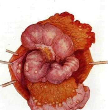
III Carcinomatosis peritonei.
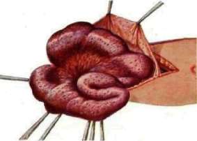
Fig. 2. Tuberculous peritonitis (wet form).
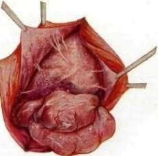
Fig. 3. Tuberculous peritonitis (dry form).
which in fish already separates from the original general body cavity a separate serous space for the heart (only in exceptional cases is there communication between them); consequently, phylogenetically the heart very early receives a pericardial sac. But the lungs for a long time remain in a cavity common with the digestive organs; only in mammals do the pleural cavities separate from the peritoneal cavity, and four non-communicating serous cavities are formed: the cavity of the pericardial sac, the two pleural cavities, and the peritoneal cavity. The latter can open to the outside in three ways: 1) in many fish there are paired or unpaired openings connecting the peritoneal cavity with the external environment—abdominal pores; apparently, they serve to remove fluid from the body cavity; 2) in fish and amphibians this is achieved through funnels, nephrostomata; the latter open into the peritoneal cavity and represent the beginnings of the excretory ducts of the kidneys (mesonephros); 3) in female vertebrates (including mammals) the peritoneal cavity communicates with the external environment through the abdominal ostia of the oviducts. In mammals, for the first time, the dorsal mesentery of the stomach differentiates into the greater omentum, which apparently has important significance as a protective organ in the fight against infections. The duodenum in the vast majority of animals has a mesentery, like the other intestines (thus, the common mesentery is preserved); only in primates does the duodenum lose its mobility and, together with the pancreas, lies against the spine, being displaced by the transverse colon. All these materials provide a comprehensive explanation of the facts of normal anatomy (e.g., the deep position of the duodenum, the formation of omenta, etc.), as well as various deviations from the norm (high position of the cecum, presence of a common mesentery, etc.).
III. Thin. Diseases of the peritoneum can be reduced to five main categories, namely: inflammation, injury, new growths, tuberculosis, and ascites (see); foreign bodies of the abdominal cavity should also be included in this category. Of all these categories, inflammation should be placed in the first place from a clinical standpoint—peritonitis, which in the vast majority of cases is a secondary disease, a complication that accompanies diseases of the abdominal organs or those adjacent to it. The following forms of peritonitis are distinguished (pathoanatomically, clinically, etc.): serous, fibrinous, purulent, putrefactive; local and diffuse; tuberculous (see separate table, figures 2 and 3), postoperative perforating, etc.; for details—see Peritonitis. Traumatic injuries of the peritoneum. Isolated injuries to the peritoneum are very rare; in most cases, abdominal organs are also affected. Out of 400 cases of gunshot wounds to the abdomen during the imperialist war, there was no injury to the intestine in only 20 cases (Petermann); with stab wounds, the intestines are more likely to escape injury. In open wounds, in most cases a piece of omentum in the form of a plug blocks the wound opening and may protrude through the wound, either alone or together with the intestines. In contusion of the abdomen, the peritoneum is usually stretched, but in some cases it may rupture; rupture of the thin-walled vessels of the mesentery and omentum with profuse internal bleeding may also occur. Recognition of injury to the peritoneum in subcutaneous trauma is not easy—usually shock, tension of the abdominal walls, and peritoneal phenomena are observed. In view of the possibility of injury to abdominal organs in penetrating wounds and sometimes in blunt trauma, it is always necessary to keep in mind the presence of such injuries, and therefore, with the slightest suspicion of these injuries, immediate surgical intervention is indicated. Internal bleeding into the abdominal cavity is manifested by increasing pallor, small, rapid pulse, fainting, cold sweat. The first sign of irritation of the peritoneum—board-like abdomen, vomiting, regurgitation. Measures—suture the injured organ, stop the bleeding by ligation of the vessel or injection, clean the abdominal cavity of excess contents by the dry method or by washing with hot boiled water or saline solution. But the most reliable means is surgery no later than 12 hours, as the experience of the imperialist war has taught. In cases of pure bleeding, autotransfusion (see) of blood filtered through gauze with the addition of saline solution or sodium citrate solution (1-2%) is indicated; the abdominal cavity can be completely closed. It is more difficult to establish indications for surgery in blunt injury to the abdomen; in cases of symptoms of internal bleeding and tension of the abdominal wall, emergency surgery is indicated. A foreign body can enter the abdominal cavity in various ways: through wounds, operations, etc. It either causes no special disturbances and remains in the abdomen for a long time or erodes the abdominal wall and comes out through the coverings (through an abscess in the abdominal wall); in rarer cases, the foreign body makes its way into the lumen of the intestine and exits naturally. New growths of the peritoneum, retroperitoneal tissue, omentum, and mesentery. 1. A unique form of primary cancer of the peritoneum is formed by the so-called "mucous cancers"; followed by endotheliomas, plexiform angiosarcomas, and then pseudomyxomas—a collection of gelatinous masses in the peritoneum, which are rarely primary, more often form secondarily as a result of rupture of gelatinous tumors of the ovary, intestines, appendix. 2. Among tumors of the retroperitoneal connective tissue, lipomas, fibromas, myxomas, and mixed tumors, ganglioneuromas should be mentioned; these tumors have a very slow growth, can reach considerable size, and cause symptoms by compression of adjacent organs. 3. Among cystic tumors, first come echinococci, usually as a result of seeding after rupture of the primary cyst; then—serous cysts, chylous cysts, originating from the lymphatic apparatus or from chylous vessels, blood cysts, dermoid cysts. 4. Teratoid tumors of the peritoneum are very rarely observed; they are simple and of mixed type. 5. Then come aneurysmal tumors—first of the aorta and its branches. 6. Actinomycosis of the peritoneum. The actinomycosis fungi can penetrate into the abdominal cavity either from the intestine or from the subserous tissue; most often—from the cecum; they form extensive tumors, little sensitive, very hard, little movable, are connected with the intestines, omentum, consist of fibrinous exudate, among which are characteristic yellow granular drusae. They are diagnosed as extensive, dense, torpid exudates that develop slowly without fever and pain; the diagnosis is confirmed by the presence of drusae in the pus. Treatment—incision in the area of softened areas, curettage, cauterization of fistulous tracts; internally—potassium iodide, salvarsan, injection of iodine into fistulas; radiotherapy is useful.
III. Voznesensky.
Related articles
Mentioned in
Cite this page
“Peritoneum.” Soviet Medical Encyclopedia. English translation of Bolshaya Meditsinskaya Entsiklopediya, 1st ed. (Moscow, 1928–1936), ed. N. A. Semashko. https://sovietmedicalencyclopedia.pages.dev/article/peritoneum/