Lamina
Historical document, translated for reference. It reflects medical knowledge of the 1920s–30s and is not medical advice.
Summary
This article from the 1928–1936 Soviet Great Medical Encyclopedia defines the anatomical term 'lamina' (plate), listing numerous examples of bone, cartilage, and brain structures, and provides a biographical entry for the physician Georgy Lang.
Encyclopedia article (1928–1936)
LAMINA (plate), a term extremely widespread in anatomy. Plates can be bony, for example, the lateral and medial plates of the pterygoid process of the sphenoid bone (L. lateralis and medialis proc. pterygoidei). L. cribrosa, perpendicularis, papyracea ossis ethmoidalis—the cribriform, perpendicular, and paper plates of the ethmoid bone. L. interna, s. vitrea ossium cranii—the internal, or glass, plates of the cranial bones, so named because of their fragility. L. spiralis ossea—the bony spiral plate in the cochlea. Cartilaginous plates: L. fibrocartilaginea interpubica—the fibrocartilaginous plate in the pubic symphysis; L. cricoidea, thyreoidea—the plates of the cricoid and thyroid cartilages. The term L. is also frequently encountered in brain anatomy: L. affixa thalami—a thin, reduced wall of the cerebral hemispheres (telencephalon) located on the upper surface of the thalamus. Laminae medullares cerebelli, thalami, nuclei lenticularis—the white matter in the cerebellar hemispheres, in the thalamus (separating its nuclei), and in the lentiform nucleus (dividing it into three segments); L. septi pellucidi—thin grayish translucent plates, parts of the medial wall of the cerebral hemispheres, between which is a small cavity (cavum septi pellucidi, s. ventriculus quintus). Laminae (s. substantia) perforatae ant. et post.—the anterior and posterior perforated (by vessels) plates on the base of the brain. Laminae chorioideae epitheliales—the remaining thin epithelial layers of the walls of the brain vesicles, covering the choroid plexuses of the ventricles. L. quadrigemina—corpus quadrigeminum—the quadrigeminal plate. L. terminalis—the limiting plate, forming the anterior wall of the third ventricle, etc. L. cribrosa sclerae—the place where the fibers of the optic nerve pass through the sclera of the eyeball. L. elastica corneae anterior (Bowman's) and posterior (Descemet's, s. Dehmers') separate the connective tissue base of the cornea from the epithelium covering it in front and behind. L. mesenterii propria—the connective tissue layer laid in the thickness of the mesentery between the two serous layers forming it. L. parietalis, visceralis peritonaei, pleurae, pericardii—the parietal and visceral layers of the peritoneum, pleura, pericardium, etc.
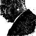
G. F. LANG (born in 1875), a famous therapist, professor of the faculty therapy clinic of the Leningrad Medical Institute and rector of the same institute (since 1929), chief physician of the Erasmian Hospital (since 1924), member of the State Scientific Council, member of the Leningrad Council of R., k. and kr. deputies (since 1929). Graduated from the Military Medical Academy in 1899. In 1905 elected privat-docent of this academy, in 1919 professor of GIMZ (State Institute of Advanced Medical Studies) at the department of diagnosis of internal diseases. Since 1922, Lang concentrates all his medical and scientific activity in the Erasmian Hospital and the Leningrad Medical Institute. Lang is one of the founders of the Leningrad Therapeutic Society (1921) and the journal "Therapeutic Archive" (M.-L., 1923), of which he is currently a co-editor; co-editor of many medical journals and editor of the therapy department of the BME (Great Medical Encyclopedia), editor (together with Pletnev) of the collective multi-volume manual on internal medicine ("Special Pathology and Therapy of Internal Diseases", M.-L., since 1927). Lang is one of the organizers of the All-Union Therapeutic Congresses. In the field of practical medicine, Lang is an implementer of the idea of creating a hospital-clinic (the hospital he heads, the Erasmian Hospital, is an example, all of whose departments are occupied by clinics of the Leningrad Medical Institute). In the field of clinical medicine, Lang represents a direction striving to apply in the clinic the achievements of theoretical medical disciplines, in particular normal and pathological physiology and anatomy. Lang has written about 40 scientific works; the majority of them are devoted to the cardiovascular area. From Lang's works, the main ones are: "Zur Methodik der Blutdruckmessung" (Deutsches Arch. f. klin. Med., B. XCIV, 1908)—the clinical method of measuring blood pressure was first experimentally verified and the frequency of its increase in heart failure was established; "Pathology of the Bundle of His" (Scientific News of NKPros, 1919); "On Hypertension" (Arch. of State Clinical Inst. for Improvement of Physicians, t. I, 1922); "Pathogenesis of Nephrosclerosis" (Proceedings of the VII Congress of Russian Therapists, M.-L., 1925), "On the so-called peripheral heart" (Proceedings of the X All-Union Congress of Therapists, L., 1929). The main direction of Lang's clinic's work is the study of the pathology of the cardiovascular system and diseases of the hematopoietic and blood-destroying apparatus.
LANG METHOD (Lange) for the treatment of sciatica was proposed by the author and described by him in 1904; it consists in the injection of large amounts of sterile warmed to 37° physiological solution mixed with an anesthetic substance. The injection is performed at the place of the nerve's exit from the pelvis between the ischial tuberosity and the greater trochanter. The patient is placed in a horizontal position on the abdomen or on the healthy side. The diseased leg is flexed in the hip and knee joints. The buttock of the diseased leg in the lower two-thirds and the back of the leg (5 cm below the gluteal fold) are carefully disinfected (alcohol, iodine). Then a line is drawn from the greater trochanter of the hip joint to the ischial tuberosity and divided in half. The point of division anatomically corresponds to the sciatic nerve. Pressure at this point gives pain sharply different from the pain when pressing on other areas of the buttock. Marking the point, a sterile needle 8-10 cm long (a needle with a stylet is desirable) is inserted to a depth of 6-8 cm (depending on the patient's build). Touching the needle to the perineurium gives pain radiating along the entire nerve to the heel. Such pain is proof that the needle is in the perineurium. Having confirmed this, 5-10 cm3 of solution is injected under high pressure into the perineurium, after which the needle is directed away from the nerve, and injection of the solution in an amount of 80 to 100 cm3 is performed in all four directions (up, down, right, left). The direction of the needle is changed without removing it. The injection is performed with a large syringe (if unavailable, a 10 cm3 syringe can be used). All instruments and material must be absolutely sterile (needle, syringe, gauze, cotton); the operator and assistant prepare their hands according to all surgical rules. After the injection, the injection site is covered with a sterile gauze pad and covered with collodion. The patient continues to remain in the same position as was given during the injection for 2-3 hours. The patient remains in bed for 2-3 days. At present, pure physiological solution without the addition of eucaine, novocaine, etc., is most often injected. Complications after the injection are almost not encountered, if one does not count the sometimes occurring, quickly passing, slight rise in temperature to 37.2-37.5°. The author theoretically justifies his method by the mechanical effect of the physiological solution injected into the perineurium and around the sciatic nerve. The latter causes the destruction of endoneural adhesions and the fusion of the nerve with surrounding tissues. Other authors consider that the solution causes infiltration in the nerve and its surroundings, which leads to the more rapid removal (washing out) of toxins and products of inflammation. Russian authors (Darkshkevich, Shambov, etc.) assume that a number of factors act together in the indicated method, the basis of which lies in the physicochemical processes occurring in the nerve and around it (osmosis and diffusion processes, etc.). The method has found wide application and gives good results, the duration of treatment for sciatica being 2-3 weeks. Due to the simplicity of the method, the possibility of its wide application in the most primitive conditions (even at home with the patient) and the results of therapy, the method deserves recommendation. Along with this method, the technique of epidural injections according to Cathelin was also developed and introduced for root sciatica.
Related articles
Mentioned in
Cite this page
“Lamina.” Soviet Medical Encyclopedia. English translation of Bolshaya Meditsinskaya Entsiklopediya, 1st ed. (Moscow, 1928–1936), ed. N. A. Semashko. https://sovietmedicalencyclopedia.pages.dev/article/lamina/