Inguinal Region
Historical document, translated for reference. It reflects medical knowledge of the 1920s–30s and is not medical advice.
Summary
The inguinal region is located in the lower abdomen and forms a rectangular triangle bounded by Poupart's ligament, the interspinous line, and the lateral edge of the rectus muscle. It contains the inguinal triangle and canal, with specific anatomical features including skin, fascial layers, blood vessels, and muscles.
Encyclopedia article (1928–1936)
INGUINAL REGION (regio inguinalis) is located in the lower abdomen and forms a rectangular triangle, the sides of which are below—Poupart's ligament, above—the part of lineae interspinarig sup., internally—a line running along the outer edge of m. recti. Within these limits, the inguinal triangle (see Inguinal Canal) is most important and best studied, separated from the thigh by a deep fold called the groin. The skin of the ing. reg. is delicate, movable, covered with hair, beginning from the middle of the inguinal fold and extending to the pubis; in men, the hair extends beyond the ing. reg., rising to the navel. The hairy part of the skin is abundantly supplied with sebaceous and
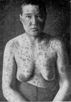
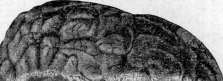
Figure z.
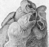
Figure l.
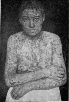

Figure 4.
iffйk
Figure 2.
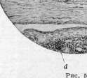
Figure 1. Pemphigus foliaceus (initial stage). Figure 2. Pemphigus foliaceus (same case several years later). Figure 3. Pachion granulations. Figure 4. Pericarditis-cor villosum. Figure 5. Infiltration of the renal pelvis wall (a) by leukocytes in pyelitis; b-epithelium, desquamated in places and accumulated in the lumen (c); d-
part of the renal parenchyma. sweat glands. The subcutaneous fascia consists of two layers, which are well separated from each other in individuals with well-developed subcutaneous fat. The first layer is the subcutaneous fascia of the abdomen (fascia subcutanea), continuing into the subcutaneous fascia of the thigh and scrotum, where it receives smooth muscle fibers (tunica dartos); within it lies the subcutaneous vein of the abdomen Haller's (v. subcut. abdom. Halleri), running from above and internally and flowing into v. saphena. The second fascial layer is the deep lamina (lamina profunda fasciae subcut., s. lam. Thompsoni), separated by loose cellular tissue from the aponeurosis of m. obliq. ext. Within it, and sometimes between it and the aponeurosis, passes a. epigastrica superficialis, anastomosing with branches of a. circumfl. ilei ext. and perforating branches of a. epigastr. inf. In the lower-middle part at the level of the lower edge of the external inguinal ring passes almost transversely a. pudenda ext., easily injured during herniotomy with a low incision. The arteries are accompanied by corresponding veins, flowing into v. saphena. The thickness of the subcutaneous fat layer reaches 4-5 cm in obese people; it is especially developed in the area of the inguinal triangle, closer to mons veneris, and sharply thins toward the inguinal fold. The fatty tissue separates both fascial layers and the aponeurosis; during herniotomy, this must be kept in mind, as the deep lamina can easily be mistaken for the aponeurosis of the external oblique muscle. Lymph, glands are absent, lymph, vessels flow into the inguinal glands, located in Scarpa's triangle. Some authors recognize the presence of 2-3 small lymph, glands above Poupart's ligament. The skin is innervated by branches of n. intercost. XII, ilio-inguinalis and genito-cruralis, without precise localization. The muscular part of the abdominal wall of the ing. reg. consists of the lower ends of the external and internal oblique and transverse muscles. The external oblique muscle from the level of spina os. ilii sup. ant. passes into a tendinous expansion, the fibers of which run obliquely from above downward and from outside inside; folding into bundles-tapes of varying thickness and width, they are closely adjacent to each other in places, while separated by layers of connective tissue in others, whereby the aponeurosis has the appearance of a perforated plate. The bundles are secured by arcuate, transversely running fibers, forming a continuation of the aponeurosis of the external oblique muscle of the opposite side (fibrae collaterales, f. arcuatae). Depending on the combination of these fibers, the aponeurosis may be strong, dense, poorly developed, fan-shaped, or divergent-fibrous. In women, the aponeurosis is denser than in men; the tendon tapes are wider and thicker, closely adjacent to each other, with more arcuate fibers. The lower edge of the aponeurosis turns inward and upward in the form of a gutter, and, merging with fascia lata, forms the inguinal ligament (lig. inguinale, lig. Pouparti), which is attached on one side to spina os. ilii sup. ant., and on the other-to tuberculum pubis, where it expands in the form of a triangle, one side of which goes deep to the bone (lig. lacunare Gimbernati), limits with a sharp edge lacuna vasorum and passes into lig. ilio-pubicum Cooperi; the other side goes forward along the anterior surface of os. pubis to the midline; the third side forms lig. inguin. reflexum, s. lig. Collesi (fig. 1). In the area of the internal end of Poupart's ligament, directly above it, the fibers of the aponeurosis split, forming a slit-the external, or subcutaneous opening of the inguinal canal (annulus ext., s. subcutaneus canalis inguin.), through which the spermatic canal
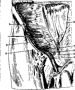
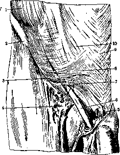
Figure 1. Muscles of the abdominal wall in the area of the inguinal canal:" 1 and 7-m. obliquus int.; 2-m. obliquus ext.; 3-m. cremaster; 4-lig. reflexum Collesi, 5-m. rectus; 6-pars pubica m. obliqui int.; 8-aponeurosis m. obliqui ext.
ligament or round ligament. The tendon bundles forming this aperture constitute the external, or lower pillar (cms, s. columna lat.) and the internal, or upper pillar (cms, s. columna int.). The lateral pillar is stronger and thicker, and, fusing with the Poupart's ligament, is attached partly to the pubic tubercle, and partly continues forward to the pubic symphysis. The upper edge of this pillar is rather sharp and in its lower part slightly concave; some of its fibers, turning under the spermatic cord, pass into the fibers of lig. ing. reflex. Collesi. The internal pillar is wider than the lateral one, attaches to the pubic bone at varying distances from the pubic tubercle (sometimes up to 2 cm), and, continuing downward and medially, enters into the composition of the superior penile or clitoral suspensory ligament (lig. suspens. sup. penis s. clitoridis). In this pillar, a long slit sometimes forms when the aponeurosis is weak, through which the g. hypogastricus n. ilio-hypog. passes; sometimes this slit continues downward to the os. pubis, and then two subcutaneous inguinal openings form—lateral and median, each of which can serve as a site for hernia protrusion. The third, or posterior pillar, also called the inguinal reflex ligament (lig. ing. reflex. Collesi, s. lig. Collesi), begins at the point where Poupart's ligament fuses with the lateral pillar, the fibers of which, turning, hide under the spermatic cord, bounding the inguinal ring posteriorly (fig. 1). Starting from the outer edge of Poupart's ligament, it runs along its groove, lying on lig. Gimbernati, thus having a common part with it; it then goes backward and upward to the anterior wall of the rectus muscle sheath and on the median line passes to the aponeurosis of the opposite side. Its lateral edge gradually fuses with the posterior wall of the inguinal triangle; it is partially covered by the internal pillar of m. cremasteris, and it can be clearly traced when the ring is wide, if the spermatic cord and m. cremaster are lifted. The pillars bounding the canal, as well as the aponeurosis, are secured by arciform fibers coming from the aponeurosis of the opposite side. These fibers (fibrae arciformes, f. intercrurales) run in a transverse direction and round the angle at the place where the pillars diverge, giving the ring various shapes (see Inguinal Canal). Not limited to the area of the pillars, they cross the entire slit in the form of a plate (fascia intercommunis), which, as the spermatic cord passes through, is stretched and spreads over it, forming Cooper's fascia (fascia cremasterica Cooperi), which constitutes the first covering of the spermatic cord. Some anatomists believe that fascia Cooperi represents a continuation of the deep plate of the subcutaneous fascia, which, attaching to the edges of the external inguinal opening, spreads over the spermatic cord when the testis descends into the scrotum. M. obliq. abd. int. and m. transversus begin from the groove of the outer two-thirds of Poupart's ligament; the transition into the tendon expansion along linea Spigelii occurs in some almost at the very edge of m. rectus, in others—2-4 cm short of it. Intertwining with each other, both muscles, approaching the inguinal triangle, pass over the spermatic cord, and m. obliq. int. with its lower edge (pars pubica) tightly surrounds it, so that the space not occupied by muscles will be only the place where the spermatic cord passes (fig. 2). From the internal oblique, muscle fibers depart—m. cremaster, s. levator testis, which continues downward onto the spermatic cord and forms a muscular sheath for it and the testis. Sometimes it runs as a continuous layer from the edge of m. obliq. int., sometimes it begins with two pillars: external—from the groove of Poupart's ligament and the edge of t. obliq. int., and internal—from the anterior wall of the rectus muscle sheath and the covering falx aponeurotica and part of lig. reflex. Collesi (fig. 2). The transverse muscle passes the spermatic cord or round ligament through itself; its muscle fibers are well developed up to the bend over the spermatic cord; further they pass into the tendon expansion, participating together with the edge of m. obliq. int. in the formation of the upper edge of the inguinal triangle. But below this edge it continues in the form of a thin aponeurotic plate, which in the middle fuses with the outer edge of the rectus muscle sheath, below—with the posterior lip of Poupart's ligament, outwardly it bounds the medial edge of the internal inguinal ring and thus constitutes the posterior wall of the inguinal triangle and canal. This plate has varying thickness in different places. In the place where it bounds the internal and lower edges of the internal inguinal opening—the slit in the transverse muscle (Tandler considers this slit the internal inguinal opening)—the plate is reinforced by a ligament (lig. interfoveolare Hesselbachi, s. lig. Hesselbachi), which is formed from tendon fibers
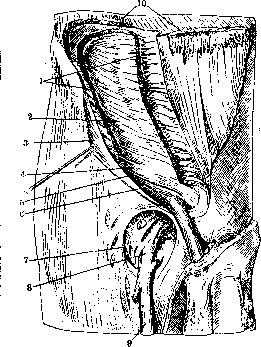
Figure 2. Muscles of the abdominal wall: 1- m. obliquus internus; 2- m. transversus; 3- m. obliquus externus; 4- pars pubica m. obliqui internus; 5- lig. inguinale; 6'- m. cremaster; 7- margo falciformis; 8- v. femoralis; 9- v. saphena magna; 10- m. obliquus externus of fibers extending from the posterior lip of the Poupart ligament; in it one distinguishes the lower leg, which limits the internal inguinal opening from below, and the ascending leg, which limits from within; above the fibers are lost in the tendinous expansion of m. transversus (see figure 4 to the article Inguinal Canal). Ligamentum interfoveolare together with the a. epigastrica inferior lying immediately behind it forms the boundary between the external and internal inguinal fossa. Behind this ligament, sometimes somewhat inward, passes a thin muscular cord (m. interfoveolaris), also beginning from the posterior lip of the Poupart ligament and disappearing in the muscular layers of m. transversus. The inner part of the posterior wall of the inguinal triangle is reinforced by tendinous fibers formed from fibers descending along the lateral edge of m. rectus, from the aponeurosis of m. transversus, as well as from fibers extending from the anterior sheath of m. rectus (falx aponeurotica inguinalis, s. lig. Henle). This ligament, ending in a sharp edge (proc. falciformis) or gradually merging with the posterior wall of the inguinal triangle, is covered in front partially by fibers of lig. reflexus Collesi and the internal leg of m. cremasteris; the lower end merges with lig. Cooperi. Between lig. interfoveolare and falx aponeurotica there is a thin plate, to which in front lies the external inguinal ring, and behind-the peritoneum of the middle inguinal fossa, through which direct inguinal hernias protrude. The strength of the posterior wall of the inguinal triangle depends on the various relationships between lig. interfoveolare, the thinned middle part and falx aponeurotica. The outer edge of the slit in the transverse muscle (internal inguinal ring) is muscular-ventral, and behind and lateral (the spermatic cord passes through this muscle obliquely)-tendinous, reinforced by fibers of fascia transversalis. Behind the muscles passes the transverse fascia (fascia transversalis), which, covering m. transversus, attaches laterally to the posterior lip of the Poupart ligament; in the area of the internal inguinal opening it is drawn by the spermatic cord passing through the abdominal wall and envelops it in the form of a common sheath for the spermatic cord and testicle (tunica vaginalis communis funiculi spermatis et testis); the place of transition is somewhat drawn into the canal and is called lig. infundibuliformis. Further inward fascia transversalis lies against the posterior aponeurotic wall of the inguinal triangle, where it is not always well expressed and represents loose cellular tissue. Tandler denies the presence of transverse fascia in this place; according to him the fascia ends at the lateral edge of the internal inguinal opening in the transverse muscle. The place of transition of fascia transversalis into fascia infundibularis is considered by most anatomists as the internal opening of the inguinal canal; according to Tandler it is located in the thickness of the transverse muscle. The preperitoneal cellular tissue is described as one or two layers. Its thickness depends on the amount of fat, which gradually increases toward the middle. In the area of the inguinal triangle three areas are distinguished: 1) the area of the abdominal opening of the inguinal canal, 2) the area of a. epigastrica inferior and 3) the prevesical space. At the level of the abdominal opening and somewhat above the preperitoneal layer is poorly developed, as a result of which the peritoneum forms here a depression (fovea inguinalis lateralis). Inward the fatty layer thickens and forms a fold protruding into the abdominal cavity, in the thickness of which passes a. epigastrica inferior and diverging elements of the spermatic cord. The preperitoneal fat of the area of the posterior wall of the inguinal triangle comes into contact with the fatty tissue of the prevesical space, being separated from it by fascia praovesicalis. The peritoneum of the inguinal region in the lateral part is even; the transitional fold is located higher or lower depending on the development of preperitoneal fat. In the medial part due to folds protruding into the abdominal cavity three depressions are formed-the inguinal fossae (see Abdominal Wall). Of the vessels of the inguinal region requires description a. epigastrica inferior, accompanied by 2 veins; it begins from the medial side of a. iliaca externa at a distance of 1-2 cm from the Poupart ligament, sometimes at its level, crosses v. iliaca externa, after which it turns upward, forming an arch. It lies in the preperitoneal cellular tissue inward from the internal inguinal opening, behind lig. interfoveolare, forms the external peritoneal fold and serves as the boundary between the external and middle inguinal fossa. From it to the spermatic cord a. funicularis branches off. The innervation occurs mainly due to the branching of nn. iliohypogastricus and n. ilioinguinicus. Both nerves above spina ossis ilii anterior superior pass between m. transversus and m. obliquus internus and then, piercing the latter, lie between the oblique muscles. N. iliohypogastricus goes along the edge of m. obliquus internus and, giving a cutaneous branch above the external inguinal ring, exits through a special slit in the medial leg of the inguinal ring (r. hypogastricus). N. ilioinguinicus, passing in the area of the inguinal canal somewhat lower than the previous branch, gives a branch to the spermatic cord; terminal branches go to the skin of the pubis and scrotum. Both nerves have many variations, replacing each other, according to Ginsburg.
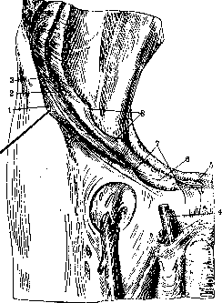
Figure 1. Abdominal wall in the area of the inguinal canal: 1- m. obliquus externus; 2- spina iliaca anterior superior; 3- lig. inguinale; 4- fascia cribrosa; 5- lig. suspensorium penis; 6- funiculus spermaticus; 7- crura annuli inguinalis subcutanei; 8- fibrae collaterales; 9- linea alba; 10- aponeurosis m. obliqui externi.
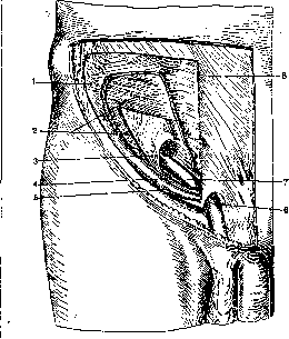
only a slit between the muscles, in which is placed the spermatic cord in men or the round ligament in women. If in a frozen corpse this formation is removed, then the slit will indeed have the appearance of a canal; the latter is also formed when the processus vaginalis does not close. Therefore in the description of the inguinal canal the boundaries of the spermatic cord are given. One distinguishes the external opening (annulus inguinalis externus, s. subcutaneus) and the internal, or abdominal (annulus inguinalis internus, s. abdominalis), and finally the canal itself, in which the anterior and posterior walls, upper and lower edges are described. The external ring is located in the lower-inner angle of the inguinal triangle, in the aponeurosis of the external oblique muscle, and is limited by the lateral and medial legs (crura, s. columna lateralis et medialis) (fig. 1), below and partially behind by the third leg (lig. inguinale reflexum Collesi, s. lig. Collesi) (fig. 2). The external opening has a triangular shape, but the arcuate fibers (fibrae intercrurales), passing in the aponeurosis, round the acute angle, and the opening takes the form of an oval or polygon. The transversely oval shape is observed in those cases when the free edge of the medial leg is considerably displaced from the pubic tubercle. The size of the subcutaneous opening is subject to large fluctuations. Tillaux determines the major length of the oval as 2-2.5 cm. According to Yaltinsky, the size of the transverse diameter varies from 1.2 to 3.2 cm, and the longitudinal from 2.2 to 3 cm; according to Venglovsky, the longitudinal diameter is 1.5-6 cm, the transverse-1.1-2.5 cm. In women the subcutaneous opening is much smaller, almost half; in most cases the shape is almost circular, rarely oval. The opening may be divided by a tendinous cord extending from the medial leg into 2 parts.
Figure 2. Section of the anterior abdominal wall parallel to the Poupart ligament: 1- m. obliquus externus; 2- m. obliquus internus; 3- m. transversus; 4- funiculus spermaticus; 5- m. rectus; 6- lig. reflexum Collesi; 7- annulus inguinalis subcutaneus; 8- annulus inguinalis abdominis.
The internal ring is located in the area of the lateral angle of the inguinal triangle, within the limits of its posterior wall, on the boundary of the inner and middle thirds of the Poupart ligament, 1-1.5 cm above it. Located in the thickness of m. transversus, in its dorsal tendinous part, it has the form of a longitudinal or oval slit (fig. 3 and 4), limited on the side and above by the tendinous part of m. transversus, reinforced by the fascia transversalis fused with its edge (Tandler); inward and below the opening is strengthened by the ligament-lig. interfoveolare Hesselbachi. Fascia transversalis, being drawn through the ring by the spermatic cord into the inguinal canal in the form of fascia infundibuliformis, passes into tunica vaginalis communis funiculi spermatis et testis. This funnel-shaped depression is by some anatomists also considered the opening of the canal. The inguinal canal is formed due to the fact that the internal oblique and transverse muscles are thrown over the spermatic cord, with the free edge (pars pubica) of the internal oblique
|
Figure 3. Annulus inguinalis from the front: 1- m. obliquus internus; a- m. transversus; 3- annulus inguinalis abdominis; 4- m. cremaster; 5- lig. inguinale; 6- annulus inguinalis subcutaneus; 7- funicu
|
lus; i
|
|
|
lus spermaticus; 8-aponevros m. obliqui externi. j сой мышцы при хорошо развитой мускулатуре прилегает вплотную к семенному канатику вплоть до выхода его из канала. M. transversus лежит несколько выше, отходит от ка
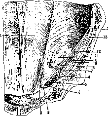
рие. 4. Annulus inguinalis abdominis сзади: 1- line'a semicircularis Douglasi; 2-falx inguinalis; 3 -lig. Gimbernati; 4-lig. Cooperi; 5-va.sa femo-ralia; 6-lig. ilio-pectineum; У-lig. inguinale; 8 и 11- crus horizontal et crus verticale lig. Hesselbachi; 9-annulus inguinalis abdominis; 10- n. femoralis; 12-fascia ilio-psoica; 13-m. ilio-psoas; 14-m. transversus.
натика и образует слабую заднюю стенку пахового треугольника. Канал имеет косое направление-сзади наперед и сверху вниз; длина его равна y мужчин 4-4,5 см. y женщин он несколько длиннее в зависимо-- 1 ве- сти от большей ширины таза. Так как внутреннее кольцо находится выше подкожного (1-1,5 см), то канал идет не параллельно паховой связке, но под нек-рым углом к ней. Границы П. к.: сперед и-fascia in-tercolumnaris в пределах наружного кольца, выше и кнаружи-апоневроз наружной косой мышцы, a дальше-мышечные волокна mm. obliq. int. et transversi; последний образует сухожильное кольцо-внутреннее паховое отверстие-в дорсальной части, в вентральной же мышечные волокна охватывают семенной канатик спереди, сбоку и сверху. Нижняя границ a-жолоб медиальной части Пупартовой связки, a по мере от-хождения семенного канатика кверху-волокна m. cremasteris и мышечные волокна т. transversi. Верхняя граница-край т. obliqui int. c отходящим от него и переходящим на семенной канатик m. cremaster. Задняя границ a-задняя стенка пахового треугольника, к-рая состоит по мнению многих анатомов из fascia transversa; нек-рые современные анатомы (Tandler, Spalteholz, Krau-se) считают, что задняя стенка образуется продолжением сухожильного растяжения т. transversi ниже ясно видимого его края, сливающимся c задней губой Пупартовой связки на всем протяжении кнутри от внутреннего пахового кольца и c боковым краем влагалища m. recti (рис. 4). Эта сухожильная пластинка имеет различную толщину в разных местах; снаружи она подкрепляется связкой, lig. interfoveolare Hesselbachi; кнутри, y внутреннего угла треугольника укреплена свя-зочно - апоневротической пластинкой - falx aponeurotica inguinalis (см. Паховая область). Средняя наиболее тонкая часть этой пластинки прилегает спереди к наружному паховому кольцу, a сзади, покрытая истонченной fascia transversa и предбрюшинным жиром,--к дну средней паховой ямки. У большинства людей c средней и слабо развитой мускулатурой, особенно при наличии грыжевого выпячивания, такое совершенное устройство П. к. нарушается тем, что край mm. obliq. int. et transversi отходит от семенного канатика, образуя различной формы и величины промежуток, также называющийся П. к., паховым треугольником, паховой щелью. Границы такого расширенного пахового канала: снизу-Пупартова связка, сверху-край mm. obliq. int. et transversi, c середины-край m. recti; передняя сторона-апоневроз m. obliq. ext., задняя--указанная выше апоневротическая пластинка, подкрепленная lig. interfoveolare и falx aponeur. или,как обычно описывают, fascia transversa; семенной канатик занимает в этом промежутке нижнебоковой отдел. От изменчивости верхней и средней границ зависит разная форма П. к. или промежутка. Ящинский различает три формы: треугольную, веретенообразную и ще-левидную; при треугольной форме боковой край m. recti является основанием, a верхушка обращена вбок; длина промежутка от середины основания до верхушки в среднем- 5,5 см, при основании в среднем 3,5 см. При веретенообразной форме средняя длина 7 см, a ширина 3-7 см. Венгловский различает вальную и треугольную формы. B большинстве случаев y здоровых людей встречается овальная форма; размеры П. к. при ней следующие: длина 1-2,2 см, высота 4,5-7,7 см. Гораздо реже наблюдается треугольная форма: высота (по боковому краю m. recti) 1,6- 4 см, длина (по Пупартовой связке) 4-6 см. Главная защитительная роль падает не на длину, a на высоту промежутка: чем меньше высота, тем крепче паховая область. У женщин паховой промежуток в большинстве случаев щелевидный или овальный, изредка треугольный, и выполнен круглой связкой, идущей в сопровождении п. ilio-inguinalis. K задней стенке П. к. прилегает fascia transversa и предбрюшинный жир; в области внутреннего пахового кольца fascia transversa отсутствует, т. к. она переходит в оболочку семенного канатика. Элементы его (vas deferens et vasa sperm.) занимают заднюю периферию канатика и воронки и расходятся по задней стенке живота (vasa spermatica вверх по m. ilio-psoas, v. deferens вниз к малому тазу, перекрещивая a. epigastrica inf.), a поэтому грыжи, выпадающие в воронку кнаружи от артерии, пойдут спереди и кнаружи от семенного канатика в общей c ним оболочке* и сохраняют эти отношения на всем протяжении. Co стороны брюшины к области внутреннего пахового кольца прилегает наружная паховая ямка (fovea inguin. lat.), лежащая кнаружи от боковой складки [см. отд. табл. (т. Ш, ст. 99-100), рис. 1], в толще к-рой проходит vasa epigastr. inf., и эта ямка служит началом косых паховых грыж. Кнутри от этой складки находится средняя паховая ямка (fovea inguinalis media), прилегающая к задней стенке пахового канала; грыжевое выпячивание (прямая грыжа), не испытывая препятствия на своем пути co стороны слабо развитой fascia transversa и задней стенки П. к., выходит в паховый треугольник, где встречает впереди себя семенной канатик, и располагается кнутри и сзади от него вне оболочек. Образование П. к. и сложные отношения между элементами семенного канатика и брюшной стенки связаны c процессом развития половых желез. При опускании их в утробной жизни плода брюшина образует вдоль проводника Гунтера (gubernaculum Hunteri) выпячивание, проникающее вместе c ним кнаружи от a. epigastrica через брюшную стенку в мошонку или большие губы,-брюшинный отросток (proc. vaginalis) y мужчин или Пук-ков канал (canalis, s. diverticulum Nucki, rudimentum proc. vaginal, muliebris)-y женщин. Яичко, спустившееся в мошонку позади proc. vaginalis, увлекает на своем пути встречающиеся слои брошной стенки, к-рые образуют общие оболочки для proc. vaginalis и семенного канатика. Fascia transversa вытягивает-CHBBHfletunica vagin. commun. funiculi sperm. et testis;m. obliq. int.-в виде m. cremasteris: на m. obliq. abdom. ext. выпячиваются fascia intercolumnaris, к-рая образует fasciacremaste-rica Cooperi. Только m. transversus как наиболее неподатливый образует полное кольцо; нек-рые анатомы считают, что и этот мускул участвует в образовании m. cremasteris (см. Яичко). Proc. vaginalis начинает облитериро-ваться в конце утробной жизни плода, превращаясь во влагалищную связку (lig. vagina-le, s. habenula). Причина облитерации-наличие поперечных волокон m. cremasteris, образующих поперечные складки (Крымов). Proc. vaginalis однако облитерируется не все-| гда и не на всем протяжений; он может остать-I ся открытым на всю жизнь целиком или на *б 5167 -ограниченном участке по ходу семенного ка---натика. Крымов различает 3 формы его остатка: первая-proc. vagin. весь открыт и сообщается c полостью собственной оболочки яичка; вторая-облитерация произошла внизу, сверху же proc. vagin. открыт на большую или меньшую длину; третья-полная облитерация сверху и открытый proc. vagin. co стороны tunica propria testiculi. Остатки его могут иметь сужения в одном или многих местах. У женщин Нукков канал, облитери-руясь, превращается в связку (ligamentum Nucki), по ходу которой при неравномерной облитерации остаются просветы-cystes Hga-menti Nucki. Лит.-см. пит. к ст. Грыжи.
Related articles
Mentioned in
Cite this page
“Inguinal Region.” Soviet Medical Encyclopedia. English translation of Bolshaya Meditsinskaya Entsiklopediya, 1st ed. (Moscow, 1928–1936), ed. N. A. Semashko. https://sovietmedicalencyclopedia.pages.dev/article/inguinal-region/