Poupart's Ligament
Historical document, translated for reference. It reflects medical knowledge of the 1920s–30s and is not medical advice.
Summary
Poupart's ligament is a fibrous cord located in the groin that forms the lower border of the anterior abdominal wall. It serves as an important anatomical landmark for distinguishing between inguinal and femoral hernias.
Encyclopedia article (1928–1936)
Poupart's Ligament [lig. Pouparti, s. Fallop-pii (Hyrtl), lig. inguinale, arcus inguinalis] represents a fibrous cord located in the groin and bordering the lower edge of the anterior abdominal wall. It is formed mainly from the gutter-like turned lower portion of the aponeurosis of the external oblique muscle and is reinforced below by blending with the fasciae latae leaf, and above by the fascia iliaca and fascia transversa (see figure). Stretched between the spina ossis ilii ant. sup. and tuberculum pubis, together with the edge of the ilium and the horizontal branch of the pubic bone, it forms a space which accommodates the m. ilio-psoas and femoral vessels (see Femoral Canal, Inguinal Region, Inguinal Canal). In muscular people, P. s. is palpable as a dense cord; in people with flaccid musculature, especially in multiparous women, it is in a relaxed state. The boundary between the inner and middle thirds corresponds to the passage of the femoral vessels. In obese people, when it is difficult to determine the hernial protrusion, P. s. or its projection can indicate whether there is an inguinal or femoral hernia. If Poupart's ligament cannot be palpated, one can be guided by the pubic tubercle: a hernial tumor located medially and upward from it is inguinal, while laterally and outward is femoral.
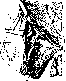
Annulus inguinalis subcutaneus, lacuna musculo-rum, lacuna vasorum: 1-lacuna musculorum; 2- m. rectus femoris; 3-m. sartorius; 4-a. femoralis; 5-fascia lata; 6-fascia ilio-pectinea; 7-fascia lata; 8-lig. Gimbernati; 9-lig. reflexum Collesi; 10- lacuna vasorum; 21-eminentia ilio-pectinea.
in the form of a dense cord; in people with flaccid musculature, especially in multiparous women, it is in a relaxed state. The boundary between the inner and middle thirds corresponds to the passage of the femoral vessels. In obese people, when it is difficult to determine the hernial protrusion, P. s. or its projection can indicate whether there is an inguinal or femoral hernia. If Poupart's ligament cannot be palpated, one can be guided by the pubic tubercle: a hernial tumor located medially and upward from it is inguinal, while laterally and outward is femoral.
Related articles
Mentioned in
Cite this page
“Poupart's Ligament.” Soviet Medical Encyclopedia. English translation of Bolshaya Meditsinskaya Entsiklopediya, 1st ed. (Moscow, 1928–1936), ed. N. A. Semashko. https://sovietmedicalencyclopedia.pages.dev/article/poupart-ligament/