Femoral Canal
Historical document, translated for reference. It reflects medical knowledge of the 1920s–30s and is not medical advice.
Summary
This article from the 1928–1936 Soviet Great Medical Encyclopedia defines the femoral canal, a space in the femoral triangle. It explains the anatomical variations in how the canal is interpreted, ranging from a broad definition encompassing the femoral vessels to a narrow definition specifically associated with the pathway of femoral hernias.
Encyclopedia article (1928–1936)
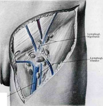
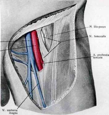
FEMORAL CANAL (canalis cruralis, s. femoralis, fossa ilio-pectinea, s. subinguinalis) is a part of the femoral triangle; it is a conventional concept and is interpreted differently by anatomists. In a broader interpretation, the femoral canal is understood as the entire receptacle for the femoral artery and vein and deep lymphatic pathways with connective tissue, bounded by the funnel-shaped divergence of the layers of the fascia lata in the upper part of the thigh (Jossel, Testut et Jacob, Bobrov, Rouviere). In a narrower understanding (see Figure 1), only a part of this receptacle corresponds to the femoral canal, specifically its inner section, which includes connective tissue with lymphatic vessels and glands, i.e., the usual pathway for the passage of femoral hernias (Henke, Merkel, Corning). The external "exit" of the femoral canal, in any case, is considered to be the opening of the fossa ovalis, which is actually closed by the fascia cribrosa (annulus femoralis ext., septum Petrequin ext.), which is usually pierced by a hernia. The internal "opening" in the broad interpretation is considered to be the lacuna vasorum (Jossel), and in the narrow interpretation, it is the opening between the lacunar ligament and the femoral vein, covered by the femoral septum, or Cloquet's septum (see separate table, Fig. 2). The entire ilio-pectineal fossa is formed by the iliopsoas muscle and the pectineus muscle converging at an angle, the inguinal ligament, and the fascia lata associated with them, which is split here.
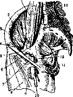
Figure 1. Fossa ovalis. Lacuna vasorum. Femoral canal in the narrow sense. 1-psoas muscle; 2-iliacus internus muscle; 3-femoral nerve; 4-inguinal ligament; 5-ilio-pectineal fascia; 6-femoral artery; 7-femoral vein; 8-sartorius muscle; 9-foramen ovale; 10-great saphenous vein; 11-lacunar ligament; 12-arrow indicates the usual path of femoral hernias, the entrance and exit "openings" of the femoral canal; 13-promontory (according to Corning).
The latter, after covering the sartorius muscle on both sides, divides at its medial edge into 2 layers: deep and superficial. The deep layer (iliac fascia) lines the iliopsoas muscle up to
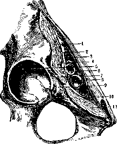
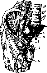
Fig. 2. Muscular lacuna and vascular lacuna: 1-iliopsoas muscle; 2-ilio-pectineal fascia; 3-vascular lacuna; 4-femoral nerve; 5-acetabulum; 6-femoral artery; 7-ilio-pectineal bursa; 8-femoral vein; 9-ilio-pectineal eminence; 10-lacunar ligament; 11-pectineus muscle (according to Spalteholz).
its attachment to the lesser trochanter, fusing at the medial edge of the muscle above with the inguinal ligament and below with the ilio-pectineal eminence of the pelvic bone, then lies behind the vessels on the pectineus muscle, attaching along the upper edge of the muscle to the pecten of the pubic bone (pectineal fascia, Cooper's fascia). The superficial layer of the fascia, in the form of a free triangular plate, is stretched in front of the vessels, attaching its upper side (the base of the triangle) to the inguinal ligament, having its outer side connected to the medial edge of the sartorius muscle; its third, inner side fuses with the deep layer on the surface of the pectineus muscle. In this way, a three-sided fascial funnel of the ilio-pectineal fossa is created, which contains the vessels and is turned with its apex downwards. One of its faces (the free plate of the fascia) faces anteriorly, and two face posteriorly: the lateral one along the surface of the iliopsoas muscle and the medial one along the pectineus muscle. The iliac fascia, with the medial edge of its outer face, divides the space between the pelvic bone
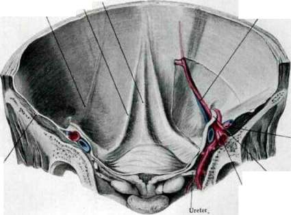
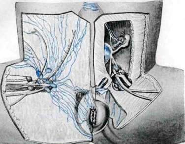
and the inguinal ligament into two sections: the muscular lacuna and the vascular lacuna (see Figure 2). Through the first passes the iliopsoas muscle and the femoral nerve; through the second passes the genitofemoral nerve (for a limited area of skin under the inguinal ligament) and the vessels. The medial angle of the muscular lacuna is reinforced by the lacunar ligament. Inside the described ilio-pectineal fossa are located the vessels: on the outside is the femoral artery, medially from it is the femoral vein; between the vein and the medial angle of the "funnel" is loose connective tissue with deep lymphatic vessels, which drain into a gland often encountered at the top between the vein and the lacunar ligament, called the Rosenmüller gland. Each of these three structures (artery, vein, and lymphatic vessels) is separated by a fascial layer, which creates three fascial beds inside the femoral canal (in the broad interpretation) - for the artery, the vein, and the glandular connective tissue. The latter bed is the most yielding and is the most frequent pathway for femoral hernias, the "femoral canal" of the proponents of its narrow interpretation (see Figure 3).
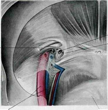
Figure 3. Schematic drawing of a femoral hernia in the femoral canal: 1-ilio-pectineal fascia; 2-femoral artery and vein; 3-hernia sac of a femoral hernia; 4-lacunar ligament; 5-hernia sac pulled medially (according to Corning).
The text fragment provided in the original source for this paragraph is garbled/OCR-corrupted and does not contain coherent anatomical description.
The text fragment provided in the original source for this paragraph is garbled/OCR-corrupted and does not contain coherent anatomical description.
The text fragment provided in the original source for this paragraph is garbled/OCR-corrupted and does not contain coherent anatomical description.
The text fragment provided in the original source for this paragraph is garbled/OCR-corrupted and does not contain coherent anatomical description.
The text fragment provided in the original source for this paragraph is garbled/OCR-corrupted and does not contain coherent anatomical description.
The text fragment provided in the original source for this paragraph is garbled/OCR-corrupted and does not contain coherent anatomical description.
Related articles
Mentioned in
Cite this page
“Femoral Canal.” Soviet Medical Encyclopedia. English translation of Bolshaya Meditsinskaya Entsiklopediya, 1st ed. (Moscow, 1928–1936), ed. N. A. Semashko. https://sovietmedicalencyclopedia.pages.dev/article/femoral-canal/