Obturator Artery
Historical document, translated for reference. It reflects medical knowledge of the 1920s–30s and is not medical advice.
Summary
The obturator artery is a highly variable vessel that typically originates from the hypogastric artery in 70% of cases and from the external iliac system in 30%. Its variations have significant surgical importance, particularly in relation to femoral hernia repair, where certain configurations can cause dangerous bleeding.
Encyclopedia article (1928–1936)
Obturator Artery, Vein. The obturator artery ("запирательная артерия", artery of the closed foramen) varies greatly in its origin, position, direction, and branching. Approximately 70% of the time it originates from the hypogastric artery (a. hypogastrica) and 30% from the external iliac system (a. iliaca ext.). In the first group, the following variants of the origin of a. obturatoriae have been noted (Yashchinsky): from the trunk of the hypogastric artery - 3.2%, from its primary posterior branch - 19.1%, from the primary anterior branch - 41.4%, from the trunk common to a. pudenda and a. glutaea inf. - 5.2%, from a. glutaea inf. - 3.2%, from a. pudenda int. - 2.9%, and from small branches - 2.6%. The second group includes cases where a. obturator arises either from the trunk of a. iliaca ext. (Fig. 1), or from a. epigastrica inf., or from a. femoralis; finally, cases of dual origin are observed - one root from the a. hypogastrica system, the other from the a. iliacae ext. system. In this case, before entering the canalis obturatorius, both arteries merge, and their common trunk enters the canal.
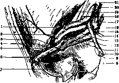
Fig. 1. Right a. obturatoria and a. epigastrica inf. of a male: 1-ductus deferens; 2-lig. Hesselbachi; 3-ramus obturatorius; 4-lig. Gimbernat; 5-ramus pubicus a. epigastricae inf.; 6-ramus pubicus a. obturatoriae; 7-vesica urinaria; 8-a. haemorrhoidalis media; 9-a. pudenda int.; 10-a. glutaea inf.; 11-a. obturatoria; 12-n. obturatorius; 13-v. iliaca ext.; 14-a. glutaea sup.; 15-a. ilio-lumbalis; 16-a. hypogastrica; 17-a. circumflexa ilium profunda; 18-a. iliaca ext.; 19-a. testicularis; 20-a. iliaca communis; 21-v. iliaca communis.
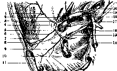
The variants of the second group (origin from the a. iliacae ext. system) have special practical significance due to the particular relationship of the artery to the femoral ring. When incising the ring strangulating a hernia, especially with the old closed method, this proximity threatens profuse, and sometimes fatal, bleeding, which is why this variant was historically called "corona mortis". According to Yashchinsky's research, among cases of this variant, which more frequently occurs on the right side and in women, a. obturatoria arises more often from a. epigastrica inf. (28.5%), less frequently from a. iliaca ext. (1.2%) (Fig. 2), and even less frequently from a. femoralis (0.4%). The relationship to femoral hernia varies greatly depending on the variant of this group; if a. obturatoria arises from a. iliaca ext., it has no relationship to the hernia; if it originates from a. femoralis, it passes behind the hernia; if it originates from a. epigastrica inf., it is located either on the lateral side of the hernia (if a. obturatoria arises from the central part of a. epigastricae) or on the medial side (if a. obturatoria branches off from a. epigastrica above the inguinal ligament); the latter occurs less frequently (Yashchinsky).
A. obturatoria may be completely absent, in which case it is replaced by branches of the femoral artery; or, alongside a well-developed main artery, there may also be an accessory artery (a. obturatoria accessoria) that anastomoses with the branches of the main one. A. obturatoria (as well as v. obturatoriae and n. obturatorius), covered by peritoneum lining the pelvic wall up to the arcus tendineus m. levatoris ani, from its origin, moves forward and downward along the linea innominata of the small pelvis and, positioned under n. obturatorius, enters the canalis obturatorius, where it divides into its branches. This direction and position of the artery occurs when it originates from a. hypogastrica. Only in the case of its very high origin (above the 2nd sacral foramen) it is positioned above the nerve, but before entering the canal, it still approaches from under the nerve (Tikhomirov). When branching from the trunk of a. iliaca ext., it approaches the canalis obturatorius, positioned medially from v. iliaca ext. and posteriorly and laterally from the femoral ring; when branching from a. femoralis, a. obturatoria penetrates the femoral canal (in front of or behind the vein), then, bending over the horizontal ramus of the pubis, approaches the canalis obturatorius, thus being positioned along the posterior semicircle of the femoral ring; when arising from a. epigastrica, a. obturatoria is positioned, as indicated, either along the outer or inner semicircle of the femoral ring and thus reaches the canalis obturatorius. The branches of a. obturatoriae - ramus pubicus, ramus internus, ramus posterior, and ramus anterior - supply the mm. ilio-psoas, quadratus femoris, levator ani, obturator ext. et int., adductor magnus et minimus, brevis, longus, pectineus, gracilis, the ilium, the symphysis, the tuber ischii, the fatty tissue of the acetabulum, the head of the femur, the lumbar lymph nodes, and the urinary bladder. Variations in the branching of a. obturatoriae mainly concern the diversity of anastomoses between it and a. epigastrica inf. (and a. iliaca ext.). The usual anastomosis between the ramus pubicus of these two arteries, if significantly developed, presents all the practically important features that occur when a. obturatoria arises from a. epigastrica inf. The obturator vein (запирательная вена), usually double, accompanies the artery of the same name, but in its variants of insertion, it does not always follow the variants of the artery's origin. It more often empties into the hypogastric vein, but usually widely anastomoses with v. iliaca ext. and v. circumflexa femoris med. It collects blood from the scrotum (labia majora) and the adductor muscles of the thigh. - Ligation of a. obturatoria is possible in the pelvic cavity or from the anterior surface of the thigh. In the first case, the access is the same as for exposing a. hypogastrica. In the second case, a vertical incision is made from the middle of the Poupart's ligament downward, and m. pectineus is exposed. By dissecting it near the pubic bone, it is pulled medially or incised. Penetrating deeper, the fascia and muscle fibers of m. obturatorius ext. are tranversely incised, thus exposing the obturator vessels and nerve; the vasa femoralia remain lateral.
Related articles
Mentioned in
Cite this page
“Obturator Artery.” Soviet Medical Encyclopedia. English translation of Bolshaya Meditsinskaya Entsiklopediya, 1st ed. (Moscow, 1928–1936), ed. N. A. Semashko. https://sovietmedicalencyclopedia.pages.dev/article/obturator-artery/