Urinary Bladder
Historical document, translated for reference. It reflects medical knowledge of the 1920s–30s and is not medical advice.
Summary
The urinary bladder is a muscular sac for storing urine periodically delivered by the ureters and expelled through the urethra. This article covers its phylogeny, ontogeny, anatomy, histology, research methodology, pathology, and surgical operations.
Encyclopedia article (1928–1936)
Urinary Bladder. Contents: I. Phylogeny and Ontogeny............119 II. Anatomy...................120 III. Histology..................127 IV. Research Methodology for the Urinary Bladder.........130 V. Pathology...................132 VI. Operations on the Urinary Bladder...............162 The urinary bladder (vesica urinaria) is a muscular sac intended for accumulating urine that periodically flows in through the ureters and is expelled through the urethra at varying intervals. I. Phylogeny and Ontogeny. The urinary bladder of fish, when present, is located dorsally to the hindgut and is formed either by the fusion of the terminal sections of the ureters (Wolffian ducts) or by an outpouching of the posterior wall of the intestine, opening to the outside separately from the anus on a special papilla or together with the openings of the sexual organs. The fish bladder is thus not homologous to the urinary bladder of other vertebrates, in which it develops from the ventral section of the terminal intestine. In amphibians, the urinary bladder is located in front of the cloaca (corresponding in origin to the allantois of amniotes), and the ureters open into its posterior wall; thus, urine enters the bladder through the cloaca. Among reptiles, only lizards and turtles have a urinary bladder; it develops from the proximal section of the allantois; in other reptiles, as well as in birds, the urinary bladder is absent in the adult state, which is connected with the properties of rapidly solidifying urine. Among mammals, monotremes and marsupials develop the urinary bladder from the proximal segment of the allantois, while in others, as in humans, it develops from the allantois and a section of the cloaca that forms the urogenital sinus. The development of the human urinary bladder begins in the 2nd month from the intraembryonic section of the allantois (see) and the upper section of the urogenital sinus (sinus uro-geni-talis), formed by the longitudinal splitting of the cloaca (see) (Fig. 1). Initially, it forms a single unit with the urethra, and the Wolffian ducts (see) open into its posterior wall, from which the ureters depart along with the rudiment of the definitive kidney (Fig. 2). Then the proximal sections of the Wolffian ducts become part of the posterior wall of the sinus; the ureters receive independent openings; the distance between them and the Wolffian ducts increases, and the constriction formed in this area isolates the urinary bladder with the ureters from the urethra with the Wolffian ducts, which here transform into ductus ejaculatorii. Thus, the epithelium of the urinary bladder has a dual origin: the larger part comes from the entoderm of the allantois and sinus, while the posterior section comes from the mesoderm of the Wolffian ducts; initially it is single-layered cylindrical, becoming multilayered transitional in the 3rd month. The urinary bladder for a long time retains a spindle shape with an elongated apex that continues into the umbilical section of the allantois; its narrow apex, called the urachus, later becomes obliterated, forming lig. vesico-um-bilicale medium. The growth of the bladder in width begins after birth. II. Anatomy. The urinary bladder lies in the pelvic cavity, immediately behind the pubic symphysis, having complex, topographically-anatomical relationships with surrounding organs and tissues that change depending on its filling. In men, behind the bladder lie the seminal vesicles, the ampullae of the vas deferens, and the rectum; in women, the uterus and the upper third of the vagina. In men, the outlet section of the bladder is located on the prostate gland (Fig. 4), in women directly on the pelvic diaphragm. These sections are most fixed. Having assumed the position characteristic of an adult, the urinary bladder is located at the level of the pubis (Fig. 3). After 40-45 years, the bladder descends further, as if spreading over the pelvic diaphragm, which in turn also descends. - The shape of the urinary bladder changes depending on its filling and the filling and position of neighboring organs. In a filled state, the bladder has an oval shape resembling a lemon, lying either vertically (more often in men, Fig. 4) or more transversely (in women). The bladder holds from 200 to 600 g of urine, and sometimes more; in pathological conditions--up to 7 liters, and then its apex is determined above the navel. When filling, the bladder stretches mainly upward, into the abdominal cavity, and less so backward and to the sides. An empty bladder is flattened from top to bottom, resembling a dish, because its upper section (hemisphere) collapses and contacts the lower one (Fig. 5 b and d); sometimes the bladder is flattened from front to back (Fig. 5 a and c), which is especially characteristic of the female bladder, on which the uterus presses from behind and above.-In newborns there is no sexual difference in the shape of the urinary bladder. When empty and contracted, it appears spindle-shaped or pear-shaped, with no base. The urinary bladder does not fit in the still very narrow pelvis; most of the bladder lies in the abdominal cavity; it is higher than in an adult. The peritoneum covers its entire posterior wall. With age, the shape of the bladder changes significantly due to its strengthening to the rectum and genitalia and its descent (descensus vesicae) into the becoming more spacious pelvis. The bladder's musculature begins to develop significantly after 6 years.-In the bladder, 1) the base (fundus vesicae), the most strengthened and therefore least movable part, 2) the apex (vertex vesicae), distinguishable only when the bladder is full, 3) the body (corpus vesicae) and 4) the neck of the bladder (collum vesicae), i.e., that section where the base of the bladder, narrowing, transitions into the urethra. Some authors, when describing the body of the bladder, distinguish the anterior, posterior, and two lateral walls that transition into one another and thus have no clear boundaries (only in the filled bladder, Fig. 4); in the empty bladder, most of its walls face upward (Fig. 3). The peritoneum covers only part of the bladder walls, namely the entire upper section (which collapses when the bladder is empty) (Fig. 4, 5 b), and on the sides and behind it descends onto the bladder lower than on its anterior surface. Topographically, its relationships are as follows: the peritoneum from the anterior abdominal wall transitions onto the bladder, forming either a more uniform (Fig. 5 b) or a more wavy line of transition (Fig. 5 a). In some cases, the peritoneum first descends onto the posterior surface of the symphysis (Fig. 5 d), and then ascends onto the bladder. In the first variant, the peritoneal fold lies very high (Fig. 5 a and c) and when the bladder is filled, it moves even higher (Fig. 4). This exposes a significant section of the bladder devoid of peritoneum, which has practical importance for extraperitoneal approaches to the bladder (see Lithotomy). In the second variant, even when the bladder is full, the free section of the bladder is very small, and during an operation, if the peritoneal fold is not detached upward, the peritoneal sac can easily be opened. The first variant is more characteristic of men and young people, while the second is characteristic of women and elderly men. From the lateral walls of the bladder, the peritoneum transitions without a sharp boundary onto the lateral walls of the pelvis (Fig. 6). Behind the bladder, in men, the peritoneum covers the vas deferens and the apices of the seminal vesicles and at varying levels transitions onto the ampullary part of the rectum; the place of transition is the lowest part of the peritoneum. The space between the mentioned organs is called the excavatio recto-vesiealis; it is often filled with loops of the small intestine, sometimes with the cecum and the loop of S-Romani. In women, the peritoneum from the posterior walls of the bladder transitions onto the body of the uterus, forming a space (excavatio vesi-co-uterina) (Fig. 5 d), incorrectly called the Douglas pouch. The level of transition also varies here, but more often the lower third of the uterine body is not covered by peritoneum. The peritoneum of the bladder is loosely connected with the underlying layer of areolar tissue, which is why it is always possible, especially in front and behind, to extract the bladder from the peritoneal sac without particular violence. The perivesical areolar tissue is especially abundant.
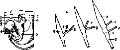
Figure 1.
Figure 2.
Figure 1. Development of the urinary bladder (diagram): 1-rectum; 2-urinary bladder; 3- allantois; 4-cloacal membrane; 5-entodermal cloaca; 6-rudiment of the definitive kidney; 7- Wolffian duct (according to Bromann). Figure 2. Diagrams showing the migration of the ureters onto the wall of the urinary bladder: 1-ureter; 2-Wolffian duct; 3-urinary bladder; 4-urethra; 5-sinus urogenitalis (according to Bromann). ducts enter into the composition of the posterior wall of the sinus^ the ureters receive an independent place of opening; the distance between them and the Wolffian ducts increases, and the constriction formed in this area isolates the urinary bladder with the ureters from the urethra with the Wolffian ducts, which here transform into ductus ejaculatorii. Thus, the epithelium of the urinary bladder has a dual origin: the larger part comes from the entoderm of the allantois and sinus, while the posterior section comes from the mesoderm of the Wolffian ducts; initially it is single-layered cylindrical, becoming multilayered transitional in the 3rd month. The urinary bladder for a long time retains a spindle shape with an elongated apex that continues into the umbilical section of the allantois; its narrow apex, called the urachus, later becomes obliterated, forming lig. vesico-um-bilicale medium. The growth of the bladder in width begins after birth. II. Anatomy. The urinary bladder lies in the pelvic cavity, immediately behind the pubic symphysis, having complex, topographically-anatomical relationships with surrounding organs and tissues that change depending on its filling. In men, behind the bladder lie the seminal vesicles, the ampullae of the vas deferens, and the rectum; in women, the uterus and the upper third of the vagina. In men, the outlet section of the bladder is located on the prostate gland (Fig. 4), in women directly on the pelvic diaphragm. These sections are most fixed. Having assumed the position characteristic of an adult, the urinary bladder is located at the level of the pubis (Fig. 3). After 40-45 years, the bladder descends further, as if spreading over the pelvic diaphragm, which in turn also descends. - The shape of the urinary bladder changes depending on its filling and the filling and position of neighboring organs. In a filled state, the bladder has an oval shape resembling a lemon, lying either vertically (more often in men, Fig. 4) or more transversely (in women). The bladder holds from 200 to 600 g of urine, and sometimes more; in pathological conditions--up to 7 liters, and then its apex is determined above the navel. When filling, the bladder stretches mainly upward, into the abdominal cavity, and less so backward and to the sides. An empty bladder is flattened from top to bottom, resembling a dish, because its upper section (hemisphere) collapses and contacts the lower one (Fig. 5 b and d); sometimes the bladder is flattened from front to back (Fig. 5 a and c), which is especially characteristic of the female bladder, on which the uterus presses from behind and above.-In newborns there is no sexual difference in the shape of the urinary bladder. When empty and contracted, it appears spindle-shaped or pear-shaped, with no base. The urinary bladder does not fit in the still very narrow pelvis; most of the bladder lies in the abdominal cavity; it is higher than in an adult. The peritoneum covers its entire posterior wall. With age, the shape of the bladder changes significantly due to its strengthening to the rectum and genitalia and its descent (descensus vesicae) into the becoming more spacious pelvis. The bladder's musculature begins to develop significantly after 6 years.-In the bladder, 1) the base (fundus vesicae), the most strengthened and therefore least movable part, 2) the apex (vertex vesicae), distinguishable only when the bladder is full, 3) the body (corpus vesicae) and 4) the neck of the bladder (collum vesicae), i.e., that section where the base of the bladder, narrowing, transitions into the urethra. Some authors, when describing the body of the bladder, distinguish the anterior, posterior, and two lateral walls that transition into one another and thus have no clear boundaries (only in the filled bladder, Fig. 4); in the empty bladder, most of its walls face upward (Fig. 3). The peritoneum covers only part of the bladder walls, namely the entire upper section (which collapses when the bladder is empty) (Fig. 4, 5 b), and on the sides and behind it descends onto the bladder lower than on its anterior surface. Topographically, its relationships are as follows: the peritoneum from the anterior abdominal wall transitions onto the bladder, forming either a more uniform (Fig. 5 b) or a more wavy line of transition (Fig. 5 a). In some cases, the peritoneum first descends onto the posterior surface of the symphysis (Fig. 5 d), and then ascends onto the bladder. In the first variant, the peritoneal fold lies very high (Fig. 5 a and c) and when the bladder is filled, it moves even higher (Fig. 4). This exposes a significant section of the bladder devoid of peritoneum, which has practical importance for extraperitoneal approaches to the bladder (see Lithotomy). In the second variant, even when the bladder is full, the free section of the bladder is very small, and during an operation, if the peritoneal fold is not detached upward, the peritoneal sac can easily be opened. The first variant is more characteristic of men and young people, while the second is characteristic of women and elderly men. From the lateral walls of the bladder, the peritoneum transitions without a sharp boundary onto the lateral walls of the pelvis (Fig. 6). Behind the bladder, in men, the peritoneum covers the vas deferens and the apices of the seminal vesicles and at varying levels transitions onto the ampullary part of the rectum; the place of transition is the lowest part of the peritoneum. The space between the mentioned organs is called the excavatio recto-vesiealis; it is often filled with loops of the small intestine, sometimes with the cecum and the loop of S-Romani. In women, the peritoneum from the posterior walls of the bladder transitions onto the body of the uterus, forming a space (excavatio vesi-co-uterina) (Fig. 5 d), incorrectly called the Douglas pouch. The level of transition also varies here, but more often the lower third of the uterine body is not covered by peritoneum. The peritoneum of the bladder is loosely connected with the underlying layer of areolar tissue, which is why it is always possible, especially in front and behind, to extract the bladder from the peritoneal sac without particular violence. The perivesical areolar tissue is especially abundant.
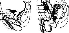
Figure 3.
Figure 4.
Figure 3. Median section through the male pelvis with empty ampulla recti: 1-urinary bladder; 2-symphysis; 3-rectum. Figure 4. Median section through the male pelvis with filled ampulla recti: ' 1-anterior peritoneal fold of the urinary bladder; 2-urinary bladder; 3-prostata; 4~symphysis; 5- ampulla recti. distinguish the anterior, posterior, and two lateral walls that transition into one another and thus have no clear boundaries (only in the filled bladder, Fig. 4); in the empty bladder, most of its walls face upward (Fig. 3). The peritoneum covers only part of the bladder walls, namely the entire upper section (which collapses when the bladder is empty) (Fig. 4, 5 b), and on the sides and behind it descends onto the bladder lower than on its anterior surface. Topographically, its relationships are as follows: the peritoneum from the anterior abdominal wall transitions onto the bladder, forming either a more uniform (Fig. 5 b) or a more wavy line of transition (Fig. 5 a). In some cases, the peritoneum first descends onto the posterior surface of the symphysis (Fig. 5 d), and then ascends onto the bladder. In the first variant, the peritoneal fold lies very high (Fig. 5 a and c) and when the bladder is filled, it moves even higher (Fig. 4). This exposes a significant section of the bladder devoid of peritoneum, which has practical importance for extraperitoneal approaches to the bladder (see Lithotomy). In the second variant, even when the bladder is full, the free section of the bladder is very small, and during an operation, if the peritoneal fold is not detached upward, the peritoneal sac can easily be opened. The first variant is more characteristic of men and young people, while the second is characteristic of women and elderly men. From the lateral walls of the bladder, the peritoneum transitions without a sharp boundary onto the lateral walls of the pelvis (Fig. 6). Behind the bladder, in men, the peritoneum covers the vas deferens and the apices of the seminal vesicles and at varying levels transitions onto the ampullary part of the rectum; the place of transition is the lowest part of the peritoneum. The space between the mentioned organs is called the excavatio recto-vesiealis; it is often filled with loops of the small intestine, sometimes with the cecum and the loop of S-Romani. In women, the peritoneum from the posterior walls of the bladder transitions onto the body of the uterus, forming a space (excavatio vesi-co-uterina) (Fig. 5 d), incorrectly called the Douglas pouch. The level of transition also varies here, but more often the lower third of the uterine body is not covered by peritoneum. The peritoneum of the bladder is loosely connected with the underlying layer of areolar tissue, which is why it is always possible, especially in front and behind, to extract the bladder from the peritoneal sac without particular violence. The perivesical areolar tissue is especially abundant.
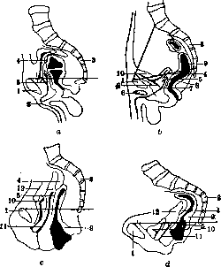
Figure 5. a-symphysipetal type of position of the urinary bladder in men (c-in women); b-sacropetal type of position of the urinary bladder in men (d-in women) (with empty bladder): /--symphysis; 2-urethra; г-sacrum; 4-cavum Douglasi posterior; 5 - aponeurosis Denonvillier; 6-corpus caverno-sum penis; 7-ductus ejaculatorius; 8-vesicula semimlis; 9-rectum; 10-vesica urinaria; 11- ,,, vagina; 12-uterus.
kovy: the peritoneum from the anterior abdominal wall transitions onto the bladder, forming either a more uniform (Fig. 5 b) or a more wavy line of transition (Fig. 5 a). In some cases, the peritoneum first descends onto the posterior surface of the symphysis (Fig. 5 d), and then ascends onto the bladder. In the first variant, the peritoneal fold lies very high (Fig. 5 a and c) and when the bladder is filled, it moves even higher (Fig. 4). This exposes a significant section of the bladder devoid of peritoneum, which has practical importance for extraperitoneal approaches to the bladder (see Lithotomy). In the second variant, even when the bladder is full, the free section of the bladder is very small, and during an operation, if the peritoneal fold is not detached upward, the peritoneal sac can easily be opened. The first variant is more characteristic of men and young people, while the second is characteristic of women and elderly men. From the lateral walls of the bladder, the peritoneum transitions without a sharp boundary onto the lateral walls of the pelvis (Fig. 6). Behind the bladder, in men, the peritoneum covers the vas deferens and the apices of the seminal vesicles and at varying levels transitions onto the ampullary part of the rectum; the place of transition is the lowest part of the peritoneum. The space between the mentioned organs is called the excavatio recto-vesiealis; it is often filled with loops of the small intestine, sometimes with the cecum and the loop of S-Romani. In women, the peritoneum from the posterior walls of the bladder transitions onto the body of the uterus, forming a space (excavatio vesi-co-uterina) (Fig. 5 d), incorrectly called the Douglas pouch. The level of transition also varies here, but more often the lower third of the uterine body is not covered by peritoneum. The peritoneum of the bladder is loosely connected with the underlying layer of areolar tissue, which is why it is always possible, especially in front and behind, to extract the bladder from the peritoneal sac without particular violence. The perivesical areolar tissue is especially abundant.
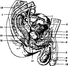
Figure 6. Relationship of the peritoneum to the bladder when viewed from the side: 1-peritoneum viscerale; 2-ductus deferens dexter; 3-os pubis; 4-m. pubo-vesicalis; 5-ligamentum pubo-prostaticum mediale; 6-pars membranacea urethrae; 7-epididymis; 8-praeputium; 9-testis; 10-glans penis; 11-glandula bulbo-urethralis; 12-musculus sphincter ani externus; 13-prostata; 14-vesicula seminalis dextra; 15-ureter dexter. (After Braus.)
in front (between the symphysis and the bladder - cavum Retzii); there is much cellular tissue on the sides and behind the bladder; somewhat less of it on the apex, while in the region of the base it is almost completely absent. The perivesicular cellular tissue communicates in women with the parametrium. The abundance of cellular tissue around the bladder has important practical significance in that when the bladder ruptures or is wounded, urine freely flows into the surrounding cellular tissue and also infiltrates the cellular tissue of neighboring organs (see below - urinary extravasations and urinary infiltrations). In men, below the excavatio recto-vesicalis, the bladder is separated from the rectum by means of the Denonvillier aponeurosis; separating it from this aponeurosis presents no particular difficulty. In women, the bladder comes into contact with the lower third of the body of the uterus, the cervix, and the vagina, being separated by a layer of loose cellular tissue, which is why the separation of the uterus occurs easily. The connection of the bladder with the vagina is very strong, and both these organs form as it were a single whole, although their blunt separation is always possible. The muscular layer of the urinary bladder consists of smooth muscle, developed to varying degrees; with sufficient persistence, 3 layers can be prepared (Testut). The outer, vertical layer of muscles is especially abundant in front of and behind the bladder. The muscle bundles begin from the pubic bone, from the pubo-vesical ligaments (lig. pubo-vesicalia) and from the neck of the bladder; in men also from the capsule of the prostate. Surrounding the bladder, the muscle reaches the base of the prostate gland, and in women - to the vagina. This muscle, the detrusor urinae, is developed to varying degrees, but is the most powerful. The second, middle layer is weakly developed in the form of circular muscle fibers. Their number increases at the place where the bladder passes into the urethra and gives rise to the sphincter muscle (m. sphincter vesicae). The latter (the sphincter of the bladder, closing the exit for urine) is located immediately under the mucous membrane in the neck of the bladder; its width is up to 1 cm. It is connected with the sphincter urethrae. In women, the sphincter is more extensible than in men; in the latter, the prostate gland prevents extension. The third, innermost muscular layer - submucous; it is very thin, its fibers lie vertically. A decrease in the number of muscle fibers and their improper arrangement create weak areas in certain parts of the bladder. Sometimes the walls of the bladder can, due to urine retention, stretch to such an extent that the mucous membrane comes into contact with the adventitia, which promotes the formation of hernias of the bladder mucosa, usually called diverticula (see Diverticulum). Sometimes with powerful development of individual muscle bundles and their hypertrophy, the formation of distinct ridges with the mucous membrane protruding between them is possible (vessie a colonnes, Balkenblase). On the floor of the urinary bladder from the side of the mucous membrane, a special area clearly protrudes, called the triangle of Lieutaud (trigonum Lieutaudi), syn. trigonum vesicae [see separate table (for Neuroglia), fig. 6], the apex of which corresponds to the internal opening of the urethra. The base of this triangle is not always a clearly expressed elevation between the two openings of the ureters (torus interuretericus). The ureters, converging, penetrate the wall of the bladder (portio intramuralis of the ureters) and slightly elevate the mucous membrane, forming an elevation that runs obliquely in the direction of the internal opening of the urethra; this elevation, a fold, not always constant, bears the name plica ureterica. The opening of the ureters lies obliquely, having the shape of an oval, a comma, or representing a slit; sometimes a fold of the mucous membrane slightly covers it. The distance between both ureteral openings (the base of the triangle) varies greatly; in women the base is wider than in men. The height of the triangle of Lieutaud also varies and in men it is greater than in women. Variants of the triangle of Lieutaud have practical significance for cystoscopy. Asymmetrical triangles are also noted, occurring more often in old people and in pathological processes in the bladder and neighboring organs. In men, in the region of the triangle, near the urethral opening, the middle lobe of the prostate protrudes, which is sometimes clearly expressed in the form of a uvula (uvula vesicae). In men, the part of the base of the bladder lying behind the torus is called 'bas fond', fossa retro-ureterica. When the bladder is filled, this part is displaced, especially in old people, and here, with weakness of the bladder muscles, urine often stagnates. This area deepens with age due to the enlargement of the prostate. The bas fond comes into contact in men with the pars ampullaris of the rectum, being separated from it by the Denonvillier aponeurosis (syn. fascia peritonaeo-perinealis, septum recto-vesicale, fig. 5 a-d). From the side of the outer surface of the bladder, the bas fond corresponds to the triangle (trigonum vesico-rectale), formed on the sides by the ampullary part of the seminal ducts, and from above by the peritoneal reflection; its apex corresponds to the prostate. In women, this area of the bladder comes into contact with the anterior wall of the vagina. When the latter prolapses, this part of the bladder wall is also drawn along, and sometimes with it a part of the triangle of Lieutaud; this condition is called cystocele. Tillo (Tillaux) believes that in women there is no bas fond, since he explains its occurrence by the presence of the prostate gland.
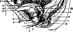
Figure 7. Arteries of the pelvis on the left: 1-vesica urinaria; 2-prostata; 3-a. dorsalis penis; 4-a. scrotalis posterior; 5-a. penis; 6-a. pudenda interna; 7-a. haemorrhoidalis inf.; 8-a. haemorrhoidalis media; 9-a. vesicalis inf.; 10-vesicula seminalis; 11-ductus deferens; 12 and 13- lig. umbilic. lat. et med.; 14-a. vesic. sup.
plexuses. In men there is a vesico-prostatic plexus, collecting blood from the bladder and prostate; it surrounds the prostate and the lateral parts of the base of the bladder. The anterior part of this plexus is called the pudendal plexus, s. praovesicalis Santorini. From the plexuses, blood is carried away through vv. vesicales sup. and inf., vv. haemorrhoidales and partly through v. obturatoria. In women, the pudendal plexus and the vesical plexus form part of an extensive venous plexus that lies above the fascia pelvis, lying
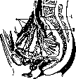
to the side of the floor of the bladder, to the vagina and rectum. - Lymphatic vessels (fig. 8) of the upper parts of the bladder partly go to the glands of the anterior abdominal wall and partly empty into gl. lymph, along the course of a. iliaca ext. (plexus iliacus), and from the middle parts into the glands along the course of a. hypogastrica (plexus hypogastricus); lymphatic vessels of the neck together with the vessels of the prostate and seminal vesicles empty into the glands at promontorium (plexus sacralis medius). Nerves of the bladder originate from the anterior-superior
Figure 8. Lymphatic vessels of the urinary bladder, flowing into the plexus of the external and internal iliac arteries: 1- lymphatic vessels and glands of the plexus of the external iliac artery; 2- same for the internal; 3- glands on the middle sacral artery. The sympathetic plexus (plexus hypogastricus inf.) consists of 2-4 trunks, which, entering the urinary bladder, go to the perivesical nodes and by separate trunks are directed to the intramural nerve nodes. For the urinary bladder, the abundance of nodes is typical, which are especially concentrated in the area of the triangle. Fixation of the urinary bladder. The bladder with its base is firmly connected in men with the capsule of the prostate gland, in women- with the superior fascial leaf of the urogenital diaphragm; this is the main fixing apparatus. Auxiliary roles are played by the processes of the lamina visceralis of the pelvic fascia. In men, the lamina visceralis is more pronounced, extending upward from the bottom of the pelvic cavity on the sides of the prostate gland. In front of it, these processes in the form of tendinous ligaments attach to the descending branch of the pubic bone, forming the so-called lig. pubo-prostaticum laterale dext. and sin. Even higher, the lamina visceralis passes to the lateral surfaces of the urinary bladder, also attaching to the pubic bone- this segment of the fascia in front of the bladder on the right and left is called lig. pubo-vesicale laterale dext. and sin. and is very inconstant. Behind the bladder, the processes of the lamina visceralis are thrown onto the lateral wall of the rectum, forming in men a little constant and weakly expressed ligament- lig. vesico-rectale. Some authors distinguish lig. pubo-prostaticum and lig. pubo-vesicale medium, an inconstant ligament, which is also a process of the pelvic fascia. In the listed ligaments, muscle fibers can sometimes be determined. In the space between the symphysis in front, the urinary bladder and part of the prostate behind, a slit is formed, the so-called spatium prae-vesicale Retzii (syn. cavum Retzii), below limited by a dense membrane, on the sides and partly below by the mentioned ligaments (lig. pubo-prostaticum and pubo-vesicale), and above communicating very widely with the retroperitoneal tissue. This space is filled with loose tissue and venous plexus- plexus venosus Santorini. Into this plexus flow the vessels penetrating here through the thickness of the urogenital diaphragm immediately under lig. arcuatum- v. dorsalis penis and v. clitoridis. In women, the mentioned pelvic fascia ligaments are expressed significantly weaker; cavum Retzii is wider but shorter than in men. A certain, albeit weak, fixing role also belongs to the vessels and finally has significance lig. vesico-umbilicale medium--a ligament stretching from the apex of the bladder to the navel and lying under the peritoneum. It is formed from the remnants of urachus, which sometimes persists throughout life, then communicating with the bladder through a narrow opening; sometimes it also connects with the navel. Lig. umbilicalia lat., remnants of obliterated aa. umbilicales, go from the lateral walls of the bladder to the navel. A large supporting role is played by the intraperitoneal pressure. In men, the empty urinary bladder contacts behind with the rectum, and above it lie loops of the small intestine; when the bladder is full, the small intestine can also lie behind it. When the rectum is full, it can push the bladder forward to the anterior abdominal wall (fig. 4). In women, behind the bladder lies the uterus; in an empty bladder, the uterus lies on it (physiol. anteflexion). When the bladder is full, the uterus rises and moves backward. The bladder is especially displaced during pregnancy. Types of bladder position. Among individual variations in the position of the urinary bladder, two types of its position can be established (Moskalenko). Symphysopetal (fig. 5 a and c) and sacropetal (fig. 5 b and d), which is determined by the position of the Denonvilliers aponeurosis ("function of pelvic topography" Moskalenko). In the diagrams (fig. 5 a-d), exact copies from sections, the features of the topography of the urinary bladder in each type in men and women are visible. With the sacropetal type of pelvic organ position, the Denonvilliers aponeurosis is tilted backward, the bottom of the urinary bladder lies below the pelvic horizontal level (fig.5 b), the prevesical fold of the peritoneum is located low (in women below the pubis) (fig. 5 d). With the symphysopetal type, the aponeurosis is tilted forward toward the symphysis (fig. 5 a and c), the bottom of the urinary bladder lies above the pelvic horizontal level, the peritoneal fold is located high above the symphysis.
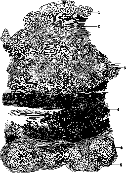
III. Histology. In the wall of the urinary bladder, the following layers are distinguished (fig. 9): 1) the mucous membrane (mucosa), 2) the submucous (submucosa), 3) the muscular (muscularis) and 4) the fibrous (fibrosa, s. adventitia), which fuses with the peritoneum covering the urinary bladder. The mucous membrane in life is pink in color due to the large number of capillaries; in the area of the trigone it is paler; in the collapsed bladder it lies in folds. Its thickness in the collapsed state is 2 mm, in the area of the trigone, where it is stretched, about 0.1 mm; with stretching, the thickness decreases significantly, reaching 0.1 mm (Kolliker). The mucous membrane consists of epithelium and propria tissue. The epithelium is multilayered cubic, or as it is called, transitional (fig. 10), in the collapsed bladder it reaches a thickness of 60- 100 μ. In it, three cellular layers are distinguished: at the base, a layer of low cylindrical cells with an elongated or, conversely, swollen apex; above it in the middle layer, cells of various shapes, polyhedral, cubic, spindle-shaped, flask-shaped (in the collapsed bladder they acquire a cylindrical character, and the number of their rows increases-4--6); on the surface a layer
Figure 9. Cross-section of the urinary bladder wall: 1- epithelium; 2- connective tissue; 3- inner muscle layer; 4- circular muscle layer; 5- outer muscle layer; 6- peritoneum. (According to Kulchitsky.)
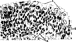
of large covering cells (Deckzellen) of cubic shape, which strongly flatten with stretching. These cells have been the subject of numerous studies (BChet-ming, Dogel, Eggeling, Danini, Flerov) and
Figure 10. Epithelium of the urinary bladder: 1- superficial layer of cubic cells; 2- deep layer of cylindrical cells; 3- connective tissue. (According to Braus.)
a number of features have been noted in them. On the free surface, a thin cuticular covering can be found, and at its boundaries with neighboring coverings, closing ridges; these formations are undoubtedly protective adaptations; many cells contain two nuclei (sometimes more), the origin of which 129
180 previously attributed to amitosis, but which probably arise as a result of abortive (sometimes multi-nucleolar) cariokinesis; in the body of the cell, two zones can be distinguished: the superficial (ectoplasm), which is homogeneous or fibrous, and the deep (endoplasm), which is light and granular, sometimes with fat droplets. Cells of different layers are connected to each other by intercellular substance, which, merging with the ectoplasm, forms a kind of spongy framework of the epithelial layer, giving it the ability to withstand considerable stretching. The propria tissue of the mucous membrane is very thin, consisting of fibrous connective tissue with a large number of elastic fibers and cells (fibrocytes); it is almost always infiltrated with lymphocytes, which in places form nodules. The existence of a special boundary membrane between the epithelium and propria is denied by most authors, while the penetration of thin outgrowths of connective tissue between the basal cells of the epithelium is described; capillaries, which form dense networks under the epithelium, sometimes penetrate together with these outgrowths into the epithelium, which gave rise to talk of its vascularization. There are no glands in the mucous membrane of the urinary bladder, but near the urethra, Kölliker described small grape-like glands that secrete a light mucous secretion; other researchers have not found them and consider them heterotopic urethral glands. Brunn's epithelial nests (see), which can simulate glands, are not found in the normal urinary bladder. The submucous membrane has a greater thickness than the mucous membrane and consists of loose fibrous connective tissue, which allows the mucous membrane to form folds. Only in the area of the trigonum is the connective tissue denser, which is why this area remains smooth. In the deep layers of the submucosa, bundles of muscle fibers appear, and it merges without sharp boundaries into the muscular layer, which forms the main mass of the wall of the urinary bladder. This layer consists of smooth muscles that form bundles of varying thickness, anastomosing with neighboring ones and forming networks, in places interrupted by elastic tendons. Between the bundles there is a fairly large amount of loose connective tissue with a large number of blood vessels; lymphatic vessels are also located here. Depending on the predominant direction of the bundles, three layers are distinguished in the muscular layer (see above). With age, there is some replacement of the smooth muscle tissue of the bladder with connective tissue.-The fibrous layer is a thin layer of fibrous connective tissue, to which the peritoneum (see) is tightly fused.-The urinary bladder is abundantly supplied with nerves (see above, as well as Vegetative nervous system); in the mucous membrane, a number of nerve endings are described: free endings in the epithelium (Retzius), various kinds of bodies and tree-like branches in the connective tissue (Mikhailov).-Physiology of the urinary bladder-see Urination. IV. Methodology for studying the urinary bladder. The study of the urinary bladder is based on questioning the patient and collecting objective data. When questioning, attention is paid to the nature of the urine, pain during and after urination, and disorders of the act of urination. Purulent or bloody urine, the presence and shape of blood clots, the appearance of blood at the beginning, end, or throughout the act of urination, the nature and time of onset of pain, increased urination, retention, incontinence of urine, changes in the stream-should be taken into account as the most important indicators of various diseases of the urinary bladder (see Urination, Hematuria). After questioning the patient, one should, if possible, personally verify with one's own eyes the changes described by the patient, by having him urinate in your presence. Then proceed sequentially to inspection, palpation, percussion, and if necessary, to catheterization, sounding, and cystoscopy.-Inspection of the area of the urinary bladder can provide valuable data. In cases where the urinary bladder is greatly distended

Figure 11. Overfilled urinary bladder ('pregnant uterus').
The urinary bladder, its area protrudes above the general level of the abdomen in the form of an elongated, rounded tumor located along the midline and sometimes reaching the navel (Fig. 11).- Palpation of the urinary bladder can provide the necessary data only when there is a large stone in the bladder or a far-advanced tumor. The palms of both hands are placed on the area being examined so that the bases of the hands are on the pubis and the fingers on the abdomen. Having the patient breathe deeply with the abdomen, during exhalation, gradually insert the fingertips deeper into the abdominal cavity; with an excessively developed subcutaneous fat layer and pliable abdominal walls, it is possible to grasp the urinary bladder from behind and partially from the sides and feel any new growth or foreign body present there. Bimanual examination provides more data, in which the index finger of the right hand is inserted into the rectum (in women, into the vagina), and the left hand palpates the area of the urinary bladder from the side of the abdominal wall. The urinary bladder should be empty during this procedure. With this method, a tumor or foreign body that comes between the two examining hands can be more easily determined.- Percussion over the pubis in an empty, healthy urinary bladder gives a tympanitic sound. When the urinary bladder is filled with urine over the pubis, a dull sound is obtained. Thus, percussion can determine the degree of distension of the urinary bladder. - Sounding, at present, is used only to detect a stone in the urinary bladder. For this purpose, a metal sound or metal catheter is inserted into the urinary bladder. The Guyon sound with a small beak and a hollow handle (stone-finder) is most suitable for this purpose, which enhances the resonance when the sound is tapped against the stone. According to general rules (see Catheterization), into the urinary bladder filled with 150-200 cm³ of liquid (warm water, boric solution, solution of Hydrarg. oxycyanati 1:5,000), a metal sound is inserted. The sign of correct insertion of the sound is the ability to turn it completely freely in all directions. The sound is advanced to the posterior wall, turned 180° with the beak downward, and then gradually withdrawn, making turns to the right and left. If a stone is present, a tapping sound is heard when the sound strikes it, and the hand feels the sensation of the sound coming into contact with a solid body, and an impression is also formed of the surface of the stone and its size. The absence of a stone on sounding cannot yet serve as a final criterion, since with this method it is impossible to detect a stone in a diverticulum and it is extremely difficult, and sometimes impossible, to find a stone lying on the deep floor of the urinary bladder with a significantly enlarged and protruding into the bladder prostate gland. The examination of the female urinary bladder with a finger by preliminary dilation of the urethra is now abandoned, as it is often followed by persistent urinary incontinence. Among the methods of examining the urinary bladder, catheterization of it (see Catheters and Catheterization) has particularly important significance and widespread use. From the direction of the stream flowing from the urinary bladder through the inserted catheter, one can judge the tone of the muscular wall of the urinary bladder. A long horizontal stream indicates good contractile ability of its walls, while a vertical or nearly vertical stream indicates atony of the urinary bladder. Failure to obtain urine through a catheter inserted into the urinary bladder after a previous injury and a long delay in urination indicates rupture of the urinary bladder. During such unsuccessful catheterization, it may happen that suddenly during one of the turns of the catheter, urine mixed with blood will flow out. This indicates that the catheter entered the rupture cavity and further confirms the diagnosis of rupture. Sometimes, such a sudden outflow of a more or less large amount of urine from the catheter after all the urine from the urinary bladder has already been released can be observed in a diverticulum of the urinary bladder, into which the catheter accidentally entered during manipulation. Catheterization, as well as sounding, is contraindicated in acute gonorrhea, in easily and heavily bleeding tumors of the prostate gland and bladder, in the presence of a fresh false passage in the urethra, and in an acute inflammatory or ulcerative process in the urethral orifice itself.- The most valuable data in the examination of the urinary bladder is provided by cystoscopy, which has recently become widespread. Cystoscopy makes it possible to examine the entire urinary bladder and recognize diseases [see separate table (pp. 143-144), Fig. 1-12], which could not be firmly established without cystoscopy, namely diverticula of the urinary bladder, stones in diverticula or lying on the deep floor of the urinary bladder behind a greatly hypertrophied prostate gland, tumors, ulcers. Furthermore, cystoscopy helps to clarify the nature of certain diseases and the extent of their spread: benign or malignant nature of the tumor, the number of stones, the nature of cystitis, etc. Therefore, work in the urological and generally in the surgical department is extremely difficult without cystoscopy. In cases where cystoscopy is impossible, X-ray examination of the urinary bladder is resorted to [see separate table (pp. 135-136), Fig. 1-4]. V. Pathology. Developmental defects. Among developmental defects, very rare malformations are: doubling of the urinary bladder cavity (vesica bi-partita), incomplete division of its cavity into two chambers (vesica bilocularis), and finally, complete absence of the urinary bladder with the ureters opening directly into the urethra.- Congenital distension and filling of the urinary bladder cavity with urine (normally the urinary bladder of a newborn contains 3-4 cm³ of urine) can be observed in urethral atresia, but sometimes it occurs without it and in this case is difficult to explain. A greatly distended urinary bladder of the fetus (more than 100 cm³ of urine) can be an obstacle during labor. Ectopia of the urinary bladder (ectopia ves. urin., ex-strophia ves. urin., inversio ves. urin., fissura ves. urin.), so-called prolapse of the urinary bladder, congenital fissure of the urinary bladder. Among all developmental defects of the urinary bladder, ectopia occupies first place in frequency and clinical significance; it occurs in approximately 1 in 40,000 of the population, and more often in boys than in girls. This malformation consists in the fact that the anterior abdominal wall in the area of the urinary bladder is as if split, and the anterior wall of the urinary bladder is also split; the urinary bladder lies as if in front of the abdominal wall and everted with its mucous membrane forward. Often, with this condition, there is a splitting of the urethra along the anterior surface of the penis (epispadias). When examining a patient with ectopia of the urinary bladder, a protruding, moist, red mass is visible above the pubis, sometimes with papillary growths; in its lower part on the sides, the openings of the ureters can be found, through which urine is excreted. As microscopic examination reveals, the mucous membrane of the everted urinary bladder is usually covered with stratified squamous, even keratinized epithelium (result of metaplasia), or sometimes with secreting mucus cylindrical epithelium. To explain the mechanism of ectopia of the urinary bladder, various theories have been proposed, of which in former times the most popular was the view that the basis of this developmental defect is an obstacle to the outflow of urine and overfilling of the urinary bladder in the fetus, due to which the separation of the fused parts of the abdominal wall occurs. However, after the research of Keibel, Enderlen and others, it is considered established that the cause of ectopia lies in the delayed longer-than-usual existence of the cloaca septum, which, as is known, also includes the area that later becomes the lower part of the anterior abdominal wall. The delayed reduction of the septum leaves the anterior abdominal wall and the urinary bladder split.- Rarely, prolapse of the urinary bladder occurs in an unsplit form (true ectopia of the urinary bladder), as well as splitting of its posterior wall with communication of the bladder cavity with the peritoneal or vaginal cavity or rectum; at the same time, anal and urethral atresia can be observed. The clinical picture comes down to constant leakage and urinary incontinence, maceration of the surrounding skin, which leads to chronic eczema. Contact of clothing with the severely irritated, sometimes covered with excoriations mucous membrane is painful. The constant irrigation of clothing with rapidly decomposing urine causes the spread of the smell of urine, making patients unbearable to those around them.- Treatment of ectopia of the urinary bladder can be conservative and surgical. Conservative measures come down to wearing various types of urine collectors that constantly collect the leaking urine, but these collectors often do not achieve their purpose and do not satisfy patients. The development of surgical technique has created a whole range of methods of surgical treatment. Plastic operations of Thiersch and Billroth are basically reduced to skin plastic surgery of the defect of the anterior wall of the urinary bladder, but they do not give particular success and due to the absence of a urinary bladder sphincter, they do not eliminate the main complaint of patients-urinary incontinence. The Trendelenburg method-approximation of the pubic bones with subsequent plastic surgery-also does not give significant success. Operations of transplanting the ureters into the skin, vagina, urethra, as well as the Makkas method-implanting the ureters into the blind gut, excluding it and creating an artificial urinary bladder with diversion of urine through the appendix, sewn into the abdominal wall,-all this did not satisfy patients, as it did not free them from the subsequent wearing of urine collectors.
Subbotin proposed forming a urinary diversion channel from the wall of the rectum; a communication is formed between the bladder and the rectum, and from the latter a separate tube is created, which does not communicate with the rectum and runs parallel to it. This operation eliminates the danger of infection entering the urinary tract, but is technically complex and dangerous, as it gives a rather high percentage of immediate mortality. In this operation in girls, the question of future sexual and reproductive activity should be taken into account, for which reason some authors resorted to simultaneous resection of the Fallopian tubes or extirpation of the uterus (Gorokhov). At present, the operation of transplanting the ureters into the large intestine is most widespread; in this case, the urine is retained by the rectal sphincter and thereby patients are freed from the most distressing symptom - urinary incontinence. To prevent the possibility of ascending infection from the intestine in this case, Maydl proposed a simpler method of transplanting the ureters together with a part of the bladder (fig. 12-14). Maydl's operation is more complex than direct implantation and gives a higher mortality. Nevertheless, kidney infection is not prevented by this method, and at present most surgeons implant the ureters individually into the pelvic colon. Patients are able to retain urine from 2 to 8 hours, in some cases emptying of the bladder occurs more frequently. The negative side of the operation is irritation of the lower part of the intestine by urine. However, individual cases have been described where operated patients lived to mature and even advanced old age. Congenital intravesical folds of the urinary bladder are rare, they
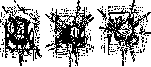
Figure 12. 1st moment. Incision of the wall of the urinary bladder with the ureters. Figure 13. 2nd moment. Incision of the sigmoid colon and implantation of the wall of the urinary bladder into the incision of the sigmoid colon. Figure 14. 3rd moment. Implantation of the wall of the urinary bladder into the incision of the sigmoid colon. ♦5 may have different directions and thereby give the bladder different shapes. A sharply expressed horizontal fold of the mucous membrane, embracing the wall of the urinary bladder all around, gives its cavity the shape of an hourglass, in which case the ureteral openings may open into both the upper and lower halves of the bladder. Such folds at the exit of the bladder may also be encountered in the form of valves and mechanically hinder the free outflow of urine. The presence of these folds is diagnosed by urethrocystoscopy. Treatment consists of incising them electro- or thermocauter endovesically or on the opened bladder. Interligamentous or intraligamentous urinary bladder is extremely rare and represents an anomaly of incorrect position (low, lateral). Diagnosis is usually clarified only during an operation undertaken for what is supposedly an intra-ligamentary ovarian cyst. Displacements of the urinary bladder may be observed in women in the form of so-called vaginal cystocele, under which is meant the protrusion of the urinary bladder into the vagina, which is usually the result of traction from the prolapsed or displaced uterus; there may also be inversion and prolapse of the urinary bladder through the urethra. Diverticula of the bladder are true and false. The first of these, which represent pocket-like protrusions in which the mucous membrane and muscular layer participate, are rare. They may be congenital and acquired; the former most often are located near the sites of ureteral insertion and, reaching large sizes [see separate table (pp. 143-144), fig. 9], can simulate a two-chambered bladder (see above). Acquired true diverticula (figure 15) usually develop as a result of increased intravesical pressure (for example in prostatic hypertrophy, stricture of the canal, stones); they vary in size, sometimes reaching a capacity of up to 1.5 liters; the opening connecting the cavity of the diverticulum with the urinary bladder is sometimes very small. False diverticula of the bladder in the form of small protrusions of a single mucous membrane between muscle bundles are most often observed on the posterior and lateral walls of the bladder in its trabecular hypertrophy. Diverticula are often multiple; this especially concerns small false diverticula. Some also attribute abscess cavities that have ruptured into the bladder and are in communication with it to false diverticula. Inflammatory processes easily develop in diverticula of the bladder, extending to the perivesicular connective tissue and the serous covering of the bladder, perforations occur into the abdominal cavity or into the connective tissue, stones form.-Upon reaching a certain
size, the diverticulum begins to play a peculiar role in the act of urination: urine flowing from the bladder during its contraction encounters an obstacle to free outflow in the above-mentioned cases, and part of it enters the diverticulum from the bladder under increased pressure, i.e., the patient as if urinates into his own diverticula (Blum). After emptying the bladder and thus reducing the pressure in it, the contents of the diverticulum, due to the contraction of its wall, again enters the bladder. Constant pressure of urine on the wall of the diverticulum causes its further expansion, atrophy of the muscle fibers. The diverticulum loses its contractile ability and can gradually turn into a large atonic sac with constant urine retention in it. Ultimately, the mechanism of diverticulum development consists of two moments: the size of the obstacle to urination and the strength of resistance of the muscular wall of the diverticulum to pressure. The presence of an atonic diverticulum in the bladder with constant urine retention in it is a predisposing factor for the development of infection; this poses a threat to the general health condition of diverticulum carriers. The danger lies not in the diverticulum as such, but in the infection that may join it. The presence of urine retention in the diverticulum, inflammation of its wall is also a favorable factor for the development of new growths, stones in it. The clinical picture in bladder diverticulum is extremely atypical and varied. Symptoms depend mainly on diseases accompanying or complicating the diverticulum (stones, prostatic hypertrophy, etc.). An uninfected diverticulum can sometimes remain completely unnoticed by the patient. The main diagnostic methods are cystoscopy and X-ray. In most cases, it is possible to see the entrance to the diverticulum in the form of a dark-colored opening with more or less pronounced radial folds of the mucous membrane on it. One should not lose sight of the fact that the sphincter of the diverticulum, when contracting, can make it invisible during cystoscopy. A more precise method of investigation is contrast X-ray and X-ray fluoroscopy, which make it possible not only to determine the topography and size of the diverticulum, but also the degree of its atony, the degree of its emptyability (see separate table, figure 3). The latter is achieved by serial films. The bladder diverticulum is filled with contrast substance through a ureteral catheter with the help of a cystoscope. Irrigating fluid is left in the bladder. The cystoscope and ureteral catheter are removed. After this, at certain intervals of time, outside and during urination, films are taken. The speed of disappearance of the shadow of the diverticulum (the speed of transition of the contrast substance from the diverticulum into the bladder), the degree of emptyability of the diverticulum during the act of urination give us an idea of the contractile ability of the diverticulum, the degree of urine retention in it. Direction of the rays in X-ray. Л ?*.'iv .'»-'
Figure 1. X-ray of a normal urinary bladder filled with contrast substance. Figure 2. X-ray of cancer of the urinary bladder. Figure 4. X-ray of a diverticulum (left)

Figure 12. 1st moment. Incision of the wall of the urinary bladder with the ureters. Figure 13. 2nd moment. Incision of the sigmoid colon and implantation of the wall of the urinary bladder into the incision of the sigmoid colon. Figure 14. 3rd moment. Implantation of the wall of the urinary bladder into the incision of the sigmoid colon. ♦5 may have different directions and thereby give the bladder different shapes. A sharply expressed horizontal fold of the mucous membrane, embracing the wall of the urinary bladder all around, gives its cavity the shape of an hourglass, in which case the ureteral openings may open into both the upper and lower halves of the bladder. Such folds at the exit of the bladder may also be encountered in the form of valves and mechanically hinder the free outflow of urine. The presence of these folds is diagnosed by urethrocystoscopy. Treatment consists of incising them electro- or thermocauter endovesically or on the opened bladder. Interligamentous or intraligamentous urinary bladder is extremely rare and represents an anomaly of incorrect position (low, lateral). Diagnosis is usually clarified only during an operation undertaken for what is supposedly an intra-ligamentary ovarian cyst. Displacements of the urinary bladder may be observed in women in the form of so-called vaginal cystocele, under which is meant the protrusion of the urinary bladder into the vagina, which is usually the result of traction from the prolapsed or displaced uterus; there may also be inversion and prolapse of the urinary bladder through the urethra. Diverticula of the bladder are true and false. The first of these, which represent pocket-like protrusions in which the mucous membrane and muscular layer participate, are rare. They may be congenital and acquired; the former most often are located near the sites of ureteral insertion and, reaching large sizes [see separate table (pp. 143-144), fig. 9], can simulate a two-chambered bladder (see above). Acquired true diverticula (figure 15) usually develop as a result of increased intravesical pressure (for example in prostatic hypertrophy, stricture of the canal, stones); they vary in size, sometimes reaching a capacity of up to 1.5 liters; the opening connecting the cavity of the diverticulum with the urinary bladder is sometimes very small. False diverticula of the bladder in the form of small protrusions of a single mucous membrane between muscle bundles are most often observed on the posterior and lateral walls of the bladder in its trabecular hypertrophy. Diverticula are often multiple; this especially concerns small false diverticula. Some also attribute abscess cavities that have ruptured into the bladder and are in communication with it to false diverticula. Inflammatory processes easily develop in diverticula of the bladder, extending to the perivesicular connective tissue and the serous covering of the bladder, perforations occur into the abdominal cavity or into the connective tissue, stones form.-Upon reaching a certain
size, the diverticulum begins to play a peculiar role in the act of urination: urine flowing from the bladder during its contraction encounters an obstacle to free outflow in the above-mentioned cases, and part of it enters the diverticulum from the bladder under increased pressure, i.e., the patient as if urinates into his own diverticula (Blum). After emptying the bladder and thus reducing the pressure in it, the contents of the diverticulum, due to the contraction of its wall, again enters the bladder. Constant pressure of urine on the wall of the diverticulum causes its further expansion, atrophy of the muscle fibers. The diverticulum loses its contractile ability and can gradually turn into a large atonic sac with constant urine retention in it. Ultimately, the mechanism of diverticulum development consists of two moments: the size of the obstacle to urination and the strength of resistance of the muscular wall of the diverticulum to pressure. The presence of an atonic diverticulum in the bladder with constant urine retention in it is a predisposing factor for the development of infection; this poses a threat to the general health condition of diverticulum carriers. The danger lies not in the diverticulum as such, but in the infection that may join it. The presence of urine retention in the diverticulum, inflammation of its wall is also a favorable factor for the development of new growths, stones in it. The clinical picture in bladder diverticulum is extremely atypical and varied. Symptoms depend mainly on diseases accompanying or complicating the diverticulum (stones, prostatic hypertrophy, etc.). An uninfected diverticulum can sometimes remain completely unnoticed by the patient. The main diagnostic methods are cystoscopy and X-ray. In most cases, it is possible to see the entrance to the diverticulum in the form of a dark-colored opening with more or less pronounced radial folds of the mucous membrane on it. One should not lose sight of the fact that the sphincter of the diverticulum, when contracting, can make it invisible during cystoscopy. A more precise method of investigation is contrast X-ray and X-ray fluoroscopy, which make it possible not only to determine the topography and size of the diverticulum, but also the degree of its atony, the degree of its emptyability (see separate table, figure 3). The latter is achieved by serial films. The bladder diverticulum is filled with contrast substance through a ureteral catheter with the help of a cystoscope. Irrigating fluid is left in the bladder. The cystoscope and ureteral catheter are removed. After this, at certain intervals of time, outside and during urination, films are taken. The speed of disappearance of the shadow of the diverticulum (the speed of transition of the contrast substance from the diverticulum into the bladder), the degree of emptyability of the diverticulum during the act of urination give us an idea of the contractile ability of the diverticulum, the degree of urine retention in it. Direction of the rays in X-ray. Л ?*.'iv .'»-'
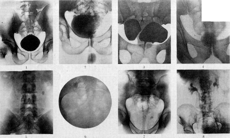
Figure 1. X-ray of a normal urinary bladder filled with contrast substance. Figure 2. X-ray of cancer of the urinary bladder. Figure 4. X-ray of a diverticulum (left)
in the urinary bladder (drawing). Figure I. X-ray of a stone (oxalate) in the urinary bladder. Figure II. X-ray of a stone in the upper part of the ureter. Figure III. X-ray of a stone in the middle part of the ureter (in the sacroiliac joint area). Figure IV. X-ray of a stone in the vesical part of the ureter. Figure V. Bilateral ureteropyelography. To ST. Urinary bladder. On the X-ray images (ventrodorsal, axial and lateral) depends on the location of the diverticulum, established during cystoscopy. While some authors are extremely radical and believe that every diverticulum of the urinary bladder should be operated on, others (Fronstein) indicate indications for surgery depending on the degree of atony of the diverticulum. The operation consists in the extirpation of the diverticulum with subsequent suturing of the defect in the bladder wall. Depending on the location of the diverticulum, it should be approached either through the anterior abdominal wall or by the sacral route. The technique is in most cases complex, and postoperative mortality ranges from 37% (Blum) to 10% (Gayet). Resection of the diverticulum must always be combined with simultaneous or prior elimination of the obstacle to the emptying of the urinary bladder. Palliative operations include: destruction of the diverticulum sphincter, creation of a bladder fistula with drainage of the diverticulum. In many cases, elimination of the cause of increased intravesical pressure leads to clinical cure of the patient, and sometimes to the disappearance of the diverticulum. Conservative treatment should be directed against infection and consist in washing out the bladder and the diverticulum cavity by means of a ureteral catheter introduced into it through a cystoscope. Atony of the urinary bladder depends on the weakening of the tone of the detrusor. Symptoms of atony are difficult urination, weak and sluggish stream, falling vertically downward, urination in drops. Atony of the urinary bladder depends most often on causes lying outside the bladder (secondary atony), less often on causes lying in the bladder itself (primary atony). Secondary atony arises from diseases that cause mechanical obstruction to the emptying of urine and from disturbance of innervation. Mechanical obstruction to the emptying of the urinary bladder occurs in strictures of the urethra, hypertrophy of the prostate, tumors located in the area of the sphincter or near it, foreign bodies (stones), etc. If the obstruction to the emptying of the urinary bladder occurs quickly (in case of blockage by a stone) and is not eliminated, atony of the bladder can develop quickly. Atony of the bladder is also observed in severe brain disorders, which is caused by the absence of normally outgoing impulses from the brain to the spinal cord centers. Such phenomena are observed especially often in apoplexy, concussion of the brain, meningitis and generally in persons who are in an unconscious state caused by an infectious disease. In such cases, atony can be temporary and with a favorable outcome of the disease, spontaneous urination is restored, but sometimes due to prolonged stretching of the walls of the urinary bladder, complete exhaustion of the bladder muscle may occur and atony of the bladder remains forever. The wall of an acutely dilated urinary bladder is usually very thin; however, if the expansion is based on a long-existing obstacle to the outflow of urine, e.g., hypertrophy of the prostate, stone or stricture of the canal, then the wall is hypertrophied. With hypertrophy of the wall of the urinary bladder, as a rule developing in the muscular layer, the inner surface of the bladder presents a very characteristic picture; hypertrophied muscle bundles protrude into the lumen in the form of a network of intersecting and as if branching beams, trabeculae or ridges (fig. 16); a urinary bladder with such hypertrophy is called a trabecular bladder (vesica trabecularis, German Balkenblase, French vessie a colonnes). More often, however, atony is central
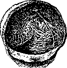
Figure 16. Muscle ridges of the urinary bladder.
Regarding its origin, it arises in diseases of the spinal cord, especially in tabes. Due to decreased sensitivity of the mucous membrane, the urge to urinate becomes less frequent. As a result of paresis of the motor nerves and weakening of the detrusor muscle, the bladder is unable to empty all the urine, residual urine appears, and subsequently complete inability to urinate independently. The urine overflowing the bladder, overcoming the resistance of the sphincter, is released drop by drop, leading to urinary incontinence (ischuria paradoxica). Lesions of the peripheral nerves can also cause atony, although it rarely occurs as an isolated disease. Atony of the bladder can result from hypotonia of the pelvic plexus, which is part of the parasympathetic nervous system. If there is no mechanical obstruction and no disease of the nervous system, then in such cases primary atony of the urinary bladder should be considered. Not all authors recognize this form and believe that in such cases there is an undiagnosed disease of the nervous system. However, the cases reported in the literature (Albarran, Walker, Fronshtein, etc.), where thorough examination of the nervous system and especially subsequent observation revealed no deviations from normal, force the recognition of the existence of this form. The etiology of primary atony has not yet been clarified. Some believe that degeneration of the bladder muscle and loss of its elasticity occur as a result of arteriosclerotic processes or prolonged infection, while others see the cause in contracture of the bladder neck. Sometimes atony of the bladder exists from childhood (congenital atony). It usually depends on underdevelopment of the spinal cord centers and, existing simultaneously with atony of the sphincter, manifests as urinary incontinence. Atony of the urinary bladder may be the initial and first symptom of tabes when no deviations from normal can yet be established from the nervous system or they are not significant. In such cases, cystoscopic examination can provide help. Nitze, Fronshtein, Walker, and others established that an early sign of central nervous system damage is trabeculation of the bladder. Some see the cause of trabeculation in hypertrophy of the bladder's muscle fibers, while others believe that it is based on passive stretching of weaker areas of the bladder wall by urine, while more powerful muscle fibers resist this pressure and appear in the form of ridges. It should be remembered that trabeculation is also observed in atony from mechanical obstruction, and therefore this symptom is significant only in the absence of the latter. Another sign of bladder disease based on central nervous system damage is Schramm's symptom. The latter consists in the fact that when withdrawing the cystoscope from the bladder with the beak downward, the field of view at the sphincter instead of sharply darkening as in normal cases remains illuminated, and the area of the posterior urethra and the seminal colliculus are clearly visible. However, studies by Schwarz, Pfister, Dunaevsky, and Mikhayev and others showed that this sign may often be absent in diseases of the central nervous system and, conversely, occurs in urogenital diseases (cystitis, bladder stones, etc.) and neurasthenia without organic lesions of the nervous system. Therefore, this symptom should be considered significant only in conjunction with other signs. Treatment of bladder atony consists of eliminating the cause that causes it. Atonies of a nervous nature and long-lasting atonies respond least well to treatment. Ruptures of the urinary bladder are not common. Predisposing factors include overfilling of the urinary bladder, which is a necessary condition for rupture. In a stretched state, the urinary bladder, adjacent to the anterior abdominal wall, is more susceptible to mechanical violence, and on the other hand, loses its elasticity due to stretching. Ruptures are usually observed between the ages of 20 and 40, and exceptionally in children. In women, due to anatomical-topographical features of the urinary bladder, ruptures are very rare and occur mainly during childbirth. The loss of elasticity of the urinary bladder walls in various pathological processes plays a significant role. A rupture of the urinary bladder can be complete or incomplete. In the latter case, only the mucous membrane is ruptured (Baradulin), while in complete rupture an opening, predominantly linear in shape with a prevailing longitudinal direction, is formed. Intra- and extraperitoneal ruptures are distinguished. The former are usually located on the apex and posterior wall of the bladder, the latter on the anterior wall. Ruptures of the base of the urinary bladder occur when pelvic bones are damaged. In intra-peritoneal ruptures, urine flows into the abdominal cavity, leading to limited or diffuse peritonitis. In extraperitoneal ruptures, urine enters the perivesical and pelvic cellular tissue, causing urinary infiltration with subsequent development of pelvic phlegmon (urinary extravasation). The first symptoms of the latter are subfebrile temperature and minor disturbances of the general condition, followed by swelling in the area of the urinary bladder, pain on palpation of the bladder, and signs of general sepsis. In most cases, shock, urinary retention, frequent urges with the excretion of a few drops of blood, and increasing pain in the lower abdomen are observed. In intra-peritoneal ruptures, peritoneal symptoms develop rapidly. Diagnosis of traumatic ruptures usually presents no difficulties, while spontaneous ones are sometimes difficult. The inability to urinate despite the presence of urges and the absence of dullness above the pubis indicate intra-peritoneal rupture. Usually, the presence of moving fluid in the abdominal cavity can be noted, and upon palpation through the rectum and vagina, a tumor can be felt, which is due to the accumulation of urine in the vesical ampulla. The main diagnostic method for extraperitoneal ruptures is catheterization, which in the presence of dullness yields not a drop of urine. Treatment of ruptures can only be surgical. The time elapsed from the moment of rupture to the operation is of enormous importance for the outcome of the surgical intervention. In all cases, the urinary bladder is first opened for orientation. In intra-peritoneal ruptures, laparotomy is performed, and after thorough drying of the abdominal cavity and freshening of the edges of the rupture, mattress catgut sutures are applied to the bladder. In fresh cases, the peritoneum is sutured tightly, while in advanced cases drainage and tampons are left for a time. For rest of the urinary bladder, a catheter is left in place for 1-2 days. In extraperitoneal ruptures, the opening is enlarged and a drain is inserted through it into the bladder to drain urine and eliminate urinary infiltration. Injuries to the urinary bladder. Punctured wounds occupy first place in frequency, followed by gunshot wounds, and comparatively rarely encountered are incised and torn wounds. Trench warfare as a rule gives injuries from artillery shells, while field warfare gives them from rifle bullets. The shape, size, and direction of the bladder wound depend entirely on the nature of the projectile and the conditions under which the injury is inflicted. The direction of the bullet channel in the wall of the urinary bladder is usually longitudinal, as it coincides with the predominant direction of the muscle bundles. A bullet passing tangentially causes tears in the bladder wall. In ricochet injuries, which occur from a bullet striking bone, large defects may form in the urinary bladder that do not correspond to those on the skin. The degree of filling of the urinary bladder plays a significant role (Kielleuthner). In a full bladder, injury to the apex is usually intra-peritoneal, to the anterior wall is extraperitoneal, while the posterior wall can be both intra- and extraperitoneal. Symptoms of injuries to the urinary bladder are not always clearly evident immediately and are usually identical to the symptoms of ruptures. At the very beginning, especially when there is significant damage to the bladder wall and abdominal organs, the picture of shock may predominate. If the injury occurred when the bladder was full, urine with an admixture of blood immediately begins to be discharged through the skin wound. The main symptoms are painful urges and hematuria. Catheterization reveals a very small amount of urine. As with ruptures, intra-peritoneal injury is accompanied by symptoms of peritonitis, while extraperitoneal injury is accompanied by urinary infiltration and phlegmon of the pelvic cellular tissue. Diagnosis of injury to the urinary bladder with minimal and combined injuries is almost impossible at first. The abdominal symptom complex indicates intra-peritoneal injury, the absence of bladder dullness in the presence of urinary retention and movement of fluid in the abdominal cavity. For extraperitoneal injury, the first diagnostic sign is urinary infiltration; as for catheter examination, it should be performed in fresh injuries with the strictest observance of asepsis. Treatment of injuries to the urinary bladder is exclusively surgical. Complicated injuries left to themselves, especially with peritoneal damage, in most cases end in rapid fatal outcome in the first 5-12 days, while less complicated ones give a mortality rate of 22-24.5%.
In view of this, an early operation, compared to ruptures, can offer better chances of a favorable outcome and should be applied even in cases where the injury is questionable (Casper) or where its intraperitoneal or extraperitoneal nature cannot be determined in advance. With developed urinary phlegmon, it is necessary to open the prevesical and paravesical spaces. For the first, an incision typical for a high section is used; for the second, a suprapubic transverse incision, and particularly convenient for the lateral parts of the cellular tissue, a pararectal incision. Pus in the posterior paravesical space can be emptied through the rectum with subsequent drainage. Operational injuries to the urinary bladder, compared to other traumatic injuries, occur quite frequently and happen during various interventions on neighboring organs. Noticed during the operation, they require careful suturing of the damaged wall and drainage of the bladder per vias naturales (permanent catheter). If not noticed in time, they give a clinical picture indistinguishable from cases of rupture of the urinary bladder and require the same treatment. Fistulas of the urinary bladder are channels or passages through which the cavity of the urinary bladder communicates directly with the external environment or with neighboring organs. In the first case, they open on the anterior abdominal wall, perineum, and inner surfaces of the thighs; in the second, into the rectum and female genital organs. They vary in length and direction and may be lined with granulations or an epithelial covering. The etiology of fistulas is very diverse. Among developmental anomalies are vesico-umbilical fistulas (fistula vesico-umbilicalis), which form due to the non-closure of the urachus. All other skin fistulas are due to traumatic injuries or bladder operations. They can also form as a result of inflammatory processes of the prostate, when an abscess simultaneously opens into the bladder and onto the perineum. Vesico-rectal fistulas form from injuries and purulent processes in the paravesical cellular tissue and prostate. Traumatic fistulas, with good care and adequate urine drainage, usually heal. Non-healing of urinary bladder fistulas depends on the fact that purulent cavities, stones, fragments of projectiles, foreign bodies, etc., form in the fistulas themselves. In the occurrence of postoperative fistulas, the condition of the walls of the urinary bladder itself and a number of factors disrupting free emptying of it are important. Vesico-vaginal fistulas result from birth trauma, when the fetal head is detained in the pelvis and presses the walls of the vagina and the urine-filled bladder against the pelvic bones, as a result of which necrosis of the vesico-vaginal septum occurs. In addition to vesico-vaginal fistulas, vesico-uterine-vaginal and vesico-cervical fistulas are also encountered, though less frequently. They can also form after gynecological and obstetric operations, after the simultaneous rupture of paravesical abscesses into the bladder and vagina, and with the growth of malignant tumors into the bladder. The size of fistulas ranges from the most insignificant to almost complete destruction of the vesico-vaginal septum. They are most often located in the upper anterior part of the vaginal wall. The main symptom is complete or partial urinary incontinence with subsequent irritation and inflammation of the skin of the external genital organs and thighs. Diagnosis is not difficult. Based on complaints and history alone, correct conclusions can be drawn. Accurate data is obtained by palpation of the bladder and vagina and by cystoscopy, which gives an idea of the location, size, and shape of the bladder fistula. Treatment. Small and non-calloused fistulas may close on their own or after the introduction of a permanent catheter into the urinary bladder and freshening of the edges of the fistula. Usually, however, it is necessary to resort to surgical intervention, which is usually undertaken no earlier than 2-3 months after the onset of the fistula. Simple freshening and suturing of the fistula edges is pointless. A necessary condition for healing a fistula is the restoration of normal mobility of the wall of the urinary bladder. This is possible only with its mobilization, which is achieved by wide splitting of the bladder and vaginal walls (Atabekov). After separating the bladder from the vaginal wall, the walls of the bladder are sutured in layers with catgut, and the vagina with silk. In the postoperative period, a permanent catheter. Inflammation of the urinary bladder-see Cystitis. Gangrene of the urinary bladder. Under this name are combined forms that are different from a patho-anatomical point of view but similar in clinical course (cystitis gangraenosa diffusa et exfoliativa, cystitis dissecans, cystitis purulenta, cystitis crouposa, diphtherica). To date, about 200 cases of gangrene of the urinary bladder have been described in world literature. Women get sick more often than men because a pregnant retroflexed uterus, pressing on the bladder, causes trophic disorders of its wall, which are assigned great importance in the etiology of gangrene of the urinary bladder. Among other etiological factors, disturbance of local blood circulation in the form of infarcts, diseases of the central nervous system, chronic urine retention and chronic cystitis of various origins, and chemical and thermal irritation of the mucous membrane of the urinary bladder are mentioned. Usually the process is diffuse in nature with a tendency to perforation of the urinary bladder. The mucous membrane is edematous, with numerous hemorrhages, covered with fibrinous films of dirty-gray, brown, or black color. The affected area is surrounded by a demarcation line, on the border of which the necrotic mucous membrane is rejected. Sometimes the entire mucous membrane of the urinary bladder separates completely, so that a kind of mold of the bladder is obtained. Clinically, the disease is expressed by a rapid rise in temperature, with chills and sharp fluctuations in it, dry tongue, vomiting, and pain in the abdomen; symptoms of the most acute cystitis, urine is turbid, alkaline, of foul odor, and contains clots of fibrin and necrotic mucous membrane of the urinary bladder. Later, urine retention may occur due to blockage of the bladder outlet by necrotic tissues. The prognosis is severe. Up to 70% of patients die from peritonitis due to perforation of the urinary bladder, ascending infection, diffuse paravesicular phlegmon, or general sepsis. Mortality among women is significantly lower due to easier drainage from the bladder. In cases ending in recovery, depending on the depth of the former lesion, either complete restoration of the mucous membrane of the urinary bladder or scars remain in the thickness of its walls, reducing its capacity, and finally, sometimes urinary incontinence is observed due to damage to the sphincter. Prevention of gangrene of the urinary bladder consists in eliminating the causes that cause it. Treatment consists of early cystostomy, which significantly improves the course of the disease and significantly reduces mortality. Stones of the urinary bladder, stones forming in the urinary organs. Formation of stones is most often observed in the renal pelves and in the urinary bladder, much less frequently in the ureters, urethra, and preputial sac. In the kidneys, only small cement stones can form inside their tubules, which, however, when they enter the pelves or further into the urinary bladder, can serve as a nucleus for the formation of larger stones. In the urinary bladder, stones often form in such a way that a small stone that has descended from the renal pelves into the urinary bladder serves as a nucleus for the development of a stone. The formation of urinary stones is based on the same conditions that play a role in the formation of stones, concretions in general. These include: 1) the appearance in urine of organic substance (precipitated protein or protein-free colloids, separated epithelial cells, blood, mucus, fibrin, pus, bacteria, parasite eggs) or more rarely -foreign bodies (e.g., fragments of catheters and needles in the urinary bladder), around which a layer of precipitated colloids forms. 2) Precipitation of salts from urine, which begin to settle on the mentioned formations as a foreign surface to urine and encrust the organic substance. The fact that the layer of settled salts again acts as a foreign surface leads to the fact that salts again precipitate from urine onto the film of precipitated colloids formed on it; such a pulsed precipitation of salts explains the layered structure of most urinary stones. Based on size, the following types of stones can be distinguished: a) urinary sand, which is like a powder from a mass of small concretions resembling sand grains; b) small stones 0.1-0.5 cm in size, often multiple; c) large stones (3-20 cm in diameter), usually found in the cavity of the pelves or bladder as single specimens or in numbers not exceeding 2-5. Stones of the last category cannot pass through the ureters and urethra. By chemical composition, stones are very diverse; it is customary to distinguish based on the main component: 1) urate stones, i.e., from uric acid or uric acid salts (Na, NH3, Mg), which are concretions of medium density, with a smooth or somewhat uneven surface, brown in color, sometimes with a yellowish or reddish tint, layered on the break.
Stones from urates form in the renal pelves and the urinary bladder with acidic urine reaction and are the most common type of urinary stones. 2) Stones from oxalates, i.e., from calcium oxalate (see separate table, figure 3), are very dense, with an uneven, small-bumpy surface that gives these stones a resemblance to mulberries, and on the break are colorless or brownish from impurities of blood pigments. They are not often found in pure form; usually oxalates are found in combination with urates either in the form of layered alternation, or more often urates form a central core and oxalates a peripheral layer. They form in the renal pelves and more often in the urinary bladder with acidic urine reaction. 3) Stones from phosphates (see separate table, figures 4 and 5), i.e., calcium phosphate and ammonium magnesium phosphate (triple phosphate), are brittle, white in color, and resemble chalk. They form only in alkaline urine. They are rarely observed in pure form; usually they are a component of combined urinary bladder stones, the central part of which consists of urates or oxalates, and the periphery is formed by phosphates that begin to precipitate when an alkaline reaction develops in the urine. In the transitional folds of the mucous membrane around the ureteral orifices. In the circumference of the trigone. Figures 1-3. Oxalate stones of the urinary bladder. 1. Initial stage of the development of an oxalate stone in the urinary bladder. The mucous membrane is thickened and uneven. 2. Oxalate stone in the background of inflammatory changes of the bladder wall. 3. Oxalate stone at the neck of the urinary bladder. 4 and 5. Phosphate stones of the urinary bladder. 4. Initial stage of the development of phosphate stones in the urinary bladder. 5. Tubercle of the urinary bladder. Tubercular ulceration around the ureteral orifices. 6. Tuberculosis of the urinary bladder with characteristic ulcers with undermined edges. 7. Ulcer of the urinary bladder (ulcus vesicae). 8. Papillomatosis of the urinary bladder. 9. Papilloma of the urinary bladder. 10. Carcinoma of the urinary bladder. 11. Sarcoma of the urinary bladder. 12. Cystitis with purulent discharge. The formation of urinary stones of one or another composition may depend on general conditions (diathesis) or on local changes in the kidneys and urinary tract (inflammatory changes, delay in urine excretion, etc.). Some note a difference in the chemical composition of urinary stones depending on geographical conditions; thus, Pfister found that in people living in Egypt and India stones from oxalates form predominantly, while in China mainly from urates. A complete qualitative analysis of urinary stones requires much time, but for practical purposes it is important to determine the type of stone, which can be done by abbreviated analysis. The stone being tested is sawn with a thin saw, and if the stone is found to be layered, its core and individual layers are removed, or powder is scraped from them and the research is conducted separately for individual layers. A small amount of the stone ground into powder is calcined on a platinum plate. If the powder does not char or burn, organic substances are absent in the stone. Charring and complete combustion of the substance indicates that the stone may consist only of organic compounds and ammonium salts. If the stone chars but leaves ash when calcined, its composition includes both organic and inorganic substances. The stone ground into powder is heated with dilute hydrochloric acid and, after cooling, is filtered. The residue on the filter is washed with water and the murexide test is performed with it (see Uric acid). The acidic filtrate is made alkaline. If a precipitate forms, the liquid is acidified with acetic acid, and if not everything has dissolved, it is filtered. The undissolved part consists of calcium oxalate; calcium and magnesium phosphates may be in the solution. In case of their presence, the filtrate upon addition of NH3 gives a precipitate of calcium and magnesium phosphates. To determine the type of stone, it is necessary to pay attention during analysis to which precipitates are obtained in large and which in small quantities (see also Concretions). To clarify the rarely occurring types of urinary stones, the stone powder can be boiled with chloroform. A blue solution indicates the presence of indigo. If the solution is colorless or yellowish and gives a purple coloration with concentrated sulfuric acid, cholesterol was in the stone. If, however, the chloroform solution when mixed with concentrated sulfuric acid remains colorless or browns, and the stone does not dissolve upon boiling (sometimes prolonged boiling is required), it is formed from fats and fatty acids. A stone insoluble in chloroform, among its rare components, may contain xanthine and cystine, both of which are soluble in NH3. The ammonia solution is evaporated to dryness and a murexide reaction is performed with part of the resulting residue. In case of xanthine content, a yellow spot is obtained, which becomes red with NaOH and upon heating with NaOH-purple-red. Another part of the precipitate is boiled with NaOH and lead sugar: in the presence of cystine, a brown or black coloration is obtained. Urinary stones containing calcium carbonate effervesce upon addition of hydrochloric acid. Clots of fibrin give color reactions to proteins and dissolve upon boiling with NaOH or hydrochloric acid. Cases of simulation of urinary stones are possible, for example, with crushed porcelain or ground sand. Such objects are insoluble even when heated with aqua regia. Symptomatology. If the mucous membrane of the urinary bladder is not inflamed, the surface of the stones is smooth and the stone itself is little mobile, then the stone causes almost no sensation. Usually, however, stones of the urinary bladder cause a number of symptoms, of which the main ones are pains, disorders of urination, and changes in the character of urine. Pains from stones in the urinary bladder arise from displacement of the stone and irritation of the mucous membrane of the urinary bladder. Therefore, pains are usually observed during walking, movements, and during urination, which in cases of stones in the urinary bladder is often painful. The pains radiate to the head of the penis, especially at the end of the act of urination, when the urinary bladder is completely empty and when the stone comes into contact with the inflamed surface of the mucous membrane of the urinary bladder. Disorders of urination are expressed by frequent urges. As a result of trauma to the mucous membrane of the urinary bladder by the stone, the character of the urine changes, i.e., microscopic or macroscopic hematuria appears. Blood is usually excreted along with urine, but can be excreted at the end of the act of urination in the form of separate drops, which depends on the trauma of the mucous membrane of the neck of the urinary bladder by the stone. The above-mentioned symptoms sharply appear after movements, physical work, after shaking the body, after horseback riding or riding on an uneven road. In a state of rest, however, pains are almost absent, urination becomes less frequent, and blood in the urine disappears. Small stones of the urinary bladder during urination can enter the internal opening of the urethra and cause interruption of the urinary stream; when the position changes, the stone rolls back, and urination is restored. When the stone enters the lumen of the urethra, complete retention of urine may be observed. A stone, being in the bladder and irritating the mucous membrane of the urinary bladder, creates favorable conditions for the development of infection. As a result of the resulting cystitis, the above-mentioned symptoms intensify, and the urine becomes purulent. It is not always possible to make an accurate diagnosis of a stone in the urinary bladder based on data from questioning and examination of urine, since these symptoms may also occur in other diseases of the urinary bladder. In most cases, it is not possible to palpate a stone in the urinary bladder. However, with large-sized stones in children, in women with relaxation of the abdominal coverings, it is sometimes possible to palpate stones of the urinary bladder by bimanual examination (per rectum or per vaginam). Instrumental examination plays a significant role in the diagnosis of stones of the urinary bladder. The most valuable method in the diagnosis of stones of the urinary bladder is cystoscopy [see separate table (pp. 143-144), figs. 2,3, 4 and 5]. In addition to the presence of stones, determining their size, quantity, mobility, and sometimes chemical composition, the condition of the urinary bladder is also determined simultaneously.
In addition, cystoscopy reveals a number of circumstances important for choosing the method of treatment, for example the presence of diverticula, enlargement of the middle lobe of the prostate, immobility of a stone (e.g. one formed on a ligature), a stone in a diverticulum or at the ureteral orifice, etc. In a number of cases (severe hematuria, marked narrowing of the urethra, small capacity of the Urinary Bladder in acute inflammation of it, etc.), where cystoscopy is not possible, one can use x-ray examination of the Urinary Bladder. For x-ray examination of the Urinary Bladder [see separate table (pp. 135-136), Fig. 4], the plate is placed under the sacrum, the tube is set with its edge slightly below the symphysis, and the central ray is directed slightly downward. On the x-ray plate, stones usually appear as a shadow, more intense and clear than bones. The clarity and intensity of the stone's shadow depend less on the size of the stone than on its chemical composition. Oxalates give the most intense and clear shadow, followed by phosphates. As for urates, they do not differ from soft tissues in their ability to retain x-rays, and therefore do not give clear images. It should be borne in mind that not every shadow in the area of the Urinary Bladder is proof of the presence of stones. Shadows can be given by so-called pelvic spots, which are phleboliths, ossified ligaments, fecal stones, etc. Unlike stones of the Urinary Bladder, these shadows are not constant. For clear imaging of stones (urates) which, by their composition, do not stand out against the background of soft tissues, and for the precise differentiation of pelvic spots and false stones from true stones of the Urinary Bladder, the method of cystography with contrast medium (air, oxygen, sodium iodide, etc.) is used. Surrounded by contrast medium, the stone stands out sharply on the plate. In clinical conditions, the simplest and most convenient method is x-ray examination, a more complex one is endoscopic. But where there is no cystoscope or x-ray (in rural settings), the diagnosis of a stone in the Urinary Bladder by sounding is the most common method of investigation. - The question of treating the stone disease of the Urinary Bladder presents great historical and practical interest. In olden times, repeated attempts were made to dissolve a stone by some internal chemical means. However, even then there was great doubt about the possibility of such dissolution, and Thompson (Thompson) on this subject definitely said that no one has yet provided proof of the possibility of completely dissolving a stone. Individual attempts are also made at present, but without success. It should be noted that once stones in the Urinary Bladder have already formed, medicinal and local treatment cannot free the patient of the stone, since the stone cannot be dissolved or reduced by medicinal means. Nor can one leave the patient with a stone in the Urinary Bladder without treatment. Hence it follows that for treating the stone disease of the Urinary Bladder only methods of surgical treatment exist, of which at present are applied the bloody - stone removal (see) and bloodless - stone crushing (see). Each of them has its own indications and contraindications, as well as its positive and negative sides. - Preventive measures against the formation of stones in the Urinary Bladder vary depending on the nature of the stone. In primary stones, prevention will coincide with the treatment of diatheses in general. As for secondary stones, it is necessary to fight against diseases of the urinary tract, which are the main factor in the formation of secondary stones, for which it is necessary to eliminate any obstacles to the proper emptying of urine by dilating the urethra or catheterizing the Urinary Bladder (stricture of the urethra and hypertrophy of the prostate), improving the quality of urine (fighting excessive acidity and alkalinity of urine) by taking large amounts of fluid and pharmaceuticals, and finally improving the walls of the Urinary Bladder (fighting inflammatory processes). It is important to observe the patient after the surgical removal of a secondary stone from the Urinary Bladder and with appropriate hygienic-dietetic medicinal measures to try to prevent the formation of a new stone and the precipitation of urinary salts, since even surgical removal of the stone does not guarantee against recurrence. Untreated stone disease of the Urinary Bladder sooner or later leads to death due to one or another complications which, as a rule, occur in such cases. Death may depend on lesions of the bladder caused by the stone (deep trauma to the wall of the urinary bladder by the stone with subsequent purulent paracystitis) or on secondary diseases caused by the stone in other organs (ascending pyelitis, pyelonephritis, septicopyemia, uremia, etc.). Ulcers of the Urinary Bladder. A simple ulcer (ulcus simplex vesicae urinariae) develops primarily on a completely healthy mucous membrane of the urinary bladder [see separate table (pp. 143-144), Fig. 10]. According to the patho-anatomical picture, a simple ulcer of the Urinary Bladder is analogous to a round ulcer of the stomach and duodenum. The disease in most cases begins suddenly. There appear pains on urination, increasing at the end of the act and often not subsiding outside it, localized in the head of the penis or above the pubis. The most characteristic symptom is periodic hematuria. Urination in most cases is not frequent; the capacity of the urinary bladder is normal. - Diagnosis can only be made by cystoscopy, when on normal mucous membrane a single ulcer with pointed, calloused edges is found. The base of the ulcer is dirty, bleeding. In individual, far-advanced cases, in the presence of alkaline fermentation of urine, salts are deposited on the base of the ulcer, which incrust it (ulcus incrustatum). A simple ulcer can be located anywhere on the mucous membrane of the Urinary Bladder, but in most cases it is found on the posterior wall or apex of the Urinary Bladder. In diagnostic respect, it is extremely important to distinguish a simple ulcer from tuberculous, cancerous, and syphilitic. Normal capacity of the Urinary Bladder, absence of tubercle bacilli, and location of the ulcer away from the ureteral openings speak against tbc. Absence of syphilis in the anamnesis, negative serological reactions exclude luetic ulceration. It is easiest to confuse a simple ulcer of the Urinary Bladder with primary cancerous disease. Infiltration of surrounding parts and of the base rather speak for cancer, but sometimes slight inflammatory infiltration can also be present in a simple ulcer if secondary phenomena of cystitis are added to it. The cause of the development of a simple ulcer of the Urinary Bladder in the majority of cases is a disturbance of the local nutrition of the tissues on the basis of thrombosis or embolism. Disturbance of the circulation causes a disturbance of nutrition in a certain area of the mucous membrane, which creates a favorable soil for the development of infection penetrating into the Urinary Bladder through the blood vessels from distant foci of the body (caries teeth, tonsils, accessory nasal cavities, etc.) or through lymphatic pathways from neighboring organs of the small pelvis, mainly from the rectum. According to their course, simple ulcers can be divided into acute and chronic. The former proceed with sharp subjective phenomena and can very soon lead to phenomena of perforation. The latter run sluggishly, slowly and little disturb the patients, have periods of exacerbation and periods of lull, during which they manifest themselves in no way. Simple ulcers of the Urinary Bladder can completely heal without leaving any traces behind, but in the presence of unfavorable conditions (retention of urine, trauma, inflammatory phenomena, etc.) they can lead to perforation. The methods of treatment of simple ulcers of the Urinary Bladder vary depending on the nature of the ulcer, symptoms, clinical course, and depth of the lesion of the bladder wall. In superficial ulcerations of the mucous membrane, good results are given by irrigations of the Urinary Bladder or instillations into it of solutions of silver salts. In deeper lesions it is necessary to give complete rest to the bladder at the same time (constant catheter). Of the methods of endovesical therapy, good results are given by electrocoagulation or fulguration of the ulcer. In case of failure, opening of the bladder, curettage of the ulcer with subsequent cauterization of it is recommended. In protracted cases, accompanied by bleeding and pains, excision of the ulcer with simultaneous removal of the entire edematous area of the wall of the Urinary Bladder is applied. Cases of perforation require immediate surgical intervention. Trophic ulcers of the Urinary Bladder develop on the basis of diseases of the spinal cord and its roots, in diseases of the perivesical tissues involving in the process the trophic nerve fibers of the Urinary Bladder, and finally in violation of these fibers in operations in the cavity of the small pelvis. Patients most often seek help on account of retention or incontinence of urine and very rarely with symptoms of frequent and especially painful urination, since simultaneously with the lesion of the trophic nerves the nerves controlling the sensory and motor functions of the bladder are also affected. Because bladder symptoms hardly disturb the patients, they often come directly to the operating table with symptoms of perforation and peritonitis. Therefore, trophic ulcers of the Urinary Bladder are diagnosed during life extremely rarely (Fronstein, Le Fur). The cystoscopic picture of them is similar to the picture of simple ulcers of the Urinary Bladder, for which reason many authors combine them into one group. Extremely characteristic for trophic ulcers is the symptom described by Fronstein.
If through a cystoscope a ureteral catheter is introduced into the urinary bladder and its end touches an ulcer, the touch of the ulcer is completely insensitive to the patient with trophic ulcers, whereas with any other type of ulcers it is painful. Consequently, both on the basis of the clinical picture of the disease and especially on the basis of the etiological factor, trophic ulcers must be isolated as a separate nosological unit. The treatment of trophic ulcers is the treatment of the underlying disease, i.e., of the spinal cord, its roots, or the pelvic organs. Locally, it is necessary to eliminate the retention of urine, which often leads to perforation. In the latter case, immediate surgical intervention is required. Disorders of blood circulation of the urinary bladder may manifest as hyperemia, which is arterial (in cystitis) and venous* congestive. In the latter type of cases, the mucous membrane of the urinary bladder has a bluish-red color, especially intense in the region of the neck and the bladder triangle, where in addition one can see with the naked eye dilated veins. Sometimes the dilation also affects larger veins, which in such case bulge in the region of the neck of the urinary bladder in the form of dark blue-red varicose formations, being a source of bleeding, and sometimes hindering urination. In cases of prolonged existence of such varicose dilated veins at the neck of the urinary bladder, they speak of hemorrhage of the urinary bladder. The dilated veins may undergo thrombosis and thrombophlebitis. Congestive hyperemia in the urinary bladder is a particular manifestation of general cardiac congestion or is associated with local obstacles to venous outflow, which occurs for example in hypertrophy of the prostate gland, in pregnancy, in thrombosis of the veins of the pelvic cellular tissue. Hemorrhages into the urinary bladder occur either in its wall, where they most often occur in the mucous membrane, manifesting in the form of small ecchymoses or larger hematomas, or directly into the bladder cavity, often leaving no traces at the site of their outflow from the mucous membrane. Hemorrhages may be the result of injuries, inflammatory or congestive hyperemia, the presence of a stone; especially often hemorrhages are given by tumors (see below); sometimes they are a particular manifestation of a general hemorrhagic diathesis. Multiple hemorrhages into the mucous membrane of the urinary bladder are called purpura vesicae urinariae. Thrombosis, besides the dilated veins of the bladder itself, is very often observed as a congestive or marantic thrombosis in the veins of the perivesical cellular tissue. Very common in these same veins are venous stones (phlebolithon).-Treatment consists in improving the blood circulation in the pelvis and the application of hemostatics. In threatening hemorrhages- ligation of the bleeding vessel or tamponade of the bladder through its high incision. Hernia of the urinary bladder, a relatively common disease, occurs on average in 1-2% of all hernias. They distinguish operative hernia of the urinary bladder, when during the separation of the hernial sac (in hernias in general and direct ones in particular) a part of the urinary bladder is drawn into the sac, which was not in this sac before the operation, and true hernia of the urinary bladder, when the latter is a component part of the hernial contents. Hernia of the urinary bladder is preperitoneal, when a part of the urinary bladder, not covered by peritoneum, is placed next to, inside and behind the hernial sac, intraperitoneal, when a part of the urinary bladder, covered by peritoneum, is placed in the hernial sac with the intestines and omentum, and extraperitoneal, when the peritoneal hernial sac is small or completely absent and the appendix of the urinary bladder is drawn into the hernial canal outside the peritoneum. Hernias of the urinary bladder are most often inguinal, less often femoral.-The etiological factor in the formation of a hernia of the urinary bladder are general causes in the form of constitutional factors, weakness of the muscles, close proximity of the urinary bladder to the hernial pathways, strong tension of the abdominal muscles, etc. At the same time, the growth of prevesical fat (lipocele praevesicalis) is of great importance, which, penetrating into the hernial opening, pulls after it the urinary bladder, the loss of elasticity by the walls of the bladder and the expansion of the bladder due to prolonged overfilling and atony of its walls, as well as adhesions of the peritoneum with the urinary bladder and organs of the abdominal cavity, which resulted from inflammatory processes, operations, etc.-The symptoms of hernia of the urinary bladder are difficult and frequent urination in the presence of a hernia, an increase in the hernial protrusion at the moment of accumulation of urine in the bladder, its decrease after the act of urination or emptying of the bladder with a catheter. In some cases the patient cannot urinate with an unreducible hernia, but this is not always a sufficient basis for recognition. Often hernia of the urinary bladder is recognized only on the operating table-usually it affects a large accumulation of prevesical fatty tissue in the operative wound, the bladder is located in the middle of the hernial sac, muscle fibers of the bladder wall may be visible in the wound; a catheter introduced into the bladder can be palpated in the hernia. Sometimes only the injury to the urinary bladder during herniotomy is the moment that allows recognition of its hernia. Often the injury is not diagnosed during the operation, but only after several hours or days-when a urinary fistula forms at the site of the operative wound, hematuria, phenomena of urinary extravasation. Sometimes when a ligature is applied to the base of the hernial sac, the wall of the urinary bladder closely adjacent to the sac is also captured.-Treatment of hernias of the urinary bladder is surgical; the part of the bladder protruding into the hernia is reduced, with the hernial sac and canal they proceed according to the general rules of radical operation. Excision of the protruding part of the bladder is permissible only in those rare cases when it is clear that a diverticulum of the urinary bladder is protruding. If the urinary bladder is injured on the operating table and this damage is recognized, it is necessary to carefully suture the wall of the bladder and close the hernial gates; in the case, however, if the injury to the urinary bladder is noticed after some time after the operation, it is necessary to open the wound, apply sutures to the bladder and leave the wound open, introducing a drain into the prevesical space. Injury to the urinary bladder in a hernia leads to serious disorders of its function, sometimes it is fatal for the patient, who dies from peritonitis due to purulent infiltration of the operative wound. Leukoplakia of the urinary bladder (see Leukoplakia) manifests as frequent and painful urination, the appearance of blood, pus, and a large number of desquamated keratinized epithelial cells in the urine.-The diagnosis of leukoplakia can be established only by cystoscopy, when on the mucous membrane of the urinary bladder white plaques of keratinized epithelium are clearly visible. Subsequently these plaques merge and can occupy extensive areas. Their edges sharply separate from the undamaged mucous membrane, the edge surface has an undulating potato-like appearance. In the initial stages of the disease, good results are obtained by electrocoagulation or cauterization of the affected areas (Baradulin), later surgical removal of them is indicated. In extensive lesions of the bladder, treatment is symptomatic, i.e., fighting the phenomena of cystitis.-Malakoplakia, the appearance on a chronically inflamed mucous membrane of the urinary bladder of yellowish color non-keratinized plaques. The disease is extremely rare. The etiology is not clarified, but according to some authors tuberculosis cannot always be excluded here. Symptoms and treatment do not differ from those in chronic cystitis. New growths. Among tumors, papillary or villous epithelial new growths are most often encountered, which belong either to benign papillomas or papillary carcinomas. These villous tumors are observed significantly more often in men than in women, and usually develop in persons of advanced (50-60 years) age. A frequent occurrence of papillomas and carcinomas of the urinary bladder is noted in workers of aniline factories (see Aniline). Papillomas of the bladder [see separate table (pp. 143-144), fig. 11], also called papillary fibro-epitheliomas, and earlier incorrectly designated fibroma villosum, most often grow in the region of the bottom and triangle of the urinary bladder and represent tumors consisting of a mass of long, thin, branching papillae, which is especially well manifested if an opened urinary bladder with such a tumor is filled with an irrigating fluid. The new growth is connected with the mucous membrane of the bladder by a movable pedicle, from which the above-mentioned papillae originate. Cases of multiple papillomas are observed. Microscopically, the papillae show a very weakly expressed connective tissue base in the form of a narrow strand, in which a thin-walled vessel passes; on this base is located a covering of multilayered polymorphic epithelium, corresponding to the epithelial covering of the urinary bladder. These tumors do not grow into the wall of the urinary bladder, however, they give persistent bleeding, explained by the fact that the delicate papillae of the tumor easily break off. Sometimes the tumor covers the exit opening of the urinary bladder and hinders urination.-Papillary, papillary or villous carcinomas macroscopically may show considerable similarity to papillomas, however, the base of the tumor is firmly connected with the wall of the urinary bladder and does not shift to the side, as in papillomas.
Microscopically, it is found that the epithelial covering of the papillae does not have the same calm appearance and regularity in arrangement as in papillomas; the cells are polymorphic; the nuclei are richer in chromatin, of varying sizes, and sometimes there are many mitoses; at the same time, there is noticeable ingrowth of the epithelium into the tissue, which is expressed by the presence in the area of the base of the tumor of cells consisting entirely of the above-described atypical epithelial cells. In late stages, the entire wall of the U. b. and adjacent tissue may be infiltrated by the tumor. Papillary cancer tends to break down, which is the basis of constant bleeding. This form of cancer does not give metastases soon; only with significant infiltration of the wall can metastases be found in the retroperitoneal lymph nodes. In rare cases, metastatic nodes were found in the liver, lungs, sometimes in the bone system.-Other forms of cancer in the U. b. are encountered much less frequently. Among them, keratinizing and non-keratinizing squamous cell cancers, medullary solid cancers, mucoid-colloid cancers, and adenocarcinomas are observed [see separate table (art. 143-144), fig. 12]; however, it should be borne in mind that some of these forms (e.g., the latter) probably do not originate from the U. b., but from elements of the prostate gland or from the mucous glands at the neck of the bladder. Cases of carcinosarcomas, chorion-epitheliomas of the U. b. have also been described. All these tumors grow in the form of nodes or diffusely infiltrate the wall of the U. b., often break down, ulcerate, and become encrusted with salts from urine; sometimes they cause perforation of the U. b. and formation of a fistula.-Other tumors in the U. b. are rare. These include adenomas, fibroadenomas, and cystoadenomas, which probably develop from aberrant prostate glands; myxomas, growing in the form of translucent, gelatinous nodes and polyps, sometimes multiple; cavernomas, leiomyomas, and rhabdomyomas; finally mixed tumors containing connective, mucous, and cartilaginous tissues, as well as smooth and striated muscle fibers, which are the result of abnormalities in the development of the urogenital apparatus. Complex teratomas of the U. b. are extremely rare. Sarcomas of the U. b. are also very rare; they are more often observed in women and at a young age; sometimes they grow, forming polypoid and papillary growths into the U. b., while in other cases they show infiltrating growth along the wall of the U. b. Histologically, sarcomas of the U. b. may belong to polymorphic-cell, spindle-cell, and round-cell forms; lymphosarcomas, myxosarcomas, and mixed osteochondrosarcomas have also been described. In addition to primary tumors of the U. b., tumors from neighboring organs can secondarily spread to it. This most often occurs with cancer of the vaginal portion of the uterus, cancer of the prostate gland, and cancer of the rectum; these cancers grow into the wall of the U. b., infiltrate it, or in the form of a node or mushroom penetrate into the cavity of the bladder; very often, when such cancers break down, fistulas form, communicating the cavity of the U. b. with the cavity of the vagina or rectum.-Metastases of tumors into the wall of the U. b. are rare; most often, implantation metastases of malignant tumors from the abdominal cavity can occur in the U. b. The most common symptom of tumors of the U. b. is hematuria. In papillomas, bleeding appears completely unexpectedly, without any visible cause, and just as unexpectedly disappears. Pain and disorders of urination, except in cases where large blood clots form in the bladder, are absent. Pain and frequent urination are observed only in cases of infiltrating growth of the tumor (cancers) or when infection has joined; the latter finds favorable ground for development both in cases of tumor breakdown and when blood clots remain in the bladder for a long time. In cancer of the urinary bladder, bleeding as a rule does not reach such intensity as in papillomas, but lasts longer.-Diagnosis of tumors of the U. b. can be established only by cystoscopy. Other diagnostic methods have to be resorted to only in cases where, for technical reasons, it is not possible to use cystoscopy (cases of urethral strictures that prevent the introduction of a cystoscope and cases of profuse bleeding that prevent obtaining the necessary transparent medium), In these cases, diagnosis can be made by contrast X-ray examination. [See separate table (art. 135-136), fig. 1 and 2.] If the tumor reaches significant size or causes strong infiltration of the bladder wall, then diagnosis is possible by bimanual palpation of the emptied U. b. Cystoscopy makes it possible not only to detect the tumor but also to determine its size, number, location, and character. Villous tumors are located on completely normal mucosa, individual villi move slightly in the fluid filling the bladder, the ends of the villi are translucent and often a vessel passing through the villus is visible; sometimes the pulsation of the entire tumor, synchronous with the pulse, is visible. Tumors sitting on a stalk cast a shadow. Papillary cancers sit on a wider base, the villi are shorter and thicker. With infiltrating growth of the tumor, as well as with dense cancer, the mucosa adjacent to the base of the tumor presents a characteristic picture of bullous edema, which is explained by the violation of lymphatic and blood circulation. Dense cancer usually sits flat on a wide base, is not covered with villi, has a fleshy appearance, and its surface is ulcerated in places. The bloody removal of papillomas by upper section of the U. b. has shown that as a result of surgical intervention, recurrences occur very quickly with faster growth and seeding, which is explained by the fact that during this the integrity of the lymphatic and blood vessels of the bladder wall is violated and tumor cells separated during the operation are easily inoculated and begin to grow intensively. New tumors form especially often at the site of the surgical scar. The circumstances mentioned have forced the search for other, more rational paths. Nitze and Casper developed the method of endovesical treatment, in which the tumor is destroyed under visual control through a cystoscope either by cauterization or is cut off with a loop. Since 1913, the technique has been significantly improved after Beer's proposal to electrocoagulate the tumor by applying high-frequency currents (diathermy). The electrode in the form of a ureteral catheter with a metal button at the end is introduced into the bladder through a cystoscope. When the tumor is destroyed, it is necessary to coagulate its base, penetrating to under the mucosa to avoid recurrence in loco. In cases where there are many tumors in the bladder or when the tumor is located in an inaccessible place for endovesical electrocoagulation, one should resort to high section and electrocoagulate through the wound. Joseph proposed the method of chemocoagulation of tumors with trichloroacetic acid, which, through a ureteral catheter under cystoscopic control, is brought directly to the tumor, as if irrigating it. Upon coming into contact with the acid, the tumor tissue necrotizes. Care should be taken to avoid the acid getting on the bladder mucosa. Chemocoagulation is applicable only in cases where the tumor is located on the floor of the bladder. With other localization of the tumor, it is not possible to bring the acid, because, being heavier than water, the acid rushes to the bottom of the bladder. In large tumors, chemocoagulation can be combined with electrocoagulation; with infiltrating growth of the tumor (papillary and dense cancers), electro- and chemocoagulation are not radical methods of treatment, since with their help it is possible to destroy only the part of the tumor protruding into the cavity of the urinary bladder and located not deeper than the submucosal layer. More radical is the wide excision of the tumor with the underlying wall of the U. b. However, resection of a part of the U. b. together with the tumor is possible only in cases where the tumor is located on the anterior or lateral walls of the U. b. In view of the fact that the tumor of the U. b. in the vast majority of cases is located in the area of the ureteral orifices or the trigone, resection of this part of the U. b. is either technically impossible or requires ureteral transplantation. A more radical operation is the total extirpation of the U. b. with preliminary transplantation of the ureters. The use of radium and X-rays has so far not yielded encouraging results.-The prognosis for benign papillomas treated endovesically is generally good (Gorash, Fronshteyn). The appearance of tumors in new places is not a true recurrence, but only a manifestation of the predisposition of the bladder mucosa of a given subject to the neoplastic process. The appearance of a tumor in the old place indicates either that the stalk of the tumor was not completely destroyed or the malignant nature of the tumor. Patients after endovesical therapy should for the next years systematically every 2-3 months undergo control cystoscopy. In malignant tumors, the prognosis is significantly worse. Partial resection gives satisfactory results relatively rarely, only in cases when the tumor together with the wall is widely excised in healthy tissues. In 80%, recurrences occur within the next months.
Total extirpation can be successfully applied only in early cases, when the tumor has not yet grown into the perivesical connective tissue and lymph glands. Epithelial tumors with atypical growth (papillary and solid cancers), if left untreated, often run a relatively benign course, for a long time not giving metastases and not causing cachexia. Tuberculosis of the bladder (tuberculosis vesicae urinariae) is never a primary disease, but as a rule accompanies tuberculosis of one or both kidneys. Tuberculosis of the urinary bladder is most often expressed by the presence in the mucous membrane of separate yellowish-gray tubercles, which are often surrounded by a red border; in case of caseous degeneration and disintegration, the tubercles turn into small flat ulcers with slightly raised edges and pale bases. The tubercles and ulcers are usually located in the lower part of the bladder in the area of the fundus and neck, often near the openings of the ureters. [See separate table (art. 143-144), fig. 6-8]. The pathologist usually encounters this form of tuberculosis of the urinary bladder as an accidental finding during autopsies of persons with tuberculosis of the lungs, intestines, or with a mild form of kidney tuberculosis, and if only one kidney is affected, the tubercles and ulcers are often located on the corresponding side of the urinary bladder near the corresponding ureter. It must be assumed that infection of the urinary bladder here occurs through the medium of tubercle bacilli entering the bladder with urine. In other, rarer cases, there is a more severe tuberculous lesion of the urinary bladder with the appearance of numerous tubercles subject to rapid caseation and disintegration, often merging with each other; in connection with this, extensive ulcers with caseous bases and undermined edges form in the urinary bladder, and sometimes almost continuous ulceration. The localization of the ulcers is generally the same as that of the small ulcers, i.e., in the lower part of the urinary bladder and near the openings of the ureters. There are cases when no traces remain from the bladder mucosa, and the entire inner surface has a uniformly curdled uneven appearance, and on a cross-section the curdled layer may be 5-6 mm thick; the surface of the curdled layer is often encrusted with salts. The cavity of the urinary bladder is dilated, its wall is thickened due to collateral inflammatory edema; the cavity contains cloudy urine with curdled flakes, and sometimes a purulent mass. Sometimes this severe tuberculous ulcerative process leads to perforation of the urinary bladder. A very rare form of tuberculosis of the urinary bladder is the complete scar transformation of its inner surface. The above-mentioned severe forms of tuberculosis of the urinary bladder are usually observed in cases of tuberculous lesions of the sexual organs (prostate gland, epididymis, seminal vesicles), as well as in severe kidney lesions. In the first case, in men, the process begins in the prostate gland or in the epididymis; from the prostate it spreads to the urinary bladder, and in cases of epididymis lesion it spreads along the vas deferens to the prostate and seminal vesicles and further to the urinary bladder. In cases of kidney lesion (or both kidneys), the process can involve the renal pelves, ureter, and from there spread to the bladder, or tubercle bacilli penetrate from the kidneys into the urinary bladder with urine, infecting the mucous membrane of the urinary bladder; such forms are called descending urogenital tuberculosis. They must be distinguished from ascending urogenital tuberculosis, in which process, having involved the urinary bladder, further ascends along the ureters due to urine stasis to the renal pelves and kidneys, causing their sequential lesions.-- Symptoms of tuberculosis of the urinary bladder are expressed in frequent and sharply painful urination at the end; terminal hematuria is very often observed. One of the early symptoms is nocturnal pollakiuria, and sometimes nocturnal enuresis. The deeper and wider tuberculosis affects the bladder wall, the more the latter loses its elasticity and pliability; the capacity of the urinary bladder gradually decreases and can reach 20-30 cm3. Urgency increases and becomes incontinent. Urine is cloudy, sometimes the color of meat washings, and when standing forms a loose sediment consisting of grayish small crumbs. Any long-lasting cystitis should arouse suspicion of tuberculosis of the urinary bladder, resp., kidney, especially when protein is present in the urine without cylinders, and pus without flora in its sediment (Fronstein). Finding Koch's bacilli in such urine finally confirms the diagnosis. It should be noted that for tuberculosis of the urinary bladder, severe pain during the introduction of instruments and worsening of subjective phenomena after washing the urinary bladder with a silver nitrate solution are characteristic. The most characteristic changes are found during cystoscopy in the area of the affected ureteral orifice. In the very early stages of the disease, the orifice is sometimes surrounded by bullous edema (Lezhnev). Later, in the area of the orifice, either separate tubercles in the form of yellowish round formations surrounded by a red ring of hyperemia and located along the course of blood vessels are visible, or separate ulcers with eaten-away edges. The orifice itself appears crater-like retracted and gaping due to infiltration and shortening of the affected ureter. Before removal of the tuberculous kidney, tuberculosis of the urinary bladder cannot be cured; but after removal of the affected kidney, tuberculosis of the urinary bladder heals by itself, and the persistence of the process after the operation depends on its extent at the time of the operation. However, complete healing of tuberculosis of the urinary bladder after nephrectomy occurs only in 40-50%, in 40-45% significant improvement is observed, and in about 10% the changes in the bladder remain the same as before the operation. As for the speed of healing, depending on the extent of the process in the urinary bladder, it ranges from several weeks to several years. In inoperable cases, as well as in persistent course of the process in the bladder, after nephrectomy, local treatment in the form of bladder washings and instillation of medicinal substances into it is indicated. Of the latter, the following are used: a solution of mercuric cyanide 1:5,000, a 5% solution of homenol, a 5% solution of guaiacol, or a 10% iodoform emulsion, rivanol 1:5,000. In recent years, instillation of a 1% solution of methylene blue into the urinary bladder in an amount of 10-15 cm3 has been proposed. Syphilis of the urinary bladder. Since the introduction of cystoscopy, syphilis of the urinary bladder is diagnosed more and more often, and at present, several dozen cases have already been described in the literature. Systematic cystoscopic examination of syphilitics in different stages of the disease (Chocholka, Petrov) showed that changes in the bladder are found in 0.5% to 4.5% of all cases of syphilis. Changes are most often observed in the tertiary period, less often in the secondary. During cystoscopy in the tertiary period, gummas are found, which externally resemble a tumor, papilloma (Asch). These gummas, due to central disintegration, can simulate an ulcer, sometimes both forms are present simultaneously. In secondary syphilis, roseolas, papular macules, and ulcers are found on the bladder mucosa. Due to the influence of urine, all syphilitic manifestations in the bladder have a tendency to ulceration, sometimes with encrustation of salts (Engelmann). Due to the accompanying inflammation of the bladder mucosa, the cystoscopic picture is extremely difficult for diagnosis. Gummas can be confused with a neoplasm. For secondary syphilitic ulcers, it is characteristic that they are sharply demarcated from the surrounding mucosa, which often has a completely normal appearance, and are surrounded by a wreath of small vessels. The edges are infiltrated or raised. The ulcers are either small or larger, usually multiple, located in different parts of the bladder, but mostly in the area of the fundus. Clinical manifestations in syphilis of the urinary bladder are sometimes weakly expressed, sometimes there are symptoms of acute or chronic cystitis (pyuria, pollakiuria). Terminal hematuria is often observed. The Wassermann reaction is of great importance for diagnosis. Confirmation of diagnosis usually has to be sought in the favorable result of antiluetic treatment. Animal parasites of the urinary bladder. Filariasis (see.), bilharzia (see. Schistosomiasis), penetrating into the cavity of the urinary bladder from the lymphatic pathways of the bladder wall, echinococcus cysts (see.), usually entering the urinary bladder through the ureter from the kidney or from adjacent parts through rupture of the urinary bladder wall, are observed in the urinary bladder. Ascaris and pinworms are found in the urinary bladder, rarely (in girls) crawling into the urinary bladder through the urethra or entering it when there is communication between the urinary bladder and intestine; Trichomonas accidentally enters the urinary bladder. Bilharziasis of the urinary bladder (see. Schistosomiasis). The disease is expressed by intermittent hematuria, more often terminal. Eggs of the parasite are found in the urine. Subsequently, secondary infection very quickly joins, from which patients quickly die. Prevention consists in using only boiled water in tropical countries. Treatment is purely symptomatic. Neuroses of the urinary bladder, disturbance of the normal activity of the urinary bladder, depending on functional disorders of the nervous system; therefore, only those cases can be attributed here where organic changes of the nervous system and urinary tract are excluded and where the composition of urine is normal.
The number of cases treated as neuroses of the urinary bladder has recently decreased significantly, as the introduction of endoscopic methods into urology has made it possible in a number of cases, previously classified as neuroses of the urinary bladder, to discover organic changes in the bladder itself (diverticula, valves of the neck, primary atony of the urinary bladder, chronic inflammatory changes in the neck of the urinary bladder in women, etc.). - In the etiology of neurosis of the urinary bladder, reflexes from other organs and the external environment, as well as the influence of the psyche, play the main role. These factors also affect the function of the urinary bladder in healthy people - for example, it is known that cold, dampness, and emotional agitation increase the urge to urinate; in the same direction, irritations from the rectum (diarrhea, hemorrhoids) or from the urethra (colliculitis, phimosis) act. The sound of falling water, the sight of a toilet, give rise to the urge to urinate, and conversely, certain factors hinder the act of urination. Thus, many people cannot urinate in the presence of others. With a neuropathic constitution, in neurasthenics, and in suggestible people, such factors can cause, through psychological trauma, a more prolonged disorder of the function of the urinary bladder and give a picture of neuroses of the urinary bladder, which thus often stand on the border between physiology and pathology. Neuroses of the urinary bladder can be divided into 3 main groups. - A. Frequent urination, often with imperative urges, with pure urine and a normal bladder (nervous pollakiuria, irritable bladder, cystospasm, cystoneuralgia, 'irritable' bladder, etc.) are observed under the influence of the above-mentioned factors - cold, agitation, etc. Pollakiuria was especially often observed during the war in winter in soldiers staying in damp and cold trenches. In severe cases, it came to minute, irresistible urges with the excretion of a few drops of urine and finally to urinary incontinence (Blum). All phenomena usually disappeared after staying in warmth, sometimes within a few hours. Observations (Dennig, Schwarz, Miiller, etc.) showed that patients with nervous pollakiuria can be divided into two groups: 1) with normal intravesical pressure, 2) with sharply fluctuating intravesical pressure. War neuroses of the urinary bladder belong to the second group. For the first group, Genouville explains pollakiuria by hyperesthesia of the neck of the urinary bladder; for the second, Schwarz accepts a special hypertension of the urinary bladder. In relation to war neuroses of the urinary bladder, the possibility of simulation must also be considered. Pollakiuria as a neurosis of the urinary bladder is sometimes found in neurasthenics, mainly in hypochondriacs, after repeated cystitis or gonorrhea, which have fixed the patient's attention on the urinary bladder. A special form of pollakiuria with pure urine is quite often observed in women and is called cystalgia (Marion). This disease cannot be considered a neurosis of the urinary bladder in the full sense of the word, as it is based on weakly expressed stagnant and inflammatory changes in the neck of the urinary bladder (cystitis colli chronica, trigonitis), caused by repeated or prolonged local circulatory disorders (pregnancy, inflammatory processes, incorrect position of the uterus). But since cystalgia is observed by no means in all women with such changes, it must be assumed that the latter cause hidden irritation of the neck of the female urinary bladder, which therefore reacts particularly sensitively to all factors that reflexively cause pollakiuria. B. Urinary incontinence: 1) occurs in very severe cases of pollakiuria on the basis of neuroses of the urinary bladder (see above); 2) with retention of urine on the basis of hysteria, ischuria paradoxa may occur; 3) nocturnal enuresis (see in detail Enuresis nocturna). - C. Reflexive retention of urine can be observed after operations on abdominal organs, genitals, and rectum (hemorrhoids), usually lasting from one to several days. The same retention of urine is also observed after childbirth. Retention of urine that can be explained only by spasm of the sphincter must be observed in the first months of pregnancy. In such cases, retention ceases under the influence of drugs that relax the spasm of smooth muscle (atropine). Toxic paralysis of the detrusor in acute infectious diseases (typhoid, pneumonia, tetanus, etc.) and paralysis on the basis of injury, for example during lithotripsy, can also lead to complete retention of urine or difficult urination with residual urine. Hysteria is often the cause of retention of urine, causing, as is believed, a strong spasm of the sphincter. Complete acute retention of urine in hysteria can pass into a chronic form of incomplete retention with residual urine and difficult urination. Difficult urination as a neurosis of the urinary bladder in its initial form is observed in many people who cannot urinate in the presence of others or outside a toilet. This condition sometimes passes into obvious pathology. Dennig reports on a patient who could only urinate if he was alone on the entire floor. Finally, there are cases of a special irritative condition - hypertension of the sphincter, causing difficulty in urination. In such cases, catheterization is sometimes difficult due to spasm of the sphincter. Mention should be made of incontinence and retention of urine in mental disorders: idiocy, schizophrenia, during epileptic seizures, etc. - The diagnosis of neuroses of the urinary bladder is often quite difficult and can only be made by exclusion. If it is possible to exclude organic diseases of the urinary bladder and the nervous system, and from questioning and examination of the patient any factor is found that, by reflex from other organs, the external environment, or the psyche, causes a disorder of the function of the urinary bladder, then the diagnosis of neurosis of the urinary bladder becomes quite real. - As a general rule in the treatment of neuroses of the urinary bladder, it should be accepted that local treatment (catheterization, bougienage) can only be used in extreme cases due to the danger of introducing infection, especially in atony. Sometimes, if pollakiuria is too tormenting, one has to resort to a permanent catheter, and in prolonged retention, to catheterization. However, all other means must first be tried. Thus, in a spastic condition of the detrusor or sphincter, good results are obtained from heat in the form of sitz baths or hot water bottles on the bladder area. If the neurosis of the urinary bladder is caused reflexively by the external environment (cold) or from other organs (hemorrhoids, diarrhea, colliculitis, urethral caruncles, etc.), then treatment consists in eliminating these factors. If the psyche plays a role - suggestion, upbringing, hypnosis. For urinary incontinence and pollakiuria, bladder gymnastics sometimes gives good results, i.e., systematic training of the patient to retain gradually increasing amounts of fluid in the bladder, introduced through a catheter. Of medicinal substances, the following are used: for spasm of the detrusor, atropine, calcium preparations, and magnesium sulfate. The latter is administered subcutaneously in an amount of 2-5 cm3 of a 25% solution. Voitashewsky used this preparation with great success in retention of urine of nervous origin. Good effect was also observed in nocturnal enuresis. For retention of urine due to atony of the detrusor - hypophysin, urotropin into the vein, pilocarpine under the skin or glycerin into the urinary bladder. Catheterization is only permissible in extreme cases (danger of infection in atony). For spasm of the sphincter, papaverine taken orally or under the skin works best. VI. Operations. Operations on the urinary bladder can be basically reduced to two large groups: so-called bloody operations and endovesical. The group of endovesical operations includes catheterization of the bladder (see Catheterization), washings and cauterizations with chemical solutions, cauterizations with electricity in one form or another, and lithotripsy. Bloody operations include: puncture of the urinary bladder, incision of the bladder wall, creation of a fistula (cystostomy), resection of the bladder, partial or complete, plastic surgery of the bladder neck. - Endovesical operations in the form of catheterization, washings, and cauterizations are very widely used in the daily work of a doctor, regardless of specialty. Washings and cauterizations are actually a continuation of catheterization. After removing the urine present in the bladder with a catheter, the operator attaches to the outer opening of the catheter a glass surgical nozzle attached to a rubber tube connected to an Esmarch's can, and pours into the bladder the liquid intended for washing. The temperature of the liquid should be 35-38°C, the can not higher than 1 m above the level of the operating table. The liquid should be introduced slowly, without causing spastic contractions of the bladder in the patient, which could cause it to expel both the liquid and the catheter. As washing liquids, physiological solution, boric acid solution (3%), antipyrine (1/2-1%, acts as an anesthetic), rivanol (1:1,000), argyrol (1:1,000), potassium permanganate (1:8,000), collargol (1:5,000), and mercuric oxycyanide (1:5,000) are used.
Upon filling the bladder (80-250 cm3 depending on its capacity), the tip is removed and the bladder is emptied, with the operator pressing on the suprapubic area to accelerate emptying. The bladder is then refilled with liquid and emptied again until the irrigation fluid becomes completely clear. In cases where it is desirable to also wash the posterior urethra, some fluid should be left in the bladder before removing the catheter, and upon subsequent emptying, the patient will thus wash the urethra. In cases where the operator wants to more vigorously wash the bladder cavity, glass piston syringes (Janet) are used instead of the pitcher. Irrigations are performed as needed, from several times a day (with an indwelling catheter) to once every 3-4 days. Irrigations are mainly used for the treatment of cystitis or as a preparatory procedure for other endovesical manipulations (electrocoagulation, lithotripsy, cystoscopy).-Chemical cauterizations of the bladder consist of introducing concentrated solutions of silver nitrate or trichloroacetic acid into its cavity using a syringe connected to a catheter. The first is used in acute inflammations of the bladder neck (see Cystitis), the second in malignant tumors of the bladder. Cauterizations are usually extremely painful and require the use of narcotics afterward in the form of suppositories or almond emulsion.* Electric cauterizations—both with a heated platinum loop and by the electrocoagulation method—are used in cases of ulcerative and neoplastic processes and are performed by introducing instruments through a cystoscope (see).-For lithotripsy, see Lithotripsy. - Puncture of the urinary bladder is used in cases of acute urinary retention and inability to pass a catheter into the bladder due to traumatic injury to the urethra (rupture, crushing, and false passages), its severe narrowing, or significant hypertrophy of the middle lobe of the prostate as an emergency surgical intervention. After removing hair from the pubic area and disinfecting the skin, along the midline, just above the pubic symphysis, a long needle is inserted into the filled bladder, and urine is aspirated with a syringe. The danger of injuring the peritoneum is excluded in this case, as it is usually pushed upward by the distended bladder. Some authors point out the drawback of this surgical procedure, which is that after removing the needle, urine may leak through the puncture hole into the prevesical space and cause phlegmon formation here. Usually this does not happen, as the contractions of the muscular wall of the bladder are sufficient to hermetically seal the puncture defect. However, the possibility of such a serious complication and only the symptomatic temporary effect from puncture necessitate limiting the indications for it to the impossibility of major surgical intervention. Before presenting the indications and methods of other bloody operations on the bladder, we should consider the approaches to the bladder. At present, only the suprapubic route (sectio alta) or the vaginal route in women (sectio vaginalis) are in use. The perineal route (sectio mediana) is now completely abandoned (see Lithotomy). The surgical intervention is performed under local, spinal, or sacral anesthesia. Inhalation anesthesia should be avoided during operations on the urinary tract in order to exclude the toxic effect of anesthesia on kidney function. During cystostomy, a thick-walled rubber drain is sutured into the bladder incision, and the edges of the incision are sutured to the muscle-aponeurotic incision with catgut sutures. The drain is connected by a rubber tube to a urine collector, thus ensuring free outflow of urine from the bladder. Patients can exist with a bladder fistula for a very long time and usually quickly learn to care for it. The creation of a bladder fistula is indicated as a palliative operation for tumors of the bladder, for far-advanced ulcerative processes in it, and for malignant tumors of the prostate to relieve patients of the painful tenesmus caused by constant stretching of the bladder by accumulating urine. Furthermore, cystostomy is performed as the first step in the operation for prostate removal in senile enlargement. -Partial resection of the bladder is possible only when the pathological process is localized on the anterior or lateral walls of the bladder. After opening the bladder, the area to be resected is outlined, and the defect in the bladder wall is sutured in layers with catgut. Resection is indicated for neoplastic and ulcerative processes in the bladder. Complete resection of the bladder has a significant postoperative mortality rate, which is explained on one hand by the pathological process that prompted the operation, and on the other by the severity of the operation itself. Therefore, at present, it is proposed to divide the operation into two stages: in the first stage, urine is diverted from the kidneys (ureteral transplantation); after 1-2 months, when the patient has recovered from the first operation, the bladder is then extirpated. The incision in the anterior abdominal wall is made transversely. The rectus abdominis muscles are severed at their attachment to the pubic bones. The peritoneum is first bluntly dissected from the bladder wall and then incised. After protecting the peritoneal cavity with tampons, the bladder is separated from the rectum. At this time, fairly severe bleeding occurs from the venous plexuses, which is stopped by tamponade. Pulling the bladder to the side, the vessels passing into it from the sides are ligated. The neck of the bladder is severed with a thermocauterizer and sutured with catgut. Suturing of the peritoneum, drainage, and tamponade of the operative cavity.-Plastic operations on the bladder neck are performed for partial or complete urinary incontinence caused by weakening of the sphincter apparatus and consist in creating an artificial barrier that prevents free outflow of urine. They are most widely used in female urology. The simplest method is suturing the area of exit from the bladder: an incision along the lower wall (vaginal) of the urethra, dissection of the latter, and placement of a series of tightening sutures on the surrounding tissues. Layered suturing of them. The most complex is the muscle plastic operation according to Göbel-Stoeckel: two fibromuscular strips are fashioned from the pyramidal and rectus muscles and the aponeurosis of the abdomen, which are left in connection with the pubic symphysis. From a vaginal incision in the area of the bladder neck, these strips are brought down with a forceps and sutured together. A muscle-fibrous ring is formed, which pulls the bladder upward. In men, a number of methods of muscle plasticity of the perineum have been proposed (Brzhozovsky, Froenstein, Fayerman) to strengthen the sphincter apparatus of the bladder.
Related articles
Mentioned in
Cite this page
“Urinary Bladder.” Soviet Medical Encyclopedia. English translation of Bolshaya Meditsinskaya Entsiklopediya, 1st ed. (Moscow, 1928–1936), ed. N. A. Semashko. https://sovietmedicalencyclopedia.pages.dev/article/urinary-bladder/