Urine
Historical document, translated for reference. It reflects medical knowledge of the 1920s–30s and is not medical advice.
Summary
An overview of urine, its physical properties, color, transparency, odor, taste, reaction, and various types of fermentation as described in the 1930s Soviet medical literature.
Encyclopedia article (1928–1936)
URINE (urina), the fluid separated by the kidneys and excreted from the organism outward through the urinary tract. Urine removes from the organism almost all nitrogenous metabolic products (with the exception of small amounts excreted in sweat and into the intestine), as well as a significantly larger portion of the inorganic salts excreted from the organism, about half of the total amount of water excreted by the organism, and also a part of carbonaceous, non-nitrogenous metabolic products. Medicinal substances and those accidentally entering the organism, or products of their alteration within the organism, are also excreted to a greater or lesser extent with urine. Numerous inorganic and organic substances are found in urine in the form of an aqueous, partly colloidal solution; the number of known constituents of urine at the present time, both normal and pathological, excluding accidental ones, exceeds 150. Besides dissolved constituents, sediments are also encountered in urine. I. Physical properties of urine. The color of normal human urine presents various shades of yellow, from pale yellowish to saturated reddish-yellow; most often it is amber-yellow. The coloration of normal urine depends on the content of various pigments, some of which are still very little investigated (urochrome, urobilin, coproporphyrin, uroerythrin, and others). Until recently, urochrome was considered the main pigment of normal urine, but Weiss's studies (M. Weiss) established that only approximately 1/4 of the total coloration of urine is accounted for by yellow pigments of the urochrome group, while the main part is accounted for by the urobilin-like fraction. Prolonged excretion of pale, even almost colorless urine is observed in diabetes mellitus and diabetes insipidus, in a contracted kidney, etc. [see separate table (p. 99-100), fig. 5]; heavily pigmented urine is excreted in febrile diseases, etc. Depending on the content of blood coloring agents, urine is colored in various shades of red, sometimes becoming almost black. Urine containing bile pigments is colored saffron-yellow, brown, greenish-brown, almost green. Urine in patients suffering from melanotic tumors is colored by melanin (see) dark brown or black; more often, freshly passed urine does not have a dark color, but darkens upon standing in the air due to the transition of melanogen into melanin. Darkening of urine in the air is observed in alkaptonuria (see), and also after taking aromatic medicinal substances. After taking chrysophanic acid and santonin, the coloration of urine resembles that of jaundiced urine, but becomes red upon the addition of alkali; reddening by alkali is also observed in urine excreted after taking phenolphthalein (purgen). The coloration of urine to yellow-red or red is encountered after taking antipyrin, antifebrin, sulfonal, and others. A case of lysol poisoning has been described where urine was colored blue by the acid and red by the alkali. Plant pigments and certain coal-tar dyes (e.g., methylene blue, eosin) can pass into urine. Urine can be dirty blue or violet from the formation of indigo. Urine is milk-white from an admixture of a large amount of pus and in chyluria. Transparency. Normal freshly passed urine is transparent and only slightly fluorescent. Upon standing, a translucent cloud (nubecula) separates from it, consisting of mucin, epithelial cells, and mucous corpuscles, sometimes with an admixture of crystals of uric acid and calcium oxalate. Upon standing, urine may become turbid due to the formation of sediments in it. Under various pathological conditions, urine is already excreted turbid from the urinary bladder. As a result of the development of microorganisms, urine may opalesce. Odor of freshly passed urine is weak, somewhat resembling the odor of broth. Various substances introduced into the organism can impart a peculiar odor to urine: valerian, garlic, onion—their inherent odor; turpentine—violet-like; asparagus—putrid (from the presence of methyl mercaptan); cubebs, copaiba balsam, saffron, menthol impart an aromatic odor to urine. In alkaline fermentation, urine has a sharp ammoniacal odor. During putrefaction of urine containing protein, blood, or pus, and also sometimes in cancer of the urinary bladder or kidneys, urine may have the odor of spoiled meat. In rare cases, hydrogen sulfide is found in urine, imparting its odor to it. The presence of acetone in urine imparts a fruity odor to it. Urine has a liquid consistency and foams easily, especially when it contains protein. In cystitis, as well as upon the multiplication of B. viscosus in it, urine can be viscous or gelatinous. Taste of urine is salty (from NaCl) and slightly bitter (from urea). When it contains a large amount of sugar, urine has a sweet taste. Normal urine very weakly rotates the plane of polarization of light rays to the left (-0.01° to -0.05° in a tube 10 cm long). Reaction of freshly passed urine is acidic or weakly acidic to litmus, depending on the content of acid-reacting salts, mainly NaH2PO4; the content of monometallic phosphates averages 57% of the total amount of urinary phosphates. Of the free acids, only carbonic and organic acids can be found in urine. Along with monometallic phosphates, urine also contains dimetallic ones: Na2HPO4. When the content of monometallic phosphates drops to 35% of the total amount of phosphates, urine begins to react amphoterically, and when it drops to 20%, the reaction of urine becomes alkaline. The active reaction (see) of urine is expressed by pH = 5-7, on average 6.0. The potential (titratable) acidity of urine, calculated as free HCl, amounts to 1.5 to 2.3 g of HCl per day. Potential acidity is highest in morning urine. After food intake, corresponding to the time of maximum secretion of gastric juice, the acidity of urine decreases, and the reaction of urine may even become alkaline. Later, depending on the secretion of alkaline intestinal juices and the absorption of HCl, acidity increases. A decrease in urinary acidity is observed with profuse sweating. With a vegetable diet or upon the introduction inside of large quantities of alkaline-reacting salts, the reaction of urine can change to alkaline. Urine becomes alkaline during its alkaline fermentation. Urine can undergo various types of fermentation. In so-called acidic fermentation, urine darkens somewhat and a small sediment of uric acid separates from it. The name "acidic fermentation" is incorrect, since the acidity of urine during this fermentation mostly decreases due to the transition of NaH2PO4 into Na2HPO4: C5H3NaN4O3 + NaH2PO4 = C5H4N4O3 + Na2HPO4 (acidic urine, monosodium urate).
Author: A. Sokolov
acid. Only in rare cases is the acidity of the urine increased due to the formation of acids from carbohydrates. The chemistry of the development of acid fermentation has not yet been elucidated. Upon standing for a sufficiently long time, the urine undergoes alkaline (ammoniacal) fermentation: the urine pales, opalesces from the development of microorganisms, becomes covered with a film, and deposits a sediment [mainly Ca3(PO4)2, Mg(NH4)PO4 + 6H2O and acid ammonium urate, less commonly CaCO3]; the reaction becomes alkaline, and the odor ammoniacal. Such a change in urine depends on the decomposition of urea under the influence of urease (see): CO(NH2)2 + 2H2O = CO(O.NH4)2. As a result of the hydrolytic dissociation of aqueous solutions of the resulting ammonium carbonate salt, an ammoniacal odor and an alkaline reaction develop. Because of the appearance of an alkaline reaction, those components of urine that are insoluble in an alkaline reaction are precipitated. In catarrh of the bladder, urine may be secreted in a state of alkaline fermentation already from the bladder. Pneumaturia is rarely encountered, when fermentation occurs inside the urinary bladder, accompanied by the evolution of a significant amount of gases (CO2, H2, CH4). The specific gravity of urine, determined by a urinometer, normally fluctuates within significant limits (1.002-1.035); in men, it is most often equal to 1.015-1.020 in winter and 1.020-1.025 in summer, and is lower in women and children. Fluctuations in the specific gravity of normal urine depend on the amount of fluid consumed, perspiration, and the dietary regimen. Under pathological conditions, the specific gravity of urine can be significantly reduced for a long time (contracted kidney, diabetes insipidus). An increase in specific gravity occurs particularly in diabetes mellitus, when it can reach 1.060. A case has been described where the specific gravity of urine was 1.112. Fluctuations in specific gravity go in the same direction as fluctuations in the color intensity of the urine and in the opposite direction to fluctuations in the daily amount of urine; an exception is diabetes mellitus: a large amount of pale urine, but with a high specific gravity; in uremia, the specific gravity can be low despite a small amount of urine. The daily amount on average is 1,100-1,600 cm3, while in diabetes it can even reach up to 40 liters. The osmotic pressure of urine, determined cryoscopically (see Cryoscopy), even under normal conditions fluctuates quite significantly (Δ = 0.08° to 3.5°, most often lying within the range of 1° to 2.5°), depending mainly on the amount of urine and on the diet (in particular on the amount of introduced proteins and NaCl). For this reason, in order to judge the working capacity of the kidneys from the results of cryoscopy, one has to use various coefficients. Claude and Balthazard (Claude, Balthazard) proposed the following coefficients for this purpose: 1) total molecular diuresis Δv/P, where Δ is the value of the depression of the freezing point of urine expressed in hundredths of a degree compared to the freezing point of water t°, v is the daily amount of urine in cm3, and P is the body weight in kg. This value, which normally fluctuates between 3,000-4,000, expresses, according to Koranyi's theory, the conventionally total number of molecules and ions that have passed per day and per unit of body weight through the glomeruli. If the part attributable to NaCl is subtracted from the total value of Δ, one obtains 2) Δ'/P = diuresis of molecules and ions produced in the organism; Δ' = Δ - 60.5p, where p equals the percentage content of NaCl in the urine, and 60.5 is the freezing point depression of a 1% NaCl solution. It is assumed that the so-called molecules and ions produced in the organism enter the urine from the blood by exchange with the urine NaCl through the walls of the tubules, molecule for molecule or ion for ion. Normally, this second coefficient lies within the limits of 2,000-2,500. According to Claude and Balthazard, a decrease in this value should indicate a decrease in molecular exchange in the tubules due to a disorder in the activity of their epithelium. Relation ,
Avvenushen etinyah dvuh koef., t. e. -p : -p , daet -3) stepen' molekulyarnogo obmena v kanal'calx -, k-ryj v norme ne vyshe 1,5, esli obshchij molekulyarnyj diurez = 3 000, i ne vyshe 1,7, esli etot diurez = 4 000. Krios-kopicheskij metod mozhet byt' poleznym dlya suzhdeniya o rabotosposobnosti pochek, but na praktike prixoditsya vstrechat'sy s bol'shimi zatrudneniyami, proistekayushimi ot togo, chto dieta i celyj ryad drugix uslovij mogut dazhe pri normal'nyx usloviyax zna- chitel'no izmenit' krioskopicheskuyu kartinu mozhi. The cryoscopic method has not justified in diagnostic and prognostic respects the hopes that were widely placed in it during the first period after its introduction into practice. More reliable results can be given by comparative cryoscopy of urine collected separately from the right and left kidney, to judge the relative performance of each kidney. The electrical conductivity of normal urine, depending mainly on NaCl, ranges within 0.013 - 0.033 reciprocal ohms. The viscosity of urine, equal to 1.02-1.14, increases from the admixture of cellular elements, casts, and crystalline sediments. The surface tension of urine, constituting 85-95% of the surface tension of water, decreases in the presence of protein substances, phenols, bile acids, and quinine. The totality of the constituent parts of urine that lower its surface tension, Bechhold calls stalagmones, and he defines the stalagmometric coefficient of urine as the ratio of the number of drops of urine flowing out of a stalagmometer before adsorption to the number of drops after treatment of the urine, mixed with acid to a neutral reaction to Congo red, with animal charcoal. This coefficient for normal urine lies within the range of 50-200 (i.e., the indicated ratio is 1.050 to 1.200), whereas under various pathological conditions it is 400 or more. The caloric coefficient of urine, i.e., the number of calories per 1 g of N of substances excreted with urine, = 7.3- 8.94 in normal conditions (Benedict), in chronic nutritional decline and cachexia rises to 12- 14.5, which is connected with the increased excretion of products of incomplete breakdown of nitrogenous substances (Fürth). The gold number (see) of urine lies within 1.5-0.25. Toxicity of urine. Since among the products of reverse metabolism eliminated from the body with urine there are also toxic leukomaines, the accumulation of which in the body causes phenomena of autointoxication, urine naturally possesses toxic properties. The toxicity of urine is also attributed to its hypertonicity, the presence of surface-active non-dialyzable colloids, and the potentiated action of the sum of individual, slightly toxic constituent parts. The toxic action of urine is expressed in the development of convulsions, paralysis, increased secretion of saliva, urine, tears, changes in the width of the pupils, and a coma state. The degree of toxicity of urine varies in different animals. Bouchard calls urotoxia the number of cm3 of urine that must be injected to kill 1 kg of rabbit weight; the urotoxic coefficient is the number of urotoxies excreted by the body per 1 kg of body weight. To determine the toxicity of urine according to Bouchard's method, a part of the mixed daily quantity of it is taken, neutralized with caustic soda, filtered, heated to 37° and injected under slight pressure and at a constant speed (3 cm3 in 1 minute) into the v. auricularis of a rabbit until death occurs. The number of cm3 of injected urine, divided by the weight of the rabbit in kg, gives the number of urotoxies. On average, it is required to inject 40-45 cm3 of urine from a healthy person to kill 1 kg of rabbit weight. In patients, the toxicity of urine changes in the opposite direction compared to the toxicity of their blood serum. A number of authors (Fr. Müller, B. M. E., vol. XIX, Ewald, von Noorden) treat Bouchard's method very critically. II. Qualitative composition of urine. The following non-exhaustive list gives an idea of the extreme complexity of the composition of normal human urine. It contains: urea, uric acid, purine bases (adenine, hypoxanthine, guanine, xanthine, 1-methylxanthine, heteroxanthine, paraxanthine, epiguanine), adenosine, nucleic acids, allantoin, oxaluric acid, guanidine derivatives (creatine, creatinine, methylguanidine, dimethylguanidine), histidine, imidazolylacetic and indoleacetic acids, hippuric acid, paired glucuronic and paired sulfuric acids, pigments (urobilinogen, urobilin, urochrome, uroerythrin, uroporphyrin, urorosein, urorubin), so-called neutral sulfur (protein acids: oxyprotein, alloxyprotein, anthoxyprotein, derivatives of cystine or cysteine, chondroitinsulfuric acid, thiocyanic acid, traces of proteins), para-oxyphenylacetic, para-hydrocoumaric acids, traces of glucose, acetone, acetoacetic acid, bile acids, acetaldehyde, lactic acid, amino acids, cholesterol (0.25 mg per day), inositol, traces of fats (?), higher fatty acids, volatile fatty acids (formic, acetic, propionic, butyric), oxalic, succinic, glycerophosphoric acids, ammonium methyl-pyridyl-hydrate (formed probably from pyridine introduced with coffee and tobacco smoke), trimethylamine, mingin C13H18N2O2 (?), gynezin C19H23N3O3, vicianin C5H14N6 and very poorly studied bases, to which a part of the toxic action of urine is attributed. Furthermore, the following were found in urine: pepsin, trypsin (?), chymosin (?), amylase, antihemolysin, sex hormones, anterior pituitary hormone (in pregnant women). Of mineral substances, urine contains ions: Na, K, Ca, Mg, Cl, SO4, PO4, SiO3, F, NO3, NO2, CO3; Fe is found in the form of organic compounds; in the form of traces there may be: Mn, Zn, Cu, etc. Urine gases: N2, CO2, O2. - Urine of newborns - see below. Under various pathological conditions, the content of some components normally found in urine in very small amounts (blood plasma proteins, glucose, acetone, acetoacetic acid, amino acids, fats, higher fatty acids, cholesterol, lactic acid, etc.) increases, and many other components appear: albumoses, peptones, nucleoalbumin, nucleohistone, histone, mucin, Bence Jones protein, fibrin, hemoglobin, oxyhemoglobin, methemoglobin, hematin, pigments of the porphyrin group, bile pigments, melanin, melanogen, urochromogen and other very poorly studied pigments or their chromogens, β-oxybutyric acid, fructose, pentoses, rhamnose, heptose, lactose, galactose, maltose, dextrins, glycogen, cystine, putrescine, cadaverine, arginine, peptides, homogentisic acid, lecithins, oxyphenyllactic acid, skatoxylsulfuric acid, skatoxylcarboxylic acid, lipase, agglutinins (see Agglutination), hydrogen sulfide, hyposulfurous acid. A number of organic bases isolated from urine in various infectious and psychiatric diseases according to the Stas-Otto method for the detection of alkaloids in forensic chemical analyses and by other methods have been described; however, not only the purity of the isolated substances, but even their very existence is doubtful. In Addison's disease, the base C5H7NO3 was found in urine. A long series of medicinal substances or substances accidentally entering the body appears in urine partly unchanged, but mainly in the form of products of very diverse and interesting changes to which the introduced substances are subjected in the body. Most of these kinds of changes reduce the toxic properties of the introduced substances. The study of the chemical changes to which foreign substances are subjected in the body has greatly contributed to clarifying the details of the chemical dynamics of the body. Partially excreted unchanged with urine are: chloroform (in very small amounts), ethyl alcohol (0.6-2.4% of the introduced amount), paraldehyde, chloral hydrate, acetone, salicylic acid, saccharin, methylene blue, some alkaloids, and many others. Some substances pass into urine in the form of oxidation products: benzene - in the form of phenol, hydroquinone, and pyrocatechol; toluene - benzoic acid, naphthalene - naphthols, methyl alcohol - formic acid. Products of the reduction of substances introduced into the body are found in urine much less frequently; e.g., nitrophenol is excreted partly in the form of aminophenol. Frequently, products of hydrolysis of introduced substances are excreted with urine; e.g., upon the introduction of arbutin, hydroquinone appears in urine in the form of ethereal sulfuric acid, phenacetin C2H5·O-C6H4-NHCO-CH3 partly passes into acetyl-aminophenol HO-C6H4-NH-CO-CH3. On the other hand, some substances pass into urine in the form of their anhydration products; for example, ammonium benzoate is excreted partly in the form of benzamide C6H5CONH2.
Hydration reactions also include very numerous cases of excretion with the urine of various paired compounds formed in the urine by the synthesis of introduced substances with various residues: sulfuric, glucuronic (very many medicinal substances are excreted with the urine in the form of paired glucuronic acids), acetic acid, ornithine, glycocoll (e.g., the conversion of benzoic acid into hippuric, salicylic into salicyluric C6H4(OH)-CO-NH-CH2-COOH). This also includes the combination of introduced substances with the CO-NH group, i.e., the formation of uramino acids [e.g., sarcosine NH(CH3)-CH2-COOH is excreted in the form of methylhydantoic acid NH2-CO-N(CH3)-CH2-COOH]. Both products of methylated introduced substances can be excreted with the urine, for example, glycocyamine, HN=C(NH2)-NH-CH2-COOH, passes into creatinine, and, conversely, products of the cleavage of methyl groups (caffeine is excreted in the form of substances containing 1, 2, and 3 methyl groups less than caffeine itself, and moreover unequally in various animals). A remarkable example of synthesis is the excretion in the urine of bromobenzene C6H5Br in the form of bromophenylmercapturic acid C6H4Br-S-CH2-CH-(NH-CO-CH3)-COOH. Mineral substances introduced into the body are excreted with the urine partly in a free state and partly in the form of organic compounds. Urine of various animals. In the urine of various invertebrates or in the fluid replacing it, urea was either not found at all or was found only in small quantities; the place of urea here is taken by uric acid or guanine. In addition, in the urine of invertebrates were found one or another of the following constituents: purine bases, creatinine, pigments, hippuric acid, leucine, oxalic acid, volatile fatty acids, leucomaines, mineral salts. In some invertebrates, the urine contains a very large amount of ammonium salts: in the leech, for example, NH3 accounts for up to 75% of the total N. The urine of the octopus contains 0.9% protein. A special (carcinuric) acid was found in the crayfish. The urine of the fish Scyllium catulus is a transparent, slightly yellowish liquid of acidic reaction, with a specific gravity of 1.027-1.035; it contains urea and ammonium salts; uric acid and creatinine are absent. The urine of freshwater fishes is very poor in dry residue, while the urine of marine fishes contains many mineral salts. The urine of frogs is transparent, almost colorless, of weakly acidic reaction, specific gravity 1.002, contains urea (0.06 g per 1 l), uric acid is absent. The urine of snakes and lizards shortly after excretion solidifies into a gruel-like mass of yellowish-white color with a high content of uric acid and its salts; in the urine of the python was found: 46% water, 46.3% uric acid, 0.9% NH3, 1% protein. The urine of the turtle is a mucous liquid; the main product of protein metabolism here turns out to be urea. The urine of birds can be of both liquid and gruel-like consistency depending both on the species of bird and on the food; the main organic constituent of urine here is uric acid. In the urine of Echidna aculeata there was no uric acid and purine bases, urea makes up 81.4% of the total amount of N, ammonium salts 7%. The excrement (urine) of the Egyptian fruit bat represents wax-yellow tubercles. In addition to the constituents common with human urine, in the urine of the dog have been found: urocanic acid, kynurenic acid, which is also found in the urine of the wolf, as well as the rabbit (when fed with tryptophan), further cynosin C13H26N4O4, mannitol, carbamic acid, ethyl sulfide. In the urine of dogs poisoned with phosphorus, Takeda found the bases: butyrobetaines N(CH3)3-CH2-CH2-CH2-CO (as a product of reduction of carnitine), C13H26N2O3, C13H26N2O5. The urine of the horse and rabbit is frequently of mucous consistency. The urine of herbivores often has an alkaline reaction and then is turbid. In cow's urine, there is comparatively much skatolecarboxylic acid. Salts of thiosulfuric acid are constantly found in the urine of cats, usually in the urine of dogs, and sometimes in the urine of rabbits. In horse urine, among other things, enanthyl-glycocoll C6H13CO-NH-CH2-COOH has been found. The urine of many animals rotates to the left stronger than human urine: cow urine up to -0.3°, horse urine up to -0.2° (tube length 1 dm). III. Quantitative composition of urine. The percentage composition of urine is subject to such large fluctuations depending simply on the magnitude of diuresis that it is better to express the composition of urine not in percentages, but in the form of the excretion of its individual constituents in g per 24 hours; having determined the content of one or another substance in a certain volume of urine taken for analysis, the result is converted to the daily volume of urine (Tables 1 and 2). Such tables, in view of the very significant fluctuation of urine even under normal conditions, can give an idea only of a certain average quantitative composition of the urine of healthy animals. Regarding fluctuations in the daily excretion of urine constituents, see the corresponding words and Metabolism. IV. Analysis of urine. Collection of urine. Urine is collected in a clean and dry vessel with a lid. For qualitative analysis, morning urine is usually taken; in some cases, it is necessary to examine individual portions of urine excreted at different periods of the day, or several portions of the same urination (e.g., in diseases of the urethra, prostate). For the detection of easily decomposable substances in urine (e.g., acetoacetic acid), the examination must be carried out with freshly voided urine. The daily amount of urine is collected as follows: at a definite hour (e.g., 8 a.m.) the subject voids all urine from the bladder not into the vessel intended for collecting urine, and from this time all urine is collected into the designated vessel, and all urine from the bladder is voided into it for the last time at 8 a.m. of the following day. Before analysis, the urine is thoroughly mixed. To protect urine from putrefaction, especially in summer, urine can be kept on ice or antiseptics can be added to it, which of course must not interfere with the course of the analysis; formalin, for example, is generally unsuitable for this reason. Powdered camphor or thymol can be added, or the urine can be shaken with chloroform (5 cm3 per 1 l of urine) or toluene poured onto the surface of the urine; chloroform is unsuitable if reduction reactions for sugar are to be carried out. Except for cases of Ca determination, sodium fluoride is applicable (6 g per 1 l). 0.2 g of mercuric cyanide Hg(CN)2 per 1 l of urine can be added. Acidity of urine. The active reaction of urine is determined by the gas cell method or colorometrically (see Active reaction, Hydrogen ions, Colorimetry). For the determination of titration acidity, Folin's method is used: 20 g of potassium oxalate ground into a powder, which must have a strictly neutral reaction, are added to 25 cm3 of urine, 2 drops of a 1% alcohol solution of phenolphthalein are added, shaken for two minutes and, continuing the shaking, titrated with n/10 NaOH to a faint, but clear pink coloration. Dry residue (Salkowski). 5 cm3 of urine in a weighed flat dish is evaporated at room t° in a vacuum desiccator over sulfuric acid, weighed after a day, and drying in a vacuum and weighing are repeated a day later until the last two weighings give the same result. To facilitate evaporation and drying of very concentrated types of urine, calcined pieces of pumice stone, which are weighed together with the dish, can be preliminarily placed in the dish. Ash - see Incineration. Normal constituents of urine. Total amount of nitrogen is determined by Kjeldahl's method (see) in 5 cm3 of urine. For the determination of urea (see), a large number of methods have been proposed; some of them, giving more reliable results, are described below. 1. Folin's method is based on the fact that when urea is heated with MgCl2 and HCl, it quantitatively converts into NH4Cl. In a Kjeldahl flask with a capacity of 500 cm3, 5 cm3 of urine, 20 g of magnesium chloride, 5 cm3 of concentrated HCl, a piece of paraffin the size of approximately a bean (to prevent foaming during boiling), and several drops of red alizarin solution are placed. The flask is heated on such a flame that the excess water evaporates within 15 minutes. Heating is continued for another 1 1/2 hours, regulating the flame so that water no longer boils away and acid does not volatilize; if the color of the indicator begins to indicate a lack of acid, 2-3 drops of concentrated HCl are added; to prevent evaporation of water, the flask should be connected to a short condenser. Upon completion. Table 1. Composition of human and various animal urine. Constituents of urine. Human (Folin) Average. Min. Max. Daily amount in cm3... Dry residue... Organic substances... Inorganic substances... Total amount of N... Urea...
Uric acid ........ Purine bases . . . Creatine .......... Creatinine ......... Allantoin ......... Hippuric acid ...... Phenols ........... Indoxyl .......... Kynurenic acid ..... Protein ............ Oxalic acid ...... Na* .............. Ca" ............. Mg" ............. NH4" ............ Cl' .............. Total amount of S . . . SO4" inorganic ....... SO4" ethereal ....... "Neutral" S ......, PO4'" ............ Glycerophosphoric acid CO3" ............ 67.2 1.4 2.16 2.96 2.75 0.24 0.17 3.82 5.60 4.60 20.6 0.25 0.46 2.91 1.09 0.24 0.08 5.00 1.52 2.22 1500 72.00 51.00 21.00 33.18 0.55 0.005 0.91 0.40 0.025 1>,09 2.50 0.26 0.21 0.77 0.015 28.72 50.03 1.23 0.18 2.79 2.10 5.30 60.3 35.2 25.1 30.0 0.7 1.5 0.7 5.9 2.7 0.2 0.3 0.7 9.12 2.5 3.3 16.0 29.8 0.37 1.55 0.90 6.1 1.32 3.50 0.264 0.068 0.52 14.8 27.3 0.24 1.36 0.71 5.6 1.25 3.20 0.228 0.052 0.46 18.2 34.7 0.46 1.77 1.09 6.9 1.49 3.90 0.30 0.00 0.60 3.60 4.72 0.27 1.61 0.54 0.264 0.422 53.8 114.3 2.4 96.38 24.42 30.92 traces 7.59 1.19 5.19 1.99 8.01 [ 1.89 0.14 7.5 0.27 11.85 0.15 0.03 0.008 4.86 0.57 0.57 0.06-0.1 7.49 0.79 0.25 0.028(?) 2.252 0.306 0.615 0.056 1.437 0.007 0.024 0.016 0.005 0.026 0.012 0.091 0.256 1) Average from praeternaturalis. 30 analyses of 6 individuals. Mixed diet: 119 g proteins, 148 g fats, 225 g carbohydrates. 2) Almost nitrogen-free diet: carbohydrates, cream. 3) Anus Table 2. Distribution of N among the constituents of urine of various animals (in percentage of total N). N as Human *) (Folin) Average Min. Max. h M« Capon Duck (Szalagyi and Kriwuscha) to B" about S3 O) " And p->a f^nS PcH f-lH Salaskin and Kovalevskaya ,-. I
tQ
... [Table of values] ... heating, 350 cm3 of water is added, and after adding 15 cm3 of 10% NaOH, the formed NH3 is distilled off for 1-11/4 hours, as in the determination of N by the Kjeldahl method. 1 cm3 of n/10 acid or alkali corresponds to a content of 0.060047% urea. Since this method determines the ammonium salts of urine together with urea, it is necessary to determine the content of preformed NH3 of urine in another portion and subtract the found value from the amount obtained in the first analysis. Although this method also determines allantoin N, the method yields good results for human urine, which contains only very little allantoin. The presence of sugar in the urine can yield a result significantly lower than the true one. - II. Mörner's method (K. H. Mörner). To 5 cm3 of urine, 2 g of powdered barium hydroxide is added, shaken multiple times, 100 cm3 of an alcohol-ether mixture (2 : 1) is added, filtered the next day, the precipitate is washed with the alcohol-ether mixture, and after adding magnesium hydroxide to the filtrate, it is evaporated at 50°. The residue in the dish is dissolved in water, 2 cm3 of HCl is added, evaporated after transferring to a Kjeldahl flask, 20 g of MgCl2, 5 cm3 of concentrated HCl are added, and further procedures are carried out as in the determination by method I. By Mörner's method, NH3 is also removed from urine along with other nitrogenous substances—the determination of which is therefore unnecessary—and sugar is also removed. - III. Marshall's urease method as modified by van Slyke and Cullen is based on the determination of ammonia formed during the decomposition of urea by urease (see). Preparation of urease: 100 g of soy flour is triturated with 500 cm3 of water, left to stand for 2 hours with frequent shaking, centrifuged, and the liquid is poured into a 10-fold volume of acetone. The settled precipitate is filtered off, pressed, redissolved in water, and precipitated with acetone. The resulting preparation is dried in a desiccator over P2O5 at a pressure below 1 mm. The preparation is stable, but urease solutions are very sensitive to sunlight and traces of heavy metals. It is used as a 10% aqueous solution. It must be borne in mind that not every soy flour contains sufficiently active urease; the suitability of the urease preparation is determined by its ability to decompose urea. Analysis: 1 cm3 of urine is placed in cylinder (A) of a pre-assembled Folin apparatus for NH3 determination (see below; cylinder dimensions approximately 3 × 26 cm), 2 cm3 of buffer solution (4.60 g NaH2PO4 + 23.88 g Na2HPO4 · 12H2O per 100 cm3 of water) and 2 drops of octyl alcohol (to eliminate foaming) are added here as well; 10 cm3 of n/10 sulfuric acid and 15 cm3 of water are placed in the absorption cylinder (B); for certainty, it is better to place a second identical cylinder; a cylinder with diluted sulfuric acid is placed before cylinder A to retain NH3 that may be contained in the air. Left to stand for 20 minutes at 15° or 15 minutes at 20°, then air is sucked through the apparatus for 1 minute, 10 g of powdered soda is quickly poured into cylinder (A), and while sucking a strong stream of air (600 liters per hour) using a pump for 1/2 hour at 20-25°, the formed NH3 is transferred into the acid of cylinder (B), where it is determined by titration (see method I). In parallel, the amount of ready-formed NH3 is determined in the same urine, and the obtained value is subtracted from the result of the first determination. In the urease method, other nitrogenous constituents of urine, apart from urea, do not yield NH3. - IV. Fosse's xanthydryl method is based on the ability of urea (see) to form very sparingly soluble compounds with xanthydrol. 10 cm3 of urine, previously diluted 10-fold, is measured into an Erlenmeyer flask, 35 cm3 of glacial acetic acid and 5 cm3 of a 10% methyl alcoholic solution of xanthydrol are added in portions of 1 cm3 at 10-minute intervals, mixing with circular motions each time. Left to stand for 1 hour, the precipitate is collected on a Jena crucible (see), washed with methyl alcohol, dried, and weighed. The found weight of dixanthylurea multiplied by 142.857 gives the urea content in 1 liter of urine. Other constituents of urine do not form a precipitate with xanthydrol, with the exception of certain accidental ones (antipyrine, barbituric acid derivatives). If urine contains protein, it must be removed beforehand. For this purpose, urine having an acid reaction or brought to a weakly acid reaction with diluted acetic acid is boiled in a dish; if good protein flakes do not form, a few more drops of diluted acetic acid are added and boiled again. Filter into a 100 cm3 measuring cylinder and wash the dish and filter with small portions of water until the volume of the cooled filtrate reaches 100 cm3. - V. Borodin's method is suitable for clinical purposes (see). - VI. Many different instruments have been proposed that serve for the clinical determination of urea by measuring the volume of gas released during the decomposition of urea, and are called ureometers. Determinations by these instruments are less accurate than determinations made by the first 5 methods. For the determination of uric acid (see), many different methods have been proposed. I. The method of Salkowski and Ludwig is based on the precipitation of uric acid as a double silver-magnesium salt, decomposition of the precipitate with hydrogen sulfide, and weighing of the liberated uric acid. Reagents: a) ammoniacal silver oxide solution: 26 g of AgNO3 is dissolved in water, NH3 is added until the precipitated brown precipitate dissolves, and diluted with water to 1 liter; b) magnesia mixture: 100 g of MgCl2 and 200 g of NH4Cl are dissolved in water, NH3 is added, and diluted with water to 1 liter. To 200 cm3 of urine, which must be free of sugar, proteins, peptones, etc. (for protein removal see below) and the specific gravity of which must not be higher than 1.020 (dilution with water if necessary), add a mixture of 20 cm3 of solution a and 20 cm3 of solution b, to which NH3 has been added until the precipitated AgCl precipitate dissolves. After standing for one hour in a dark place, the precipitate is filtered off, washed with water slightly alkalized with ammonia, washed into the beaker where precipitation took place (the liquid volume should be about 250 cm3), 10 cm3 of a 1% CuSO4 solution and a few drops of HCl are added, a stream of hydrogen sulfide is passed through, boiled for a few minutes, filtered, and washed with hot water. The filtrate is evaporated to a volume of several cm3, 5 drops of HCl are added, and left overnight. The precipitated uric acid precipitate is collected on a small filter dried to constant weight, washed with as small portions of water as possible, then with alcohol, carbon disulfide, and ether, dried at 105°, and weighed. In view of the fact that uric acid is soluble in water, albeit with difficulty, a correction is made: for every 10 cm3 of filtrate with washings, 0.00048 g is added to the found weight of uric acid. The method yields accurate results, but is somewhat cumbersome. - II.
The Krüger-Schmid method (Kröger, Schmid) is based on the precipitation of uric acid together with purine bases in the form of their compounds with monovalent copper, decomposition of the precipitate with sulfurous alkali, and precipitation of uric acid with hydrochloric acid (purine bases remain in solution). 400 cm3 of protein-free urine is mixed with 25 g of glacial acetic acid, 24 g of sodium acetate, and 40 cm3 of a 10% sodium bisulfite solution, heated to boiling, 60 cm3 of a 10% copper sulfate solution is added, and boiling is continued for at least 3 minutes. The precipitate consisting of compounds of purine bodies with monovalent copper is filtered off, washed with hot water, and placed together with the filter into the same flask where the precipitation took place. After adding approximately 200 cm3 of water, it is shaken to grind the filter, heated to boiling, and the precipitate is decomposed by adding 30 cm3 of a sodium sulfide solution (a 10% NaOH solution is saturated with a current of hydrogen sulfide, after which an equal volume of a 10% sodium hydroxide solution is added). If after this the liquid does not color brown paper moistened with a lead acetate solution, more sodium sulfide is added. It is acidified with a 10% acetic acid solution and boiled until the precipitated S coagulates, the hot liquid is filtered (with suction on a suction funnel), washed with hot water, 10 cm3 of 10% hydrochloric acid is added to the filtrate, and it is evaporated in a small dish to 10 cm3. After standing for 3 hours, the precipitate is collected on a small filter, washed with cold water acidified with sulfuric acid until the volume of the filtrate with wash waters is 75 cm3. (The filtrate and wash waters serve for the determination of purine bases; see below.) The precipitate together with the filter is placed in a Kjeldahl flask and the N content is determined by the Kjeldahl method. The found amount of N multiplied by 3.000 and added to 0.0035 (correction for the solubility of uric acid in 75 cm3 of the filtrate) gives the uric acid content in 400 cm3 of urine. This method is accurate, relatively fast to perform, and allows the simultaneous determination of purine bases (see below). III. The Hopkins method as modified by Folin and Shaffer is based on the precipitation of uric acid in the form of ammonium biurate and the determination of uric acid in the precipitate by titration with potassium permanganate. To remove urine colloids, they are precipitated with an ammonium salt in an acidic reaction, and the filtrate upon alkalinization yields a precipitate of ammonium biurate. To facilitate the filtration of the first precipitate, the formation of a uranyl phosphate precipitate is induced simultaneously with it. 300 cm3 of protein-free urine is mixed with 75 cm3 of a solution containing, in 650 cm3 of water, 500 g of pure ammonium sulfate, 5 g of uranyl acetate, and 60 cm3 of 10% acetic acid. After stirring the liquid, it is left to stand for five minutes, filtered, and two 125 cm3 portions are measured from the filtrate. Each portion is separately mixed with 5 cm3 of concentrated ammonia and left overnight. Filter, wash several times with a 10% ammonium sulfate solution. The precipitate is transferred into a beaker using a spatula and a stream of water. To the turbid liquid, the volume of which should be about 100 cm3, 15 cm3 of concentrated sulfuric acid is added, and the warmed liquid is immediately titrated with an n/20 potassium permanganate solution until the first appearance of a rapidly disappearing pink coloration throughout the liquid. The result is verified by titrating the second prepared portion. 1 cm3 of the consumed n/20 potassium permanganate solution indicates a content of 0.00375 g of uric acid. To the obtained result, another 0.003 g must be added (correction for the solubility of uric acid and ammonium biurate). This method gives results as good as I and is simpler to perform.-IV. The Hopkins method as modified by Wörner. Take 150 cm3 of protein-free filtered urine, the acidity of which in this volume must not exceed 3 cm3 of n/10 hydrochloric acid (otherwise it must be neutralized). At 40-45°, 30 g of NH4Cl is dissolved, after an hour it is filtered by passing first the turbid liquid through the filter, and then transferring the precipitate onto the filter as well, after which the liquid is filtered again through the same filter and in this way a completely transparent filtrate is obtained. The precipitate is thoroughly washed with a 10% ammonium sulfate solution until Cl is completely removed, dissolved on the filter in a hot 2% NaOH solution, and the filter is thoroughly washed with hot water. The filtrate and wash waters are evaporated in a dish on a water bath until NH3 is completely removed, and N is determined by the Kjeldahl method. 1 cm3 of n/10 sulfuric acid corresponds to 0.0042 g of uric acid. V. Colorimetric method of Folin and Wu. Uric acid is precipitated with milk-silver salt, the precipitate is decomposed with HCl, and determined by the intensity of the blue coloration that uric acid causes in an alkaline solution of phosphotungstic acid. Solutions: 1) 5 g of milk-silver salt is dissolved in 100 cm3 of a 10% lactic acid solution and diluted with water to 1 l; 2) 5% NaCN solution (poison!); 3) saturated Na2CO3 solution; 4) 100 g of sodium tungstate, 80 cm3 of 85% H3PO4 (sp. gr. 1.71), and 700 cm3 of water are boiled in a flask with a reflux condenser for at least 2 hours. If the liquid is colored, it is decolorized with bromine, which is then removed by boiling. Finally, dilute with water to 1 l; 5) 1 g of uric acid is dissolved in 150 cm3 of a 0.4% lithium carbonate solution and diluted with water to 500 cm3. 50 cm3 of the solution is poured into a 1-liter volumetric flask, 300 cm3 of water is added. 500 cm3 of a freshly prepared 20% sodium sulfite solution is brought to the mark, thoroughly mixed, and poured into a bottle whose stopper is sealed with paraffin; the same is done with the remaining part of the initial uric acid solution. 10 cm3 of the prepared standard solution contain 0.001 g of uric acid. The solution keeps in a dark place for a very long time without change, but if the bottle has been opened, the solution lasts no more than 1 month; 6) 10% freshly prepared sodium sulfite solution poured to the top into small bottles whose stoppers are paraffined. For analysis, measure with a small precise pipette 1 cm3 of urine into a centrifuge tube, add 5 cm3 of water and 2 cm3 of solution 1), mix and centrifuge vigorously. If a drop of the transparent liquid gives a turbidity with a drop of solution 1), add another 2 cm3 of it, mix, and centrifuge. The transparent liquid is decanted, the precipitate in the same tube is dissolved by stirring in 4 cm3 of solution 2) (from a burette, not from a pipette: poison!), quantitatively transferred to a 100 cm3 volumetric flask, rinsing the tube with water, add 5 cm3 of solution 6) and about 50 cm3 of water. In another 100 cm3 flask, place 5 cm3 of standard solution 5), 4 cm3 of solution 2), and about 50 cm3 of water. To both flasks add 20 cm3 of solution 3), 2 cm3 of solution 4), bring to the mark with water, and after 5 minutes colorimeter, comparing the color intensity of the 2 prepared solutions; the height of the column of the standard solution should be set at 20 mm. Calculation: percentage of uric acid in urine = (H1 / H) * 0.001 * (100 / 1) * ... mg, where H1 is the height of the standard solution column during colorimetry, H is the height of the column of the analyzed liquid. The method yields results suitable for clinical purposes.-VI. Ronchese method - a clinical method based on the oxidation of uric acid with iodine. To 100 cm3 of urine, 15 cm3 of ammonia and 15 g of ammonium chloride are added. After an hour, the precipitate is collected on a filter, washed with a solution containing 15 g of NH4Cl per 100 cm3 of 15% ammonia. The precipitate is washed off with water, the volume is brought to approximately 300 cm3, and acidified with 10% acetic acid. Borax powder is added to the liquid until a clearly alkaline reaction is achieved, and the uric acid content is determined iodometrically (see Iodometry). Since 2 atoms of J correspond to 1 molecule of uric acid, its content in 1 l of urine is determined by the formula: x = n * 0.08403 g + 0.01 g, where n is the number of cm3 of n/10 iodine solution spent in the analysis, 1 cm3 of which indicates a content of 0.008403 g of uric acid, 0.01 g is the correction for the solubility of ammonium biurate. The determination of purine bases according to the Krüger-Schmid method is combined with the determination of uric acid according to the method of the same authors (see above). From the filtrate and wash waters collected after the precipitation of uric acid, purine bases are precipitated either in the form of silver compounds or in the form of compounds with monovalent copper.
Both methods yield mutually consistent results. - Precipitation as compounds with monovalent copper. The liquid is made alkaline with caustic soda, slightly acidified with acetic acid, heated to 70°, 1 cm3 of 10% acetic acid and 10 cm3 of manganese peroxide suspension (prepared by heating a 0.5% solution of potassium permanganate with alcohol until the color of the permanganate disappears) are added, and the mixture is shaken for 1 minute to oxidize small amounts of uric acid present in solution. Then 10 cm3 of a 10% sodium bisulfite solution and 10 cm3 of a 10% copper sulfate solution are added, boiled for 3 minutes, immediately filtered through a Swedish filter, washed with hot water, the precipitate together with the filter is shaken in water acidified with acetic acid, decomposed with a stream of hydrogen sulfide, filtered, the filter is washed, NH3 is removed from the filtrate by heating with magnesia, and N is determined by the Kjeldahl method, which indicates the content of purine base N in 400 cm3 of the taken urine; converting N to purine bases of urine is inexpedient, since the ratio of these bases in their mixture is unknown. - Precipitation as silver compounds. The analysis proceeds first in the same way as in the just described method. The obtained acetic acid solution containing an excess of manganese peroxide is made alkaline with NH3, cooled, and mixed with 10 cm3 of an ammoniacal silver oxide solution and such an amount of ammonia water that AgCl dissolves, 10 cm3 of a 6% Na2HPO4 solution and 5 cm3 of magnesia mixture are added. After two hours, the precipitate is filtered off, washed with water, transferred with the aid of hot water into a flask, NH3 is removed by boiling with magnesia, and N is determined by the Kjeldahl method. To determine ammonia, methods based on distilling off NH3 by boiling urine with alkali are inapplicable, since urea and some other constituents of urine are thereby more or less decomposed with the formation of NH3. I. Folin's method is based on blowing NH3 with a stream of air into titrated acid. 25 cm3 of urine, 8 g of NaCl, 3 drops of octyl alcohol (to prevent foaming) or 10 cm3 of kerosene or toluene, and finally 1 g of dry soda are placed in the cylinder of a pre-assembled apparatus, the cylinder is quickly closed with a stopper, and by sucking a strong stream of air (600 liters per hour) through at 20-25° using a pump for 1/2 hour, NH3 is transferred into the acid of the second cylinder (10 cm3 of n/10 sulfuric acid and 15 cm3 of water), where it is determined by titration; indicator - Methylrot or lakmoid. 1 cm3 of n/10 H2SO4 corresponds to 0.001703 g of NH3. - II. The method of Krüger, Reich, and Schittenhelm is based on distilling off NH3 at low pressure. 25 cm3 of urine, 10 g of NaCl, 1 g of dry Na2CO3, and 3 drops of octyl alcohol are placed in a flask. The flask is immediately closed with a rubber stopper, through one opening of which passes, almost to the bottom of the flask, a separatory funnel with a stopcock and a tube drawn out into a capillary at the bottom; through the other opening of the stopper passes a Kjeldahl safety bulb (see Kjeldahl method) connected to a Peligot tube (U-shaped tube 30 x 4 cm with a spherical expansion between 2 branches), into which 20 cm3 of n/10 sulfuric acid is poured; the other end of this tube is connected to a water-jet pump. Having evacuated the air from the apparatus, 20 cm3 of alcohol is let into the flask through the separatory funnel and the flask is heated in a water bath to 43°. During the distillation that has begun, 15-20 cm3 of alcohol are poured into the flask every 10 minutes; if the contents of the flask evaporate too quickly, another 10-15 cm3 of water are let into the flask. Water droplets condensing on the connecting tube are evaporated by applying a rag moistened with hot water to the tube. Finally, another 10 cm3 of alcohol are let into the flask and distillation is continued for a few more minutes. A total of 40 minutes is required to distill off NH3. Upon completion of distillation, the amount of sulfuric acid neutralized by the distilled NH3 is determined by titration. - III. Schlosing's method as modified by Schloesing. In a thick-walled heavy crystallizer, 25 cm3 of n/10 sulfuric acid are placed, a glass triangle is placed on the crystallizer, another crystallizer 15-17 cm in diameter is placed on it, everything is placed on a plate into which mercury is poured. 10 g of NaCl, 0.5 g of dry Na2CO3, and a small amount of powdered thymol are poured into the upper crystallizer. Pouring 25 cm3 of urine here as well, it is quickly mixed and immediately covered with a glass bell jar, through the tubule of which a glass tube with a stopcock passes on a stopper. Air is sucked out through this tube so that the mercury rises slightly in the bell, the stopcock is closed, and the apparatus is left for 3 days, and in the case of concentrated urine - for 5-8 days. Then the amount of sulfuric acid neutralized by the distilled NH3 is determined by titration. This method is simpler, but it is less reliable than the first two methods. Allantoin in humans and anthropoid apes is found in urine in very small amounts (allantoin N constitutes only 2% of the total purine N amount), whereas in most other mammals allantoin is the main product of purine metabolism, so that allantoin N constitutes up to 91% of purine N + allantoin. The determination of allantoin in urine was developed by Wiechowski, the method is complex and insufficiently reliable, based on precipitating urine first with phosphotungstic acid, then with lead subacetate, then with acetic silver salt, acetic mercury salt, phosphotungstic acid, removing the reagent, treating with mercuric sulfate, and crystallizing out allantoin. The determination of creatinine (see) by Folin's method is based on the Jaffe reaction (see Creatine). 10 cm3 of urine are measured into a 500 cm3 volumetric flask, 15 cm3 of a 1.2% picric acid solution and 5 cm3 of a 10% NaOH solution are added, shaken, and left to stand for 5 minutes. Then it is brought up to 500 cm3 with water, mixed, and colorimetrically analyzed by placing in the other tube of the colorimeter a potassium dichromate solution containing 24.54 g of this salt in 1 liter. The length of the dichromate solution layer in the colorimetric tube must be equal to 8 mm. The color intensity of such a column equals the color intensity of the column 8.1 mm in height of that liquid which is obtained during the production of the Jaffe reaction under the conditions indicated above, if the taken 10 cm3 of urine contain 0.01 g of creatinine. Therefore, the percentage content of creatinine in urine is calculated by the formula: x = 0.01 · 8 · 8.1 / (h · 10) = 0.0648 / h, where h is the height of the column of the analyzed liquid found during colorimetry. If h < 5 mm, the analysis must be repeated, taking not 10, but 5 cm3 of urine into the volumetric flask; if h > 13 mm, 20 cm3 of urine are taken into the volumetric flask. When calculating in the first case, h/2 is substituted instead of h, and in the second case, 2h is substituted. All solutions during this determination must have a t° of 15 - 20°. Acetone, acetoacetic acid, and hydrogen sulfide, which interfere with the analysis, must be preliminarily removed by boiling. Urine should be as freshly passed as possible. - II. Method of Autenrieth and G. Müller. 5 cm3 of urine are placed in a 1-liter volumetric flask, 15 cm3 of a 1.2% picric acid solution, 5 cm3 of a 10% NaOH solution are added, mixed, left for 5 minutes, diluted with water to 1 liter, and this liquid (if turbid, filtered) is used to fill the cuvette of the Autenrieth colorimeter. It is immediately colorimetrically analyzed against a calibrated wedge filled with a solution containing 9.816 g of potassium dichromate in 1 liter. The wedge is calibrated by comparing the intensity of its color with the intensity of the color of the Jaffe reaction, which is obtained under the above conditions with the only difference that instead of 5 cm3 of urine, various amounts of a 0.1% solution of chemically pure creatinine dried at 110° are taken. Determination of creatine. Creatine is converted into creatinine by heating with HCl and the total amount of creatinine is determined; in another portion of the same urine, the amount of preformed creatine is determined, and by subtracting the results of the second determination from the results of the first, the creatine content is found. To determine the total amount of creatine, 20 cm3 of urine and 40 cm3 of n/2 hydrochloric acid are heated in a flask with a reflux condenser on a water bath for 4 hours. Upon cooling, the acidic darkened liquid is neutralized with n/1 NaOH, testing with litmus paper (usually 38.5-39 cm3 is consumed).
In a volumetric flask, it is brought up to 100 cm3 with water, mixed, 25 cm3 (corresponding to 5 cm3 of the originally taken urine) are measured from this into a 1-liter volumetric flask and further processed as in the determination of creatinine (see above). Determination of amino acids. I. Sörensen's method is based on formol titration after preliminary removal of phosphates, carbonates, and ammonia. 50 cm3 of urine, 1 cm3 of phenolphthalein solution, and 2 g of powdered BaCl2 are placed in a 100 cm3 volumetric flask. When BaCl2 has dissolved, a saturated solution of barium hydroxide is added until the liquid turns red, and an additional 5 cm3 of the same solution is added, diluted with water to the mark, mixed, left for 15 minutes, and filtered through a dry filter. 80 cm3 of the filtrate is taken for the determination of ammonia according to Krüger, Reich, and Schittenhelm (see above); the liquid freed from NH3 is mixed with a few cm3 of approximately n/x HCl, air free from CO2 is drawn through, the solution is transferred using freshly boiled water into a 100 cm3 volumetric flask, neutralized to litmus, brought to the mark with water free from CO2. 40 cm3 (= 16 cm3 of the original urine) are taken for titration according to Sörensen to a strong red color (stage 3). In this way, the amino acid N content is determined in 16 cm3 of urine. D. Van Slyke's method (see), based on the determination of free NH2 groups of amino acids after the removal of urea using urease and the formed NH3 by drawing air through, cannot be considered sufficiently reliable when applied to urine. Many methods have been proposed for the determination of hippuric acid. Most of them are either complex or yield not only hippuric acid, which may sometimes be present in urine, but also certain other aromatic acids of urine. The following methods are most likely to be recommended: I. The Bunge and Schmiedeberg method (Bunge, Schmiedeberg). 300 cm3 of urine is made alkaline with soda, filtered, neutralized, and evaporated on a water bath to the consistency of a syrup, which is repeatedly and thoroughly extracted with cold alcohol. The alcoholic extracts are evaporated, the resulting residue is mixed with HCl and repeatedly (at least 5 times) extracted with ethyl acetate. The extracts are washed with water, concentrated at a low temperature. The resulting residue is freed from benzoic acid and other impurities by means of repeated extraction with freshly distilled petroleum ether*. The insoluble part is dissolved in a small amount of warm water, the solution is digested with animal charcoal, filtered, and the wash waters are evaporated at a temperature not exceeding 50-60°, the residue is dried * If the petroleum ether extracts are evaporated at a low temperature, the residue is dissolved in warm water, filtered, the filtrate is evaporated at a low temperature, the residue is dried over sulfuric acid and weighed, then the benzoic acid content in the urine will be determined. and weighed. More accurate results are obtained if the part insoluble in petroleum ether (see above) is boiled for 1/2 hour in a 20% NaOH solution to convert hippuric acid into benzoic acid, acidified with phosphoric acid, and the formed benzoic acid is distilled off with steam, collecting the distillate in a soda solution. The resulting distillate is evaporated almost to dryness, the residue is acidified with hydrochloric acid, the liquid is repeatedly extracted with petroleum ether, the petroleum ether solution is evaporated at ordinary temperature, and the residue is dried over sulfuric acid. Multiplying the found weight of benzoic acid by 1.467 gives the weight of hippuric acid. - II. Volcker's method (Volcker). 300 cm3 of urine is evaporated in a thin-walled glass dish on a water bath to 1/3, then after adding 4 g of Na2HPO4 to the consistency of a syrup, gypsum is added and evaporation is continued to dryness. The residue is crushed together with the dish and extracted in an extraction apparatus for 6 hours with petroleum ether, then, changing the flask, for 10 hours with dry ether. The ether extracts are evaporated at ordinary temperature, the residue is dissolved in water, the filter is decolorized with water, the filtrate is evaporated at 50-60° to 1-2 cm3 and left to crystallize. The crystals are collected on a Gooch crucible, washed with a small amount of cold water and a few drops of ether, dried, and weighed. 0.0015 g per 1 cm3 of aqueous filtrate is added to the found weight (correction for the solubility of hippuric acid in water). Determination of peptide-bound N, i.e., -CO-NH- groups. To 50 cm3 of urine, in another portion of which the N content in the form of amino acids was determined, 5 cm3 of n/5 hydrochloric acid is added and hippuric acid is extracted 6 times with ethyl acetate. The acidic aqueous liquid is boiled in a Kjeldahl flask for 3 hours with 50 cm3 of concentrated HCl, then evaporated on a water bath, transferred using an excess of n solution of NaOH and the smallest possible amount of water into a 50 cm3 volumetric flask, 2 g of powdered BaCl2 is added, and it is brought to the mark with baryta water. The strongly alkaline liquid is mixed, and after 15 minutes filtered through a dry filter. 40 cm3 of the filtrate (= 40 cm3 of the originally taken urine) is measured into a 100 cm3 volumetric flask, slightly acidified with HCl, another 5 cm3 of n HCl is added, and it is decolorized by adding 20 cm3 of a 6% AgNO3 solution. It is brought to the mark using freshly boiled water. NH3 is removed from 80 cm3 of the filtrate according to the method of Krüger, Reich, and Schittenhelm (see above). The residue freed from NH3 is dissolved in water, transferred to a 100 cm3 volumetric flask, neutralized, and further processed as in the determination of amino acids (see above). 50 cm3 (= 16 cm3 of the originally taken urine) is taken from 100 cm3 of the liquid for formol titration. The difference in the amino group N content before and after heating the urine with HCl indicates the peptide-bound N content. The determination of oxyprotein acids reduces to the determination of N in the fraction where these acids are contained, but where other nitrogenous substances may also be present; therefore, such a determination method is not reliable. According to Sassa's method (Sassa), 200-1,000 cm3 of urine (depending on the total N content) is mixed with a small amount of acetic acid and evaporated on a water bath to a syrup. Upon cooling, 10-20 cm3 of a 10% H2SO4 solution per 1 liter of taken urine is added until Congo paper turns blue, and it is mixed with 2-3 volumes (calculated on the volume of the available evaporated liquid) of 95° alcohol, stirred, the precipitate is filtered off and washed with 70° alcohol. The combined filtrates are diluted with a double volume of water, precipitated with finely ground barium hydroxide powder, adding a certain excess of it, which is then removed by a current of CO2. Without filtering the liquid, it is strongly concentrated on a water bath, the precipitate is filtered off, and washed with water not containing CO2. The filtrate and wash waters are again evaporated on a water bath to a thick syrup, which while warm is triturated with previously calcined kieselguhr so as to obtain a moist powder, which is mixed, depending on its amount, with 300-1,000 cm3 of a mixture of ether and 95° alcohol (1:2) and left in a stoppered bottle for a day with frequent shaking. The precipitate is filtered off, washed twice with the ether-alcohol mixture, dried on a water bath, and triturated into a fine powder. The resulting mass, to free it from urea, is extracted with absolute alcohol for 20 hours in an extraction apparatus which must be closed with a calcium chloride tube. After this, the powder is dried on a water bath, triturated as finely as possible, and again extracted in the apparatus with absolute alcohol for 10 hours. The substance remaining undissolved is filtered off and extracted with warm water so that the volume of the solution is 100-200 cm3; the liquid is filtered. In 20-25 cm3 of the solution, the N content is determined by the Kjeldahl method. To another portion of 40-50 cm3, a 20% solution of basic mercuric acetate and a 10% soda solution are added alternately until a yellow or red precipitate is obtained that retains its color upon standing. The precipitate containing oxyprotein acids is filtered off, washed, and the N content is determined in it together with the filter (without the addition of catalysts, but using sulfurous alkali during the distillation of NH3). The N content is also determined in the filtrate along with the wash waters (for control purposes). Detection of urobilin. I. The method of Nencki and Sieber (Nencki, Sieber). To 15 cm3 of urine, 4 drops of hydrochloric acid and 6 cm3 of amyl alcohol are added, the mixture in a test tube is inverted several times without shaking.
To the settled amyl-alcoholic solution, which in the case of urobilin content is colored a more or less intense pink-red color, are added a few drops of a filtered solution containing 1 g of zinc chloride in 100 g of ethyl alcohol and mixed with such an amount of ammonia water that the sharp odor of the latter is audible and the initially formed white precipitate almost dissolves. In the case of the presence of urobilin in the urine, a beautiful fluorescence is obtained in the amyl-alcoholic solution: in transmitted light, the liquid has a pink-red color and is transparent, disregarding the precipitated white salt of zinc, while in reflected light it appears turbid and colored green. (For the characteristic spectrum of urobilin in this test, see Spectral analysis, Urobilin.) - II. Hildebrandt's modification of Schlesinger's method. Equal volumes of urine and a reagent are mixed, which consists of a suspension of 10 g of zinc acetate salt in 100 g of wine alcohol and must be well shaken before use. The mixture of urine and the reagent is left overnight, and the urine is decanted from the settled precipitate or filtered from it. In the case of urobilin content, fluorescence and a characteristic spectrum are obtained. In this way, 1 part of urobilin can be detected in 50,000 parts of urine. In the case of the presence of porphyrins in the urine, this test is unsuitable. In the presence of bile pigments in the urine, they are preliminarily precipitated with BaCl2 at an acid reaction. If there was a sediment in the urine under investigation, the urine must be shaken immediately before testing. - III. Florence's method: invert, without shaking, a mixture of 3 cm3 of urine and 6 cm3 of a reagent of the following composition: 50 g of pyridine, 50 g of alcohol, 50 g of chloroform, 7.5 g of zinc acetate salt. Leave to stand, whereupon two layers form; the lower layer in the presence of urobilin gives a green fluorescence, in the presence of urobilinogen the green fluorescence appears little by little. When bilirubin is present in the urine, the lower layer has a greenish color and soon begins to fluoresce. - IV. Gautier and Monod's method. To 50 cm3 of urine are added 4 drops of strong acetic acid (until clearly acidic reaction) and, while shaking, 25 drops of a 1%-strength alcoholic solution of iodine, chloroform containing thymol is added, and it is shaken vigorously. An equal volume of a filtered alcoholic solution of zinc acetate salt (500 cm3 of 93%-strength alcohol, 3 g of zinc salt, 2 cm3 of acetic acid) is poured into the chloroform solution and shaken. In the presence of urobilin, a beautiful green fluorescence is obtained. With this method, besides pre-formed urobilin, urobilin formed during the oxidation of urobilinogen is also detected. - Besides the indicated methods, several others have been proposed representing various modifications of the above-described methods. Quantitative determination of urobilin. I. Spectrophotometric methods are doubtful already because one cannot vouch for the purity of the urobilin preparations that served as standards during determination; the values given by authors for the light absorption ratio (A) diverge quite significantly (from 0.000017 to 0.031825). The spectrophotometric methods of Müller, Tsuchiya (Fr. Müller, Tsuchiya) and others are based on the preliminary removal of other pigments with an alkaline BaCl2 solution, precipitation of urobilin by saturation with (NH4)2SO4, isolation of urobilin from the obtained precipitate using sulfuric acid, dissolution in a mixture of alcohol and ether, and spectrophotometric determination of urobilin in the obtained solution. - II. G. Hoppe-Seyler's gravimetric method. Urobilin is precipitated, as in the spectrophotometric method, and extracted from the precipitate by a mixture of chloroform with alcohol; the resulting solution is washed with water, the chloroform layer is filtered, evaporated, and the residue is weighed. This method yields numbers significantly higher than the true ones. - III. Viglezio's method. Dissolve 240 g of (NH4)2SO4 in 300 cm3 of urine acidified with acetic acid. The precipitate is filtered off, washed with a saturated (NH4)2SO4 solution, dried in air, and extracted with 300 cm3 of alcohol; the resulting solution is poured into a buret. 10 cm3 of 60%-strength alcohol, 2 drops of ammonia water, and 2 drops of a 2%-strength ZnCl2 solution are placed in a small beaker. The obtained urobilin solution is poured from the buret until the first appearance of green fluorescence, and then poured further until the appearance of the absorption band characteristic of urobilin (approximately triple the amount is required for this). When calculating, it is taken into account that with a solution containing 0.01 g of urobilin in 100 cm3 of alcohol, the first fluorescence appears after the addition of 0.5 cm3 of the solution, and the absorption band appears after the addition of 1.6 cm3. The method is simple, but gives only approximate results. Very approximate results are also given by other methods for determining urobilin. Exact determination is possible only by converting urobilin into urobilinogen and determining the latter by Charnass's method (see below). Detection of urobilinogen. For this purpose, urine is used that is freshly voided or kept in the dark without access to air (to avoid the transition of urobilinogen to urobilin). I. Ehrlich's method. To 10 cm3 of urine are poured 1 cm3 of a solution of para-dimethylaminobenzaldehyde (2 g of this substance are dissolved in 50 cm3 of fuming hydrochloric acid and the solution is diluted with water to 100 cm3), a red coloration is obtained, which reaches its greatest intensity after boiling and sometimes subsequent standing for several minutes; an absorption band between D and E is visible in the spectrum. Urobilin does not give this reaction. - II. Modification proposed by Charnass. 20 cm3 of urine are mixed with a few drops of a tartaric acid solution and 30 cm3 of ether, shaken vigorously, the urine layer is drawn off, the ether is washed with water, which is also drawn off. To the ether extract are added on the tip of a knife para-dimethylaminobenzaldehyde, 5 drops of a saturated solution of gaseous HCl in alcohol (valid for only 2 weeks). Upon the addition of a small amount of water, a beautiful violet solution is obtained, giving an absorption band between D and E in the spectrum. - Quantitative determination of urobilinogen. According to Charnass's method, urine is subjected to alkaline fermentation to convert urobilin into urobilinogen. For this purpose, urine to which ammonium carbonate salt has been added until an alkaline reaction is placed for 2 days in a thermostat. The liquid (operations are carried out under artificial lighting and avoiding excessive shaking) is strongly acidified with tartaric acid and extracted with ether. The ether extract is washed repeatedly with small portions of water; if the extract is strongly colored, it is preliminarily mixed with an equal volume of petroleum ether. The volume of the ether extract is measured, and 2 cm3 of it are mixed in a graduated cylinder with 0.5 cm3 of a cold-saturated solution of para-dimethylaminobenzaldehyde and 3 drops of alcohol saturated with gaseous HCl, shaken vigorously for 2 minutes, diluted with alcohol to a definite volume, and examined spectrophotometrically in the region λ 550-570, taking A = 0.000017. The detection of paired compounds of indoxyl, indoxylsulfuric acid (see Indican) and indoxylglucuronic acid is based on the hydrolysis of these compounds with hydrochloric acid and on the oxidation of the cleaved indoxyl into blue indigo; an excess of oxidant should be avoided so as not to cause further oxidation of indigo to isatin. I. Porter's reaction. 10 cm3 of urine are mixed with 10 cm3 of fuming hydrochloric acid, 2 cm3 of chloroform, and 2 drops of a 2%-strength potassium permanganate solution. The mixture is inverted several times without shaking. In the case of the presence of indigo, the chloroform is colored blue; from the intensity of the coloration, one can approximately judge the amount of indigo formed. In the case of the presence of iodide salts in the urine, the chloroform is colored red or violet; if the upper acid layer is decanted and the chloroform layer is shaken with caustic alkali, the coloration depending on iodine disappears. - II. Obermayer's reaction. Urine is mixed with a solution of neutral lead acetate salt, avoiding its excess, to the filtrate is added an equal volume of fuming hydrochloric acid containing 4 g of iron chloride in 1 l, and shaken vigorously. The formed indigo is extracted with chloroform. - III. Jolles's reaction, see Indican. - IV. Nicolas's reaction. To 10 cm3 of urine mixed with a few drops of a saturated aqueous solution of furfural are added an equal volume of fuming hydrochloric acid and 2 cm3 of chloroform, the mixture is inverted several times, after which a green fluorescence appears in the chloroform layer. - V. The reactions of Mayar, Bowma (see below - quantitative determination) can also serve for the detection of indoxyl compounds. In comparison with Obermayer's reaction, the other reactions present no advantages (Slovtsov).
The quantitative determination of paired indoxyl compounds is based on obtaining a chloroform solution of indigo, purifying it for which various methods have been proposed, and determining the indigo content by colorimetric or spectrophotometric means, or by titrating indigo in the form of its sulfocompound with potassium permanganate, or by converting indoxyl with the help of an isatin solution into red indigo, which is determined by titration with permanganate or colorimetrically. Of the numerous proposed methods, the most reliable are given below. I. The Obermayer method as modified by Wang and Maillard. 300 cm3 of urine (with a high indican content, less urine is taken, even down to 25 cm3) is mixed with basic lead acetate until a precipitate still forms. 250 cm3 of the filtrate is taken into a separatory funnel and shaken with an equal volume of freshly prepared Obermayer reagent (see above). The liquid is extracted repeatedly with chloroform, taken in portions of 30 cm3, until the chloroform ceases to become colored. The chloroform must be purified by repeated shaking with strong sulfuric acid, then with NaOH, water, and finally distilled. The chloroform extracts are purified by washing three times with 100 cm3 of water, multiple washings with 100 cm3 of 0.1% NaOH solution and again with water, then filtered through asbestos, and the chloroform is evaporated. The residue is dried on a water bath and heated on a water bath with 10 cm3 of chemically pure strong sulfuric acid until completely dissolved. The resulting solution, after cooling, is poured into 200 cm3 of water. If instead of a blue solution a dirty green one is obtained and brown flakes precipitate, it is filtered and the filter is washed with water. The resulting solution, stirring vigorously, is titrated with a potassium permanganate solution (see Oxidimetry), which is prepared such that the initial permanganate solution, containing approximately 3 g in 1 l, is diluted 40 times with water before titration. The titer of the diluted permanganate solution is established against chemically pure indigo. 1 cm3 of such a diluted solution corresponds to about 0.00015 g of indigo. Titration is carried out until the blue color turns green and disappears, and the liquid becomes colorless or yellowish. The result obtained must be increased by another 16%, as empirically found by Ellinger. - II. Bouma's method. To 300 cm3 of urine (with a high indican content, less urine is taken, even down to 25 cm3), 30 cm3 of basic lead acetate is added. 275 cm3 of the filtrate (=250 cm3 of urine) is mixed with an equal volume of a solution containing 0.020 g of chemically pure isatin in 1 l of chemically pure, Fe-free fuming hydrochloric acid (the solution is valid for no more than 1 month), heated for 0.75 hours on a boiling water bath, cooled, and extracted 3 times with chloroform (30 cm3 each). The chloroform layer is separated, after standing for a few minutes poured into a dish without capturing settled droplets of the aqueous layer, the chloroform is evaporated, the residue is dried for 2 hours at 110° and extracted repeatedly with hot water until the combined liquid ceases to give a reduction reaction. The residue insoluble in water is dissolved in concentrated sulfuric acid, diluted with water (see method I) and titrated with permanganate until the red color changes to yellow. The titer of the permanganate solution is established against chemically pure red indigo so that 1 cm3 of the permanganate solution corresponds to approximately 0.0002 g of indigo. Since 1 molecule of red indigo is formed from 1 molecule of blue indigo and 1 molecule of isatin, the red indigo content found upon titration must be reduced by a factor of 2 to find the indoxyl content in the urine. The method gives sufficiently accurate results. - III. Colorimetric determination according to Jolles' method. Take 20 cm3 of urine and 2 cm3 of basic lead acetate. 5.5 cm3 of the filtrate (=5 cm3 of urine) is mixed in a separatory funnel with 1 cm3 of a 5% thymol solution and 10 cm3 of Obermayer reagent and proceed further as described for the determination of indican in blood (see Indican). - IV. Colorimetric determination of indican according to the Autenrieth and Funk method. Take 20 cm3 of urine and 2 cm3 of basic lead acetate. To 5.5 cm3 of the filtrate, 10 cm3 of an isatin solution in hydrochloric acid (see method II) are added, after 0.75 hours shaken with 10 cm3 of chloroform and left for 5 hours in a dark place with frequent shaking. The chloroform layer is released into a measuring cylinder, the separatory funnel is washed with chloroform so that the total volume of liquid is 10 cm3. This solution is used to fill the cuvette of the Autenrieth colorimeter (see), the wedge of which is calibrated for a red indigo solution. The red indigo content found upon colorimetration is divided in half and in this way the blue indigo content is found. - V. Bouma's indicanurometer, intended for the approximate determination of indigo for clinical purposes, is a set of 11 sealed test tubes with red indigo solutions of various concentrations (must be kept in the dark). Having obtained a chloroform extract of red indigo from the urine, as in method IV, the color intensity of the obtained solution is determined in comparison with the color of standard solutions. Paired compounds of phenols with sulfuric and glucuronic acids. Among the volatile phenols in human urine, paracresol (58%) predominates over carbolic acid (42%). Pyrocatechol is present only in traces, while hydroquinone-sulfuric acid is encountered only in carbolic acid poisoning. - Determination of phenol and cresol according to Mooser's method. 500 cm3 of slightly alkalinized urine is evaporated to 0.2 of its volume, transferred to a flask connected to a condenser, and 100 cm3 of syrupy (97%) phosphoric acid is slowly added through a dropping funnel. 100 cm3 of liquid is distilled off into a well-cooled receiver, 50 cm3 of water is added to the flask, distillation is performed again, and the addition of water and distillation are repeated until Millon's reaction (see) ceases to be obtained in the last collected 0.5 cm3 of the distillate. All distillates are transferred to a flask, some excess calcium carbonate salt is poured in, and distillation is repeated under the specified conditions, passing a stream of pure CO2 (in this way, it is freed from an admixture of acids capable of binding iodine). Bringing the volume of the liquid to 1 l, a portion of it is measured out, 30 cm3 of 0.1 N NaOH solution, free from nitrites, is added, the mixture is heated in a flask to 60° in a water bath, 50 cm3 of 0.1 N iodine solution is added to the hot liquid, immediately closed with a stopper, and shaken vigorously. Upon cooling, it is acidified with dilute hydrochloric acid and the excess iodine is titrated with 0.1 N thiosulfate solution (see Iodometry). The calculation is made for cresol: 1 cm3 of 0.1 N iodine solution bound by phenols corresponds to 1.8017 mg of cresol. - II. Ellinger and Hensel's method. Urine is processed in the same way as in method I. The collected distillates are not saturated with calcium carbonate salt, but are extracted 4 times with ether, taken at 0.25 of the volume of the distillate (the ether must be preliminarily purified by fivefold shaking with dilute NaOH solution). The ether extracts collected together are extracted by fourfold shaking with soda solution, wherein the phenols remain in the ether and, after separating it from the aqueous layer, are extracted by fourfold shaking with 4% NaOH solution, taken at 65 cm3 each. In this alkaline liquid, the phenol content is determined iodometrically, as in method I. Method II is shorter than method I. The results obtained by these two methods agree well with each other. - III. Kiesel's method. Urine is distilled with H2SO4 and the resulting distillate is redistilled with soda. In the second distillate, Millon's reaction (see) is performed and its intensity is compared colorimetrically with the intensity of Millon's reaction obtained with an n solution of a mixture of phenols (3 parts of paracresol to 1 part of phenols). The method gives approximate results. Determination of thiocyanic acid according to Edinger and Clemens' method is based on the reactions: 1) KCNS + 8J + 4H2O = H2SO4 + 6HJ + KJ + JCN and 2) JCN + HJ = HCN + 2J. As a result, one molecule of KCNS corresponds to six atoms of consumed J. - 100 cm3 of filtered, protein-free urine is mixed with dilute HNO3 and 100 cm3 of 3% AgNO3 solution. After settling of the precipitate on a water bath and testing for completeness of precipitation, the precipitate is suctioned off, washed with water acidified with HNO3, and transferred together with the filter and a certain amount of water into a liter wide-necked flask closed with a ground-glass stopper. NaHCO3 is poured in here until an alkaline reaction appears and 3 g of KJ, they are dissolved by crushing the precipitate and filter with a rod, and 0.1 N J solution is added from a burette until a clear brown color (usually about 20 cm3 goes).
After closing the flask with a stopper, it is left for 4 hours in a dark place, carefully acidified with 10% hydrochloric acid, starch solution is added, and the amount of free iodine is titrated with a n/10 solution (see Iodometry). 1 cm3 of the n/10 J solution found to be bound corresponds to 0.0016195 g KCNS. The determination of ethereal sulfuric acids by Salkowski's method is based on the preliminary precipitation of pre-formed H2SO4 salts in the form of BaSO4 and the determination of the amount of H2SO4 that is formed anew upon boiling the filtrate with HCl. 100 cm3 of urine are mixed with 100 cm3 of a solution containing, for two volumes of saturated Ba(OH)2 solution, one volume of saturated BaCl2 solution; after 1/4 hour, it is filtered through a double dry filter, 100 cm3 of the filtrate (=50 cm3 of urine) are measured out, brought to a weakly acidic reaction with hydrochloric acid, another 5 cm3 of hydrochloric acid (sp. gr. 1.12) are added, and the procedure is continued as in the determination of pre-formed sulfates of urine (see below). Determination of "neutral" sulfur: in one portion of urine, the total amount of S is determined (see below); in another, the total amount (see below) of sulfuric acid (both pre-formed and ethereal sulfur compounds). The difference between the two values found gives the content of "neutral" sulfur. Paired glucuronic acids, see Glucuronic acid. The methods proposed for the determination of paired glucuronic acids are not sufficiently reliable. I. Tollens' method is based on the fact that a precipitate is obtained from 250 cm3 of urine by the action of lead acetate and ammonia. The thoroughly washed precipitate is decomposed by distilling it with hydrochloric acid (sp. gr. 1.060), whereby furfural is formed from glucuronic acid. A solution of phloroglucinol in hydrochloric acid is added to the distillate (500 cm3) and left for 16 hours. The resulting precipitate of furfural-phloroglucide is filtered through a Gooch crucible, washed with water, dried, and weighed. 0.006 g is added to the weight of the precipitate (correction for solubility), the resulting sum is multiplied by 3, and in this way the weight of glucuronic acid lactone is obtained. The drawback of the method is that it does not yield the theoretical output of furfural. Furthermore, the exact execution of a number of minor details is necessary. II. Quick's method is based on the extraction of paired glucuronic acids from urine, acidified with H2SO4, in an extraction apparatus with ether. The residue from the evaporation of the ethereal extract is hydrolyzed with N hydrochloric acid, the liquid is neutralized, and its reducing capacity is determined using one of the methods serving for the determination of glucose by titration. Knowing to what amount of glucuronic acid 1 cm3 of the given titrated solution corresponds, the content of glucuronic acid in the urine is calculated. The drawback of this method is that ether extracts from urine not only paired glucuronic acids, but also some other reducing substances of urine. The reduction equivalent of the used titrated solution must be established for solutions of pure glucuronic acid of various concentrations. Determination of oxalic acid. I. Mac Lean and Salkowski's method. 500 cm3 of urine (unfiltered, but strongly stirred if there is a sediment in it) are mixed with ammonia and calcium chloride, strongly concentrated on a water bath (without filtering), precipitated with alcohol, the precipitate is collected on a filter as far as possible, the liquid is filtered through the same filter, washed with alcohol and once with ether. The precipitate on the dish and on the filter is dissolved in 100 cm3 of diluted hydrochloric acid, the solution is shaken mechanically with an equal volume of a mixture of nine volumes of ether with one volume of alcohol. The ethereal extract containing oxalic acid is carefully separated and filtered through a dry filter. Shaking with a new portion of ether with alcohol is repeated once more. Ether is distilled off from the combined ethereal extracts, the liquid remaining in the distillation flask is transferred to a dish, the flask is rinsed with alcohol, then with water, pouring them into the dish, and the liquid is heated on a water bath, adding a little water until the smell of ether and alcohol disappears. The remaining aqueous liquid (about 20 cm3), after cooling, is filtered through a small filter, which is washed with water. Ammonia is added to the filtrate with washing waters to an alkaline reaction, 2 cm3 of a 10% CaCl2 solution, and acetic acid to an acidic reaction. The next day, the precipitate of calcium oxalate is filtered off, washed, and either 1) strongly ignited and weighed as CaO, or 2) together with the filter, transferred into a beaker, poured over with 25 cm3 of 10% H2SO4 solution, heated to 50°, and titrated with a n/10 permanganate solution. One weight part of CaO corresponds to 1.6054 weight parts of oxalic acid. 1 cm3 of the n/10 permanganate solution indicates the presence of 4.5008 mg of oxalic acid. The method gives accurate results, but is cumbersome. II. Autenrieth and Barth's method differs from the previous one in that the urine (daily amount) is not evaporated, but directly precipitated with CaCl2, adding NH3 to a strongly alkaline reaction. There are indications that in this process a part of the oxalic acid may remain in the filtrate from the precipitation of its calcium salt. III. Albahary's method is based on the solubility of calcium oxalate in magnesium salts, whereas phosphates and urates are precipitated by these salts. The daily amount of urine is evaporated on a water bath with 50 cm3 of a 10% soda solution to 1/3 of the initial volume, 20 cm3 of a solution containing 10 g MgCl2 and 20 g NH4Cl per 100 cm3 of water are added. Washed animal charcoal is added and evaporated to 1/4 of the initial volume. The hot liquid is suctioned off, made alkaline with NH3, and left for 12 hours. A small excess of CaCl2 and acetic acid to an acidic reaction are added to the filtrate from the fallen precipitate. The collected precipitate of calcium oxalate is converted into CaSO4 by heating with a few drops of H2SO4 and ignition. 1 g of CaSO4 corresponds to 0.6612 g of oxalic acid. This method may be less accurate than the first method, but is simpler. Organic phosphorus compounds. These compounds, part of which apparently consists of salts of glycerophosphoric acid, account for 1–5% of the total amount of phosphorus in urine. The determination of organically bound P is based on the fact that in one portion of urine, phosphates are determined (see below) by means of precipitation with magnesia mixture, and in another portion, the determination of phosphates is carried out after fusing the urine with soda and saltpeter. The difference between these two determinations gives, after conversion to P, the content of organically bound P. Reducing substances. In normal urine, apart from traces of glucose, other substances are also contained that act on reagents in a reducing manner. Among such substances belong purines, creatinine, paired glucuronic acids, pigments, etc. The amount of such reducing substances under normal conditions is considered to correspond, in their reducing capacity, to a content of 0.002–0.6% glucose. Under pathological conditions or following the administration of certain medicinal substances, the reducing capacity of urine increases. The amount of reducing substances excreted in the urine cannot serve as a measure for judging the energy of the organism's oxidation processes. To determine the reducing capacity of urine, one can, according to Lavesson's first method, determine by Bang's method for glucose determination (see below) 1) the total reducing capacity directly in the urine, and 2) the same capacity after fermenting the glucose of the urine with yeast for a day; the difference of these determinations gives the content of glucose in the urine. Next, in the same urine, the content of 3) uric acid and 4) creatinine is determined. It is assumed that the reducing capacity of 10 weight parts of uric acid = 3.47 parts of glucose, and the reducing capacity of 7 parts of creatinine = 4.8 parts of glucose. By subtracting the sum of the results of the other 3 determinations (after conversion to glucose) from the results of the first determination, the total content of the remaining reducing substances, expressed as a percentage of glucose, is found. II. Helier's method. 10 cm3 of strong H2SO4 are added to 10 cm3 of urine, and it is titrated with a permanganate solution containing 6.36 g KMnO4 in 1 l until a permanent pink coloration appears. The number of spent cm3 of KMnO4 directly expresses the reducing capacity of urine if 20 g of urea are contained in 1 l of it; with a content of n g of urea, the number of cm3 of KMnO4 must be multiplied by n / 20. For normal urine, the reducing capacity = 12–15 cm3. In most chronic diseases, the reducing capacity of urine found in this way turns out to be increased. III. Richet and Cardot's permanganate number is the number of liters of n/100 KMnO4 solution that can be reduced by the daily amount of urine.
In healthy individuals this figure is 50-250, mostly 80-160. Total amount of C. During starvation, the C : N ratio in various mammals is fairly constant and equals 0.76. In humans, C : N = 0.96 on a carbohydrate diet, 0.75 on a fat diet, and 0.82 during starvation. For the horse, this coefficient on a normal diet is 1.53; for the cow, when fed exclusively on hay, it is 2.49. - To determine the total amount of carbon, 5 cm3 of urine are rapidly dried in a long porcelain boat at ordinary temperature in a vacuum desiccator, and the carbon in the residue is determined according to the rules of elementary analysis for organic substances containing Cl, N, and S.
- Determination of Na and K. I. Lehmann's method. Urine is ashed (see Ashing) with the addition of 5 g of (NH4)2SO4 per 100 cm3 of urine. The ash is dissolved in water with the addition of hydrochloric acid. The solution is precipitated by adding BaCl2, NH3, and ammonium carbonate, the mixture is evaporated, dried, and the residue is extracted with water. The solution is evaporated and the residue is gently ignited to remove NH4Cl. Water and HgO are added to the residue, stirred, evaporated, gently ignited, and extracted with water. The filtered extracts are evaporated in a weighed dish, dried, gently ignited, and weighed. In this way, the weight of NaCl+KCl is found. The weighed residue is dissolved in water, an alcoholic solution of H2PtCl6 is added, evaporated, and stirred with 80% alcohol. The precipitate of potassium chloroplatinate is filtered off after several hours, washed with 80% alcohol, and dissolved in hot water. The solution and washings are evaporated in a weighed dish, dried, and weighed. Multiplying the found weight of K2PtCl6 by 0.16086 gives the K content, and multiplying the weight of K2PtCl6 by 0.30672 gives the weight of KCl. Subtracting the weight of KCl from the previously found weight of NaCl + KCl gives the weight of NaCl, and multiplying the latter by 0.39342 gives the Na content. The method is accurate, but cumbersome and requires the use of expensive H2PtCl6. - II. This latter reagent can be replaced by perchloric acid. The mixture of NaCl + KCl weighed by the first method is dissolved in hot water, HClO4 is added, and repeatedly, adding water and then HClO4, it is evaporated over a fire until all HCl is removed and heavy fumes of HClO4 appear. 96% alcohol and 4 drops of HClO4 are added to the residue. The settled precipitate is filtered through a filter dried to constant weight, washed with alcohol, dried, and weighed. Multiplying the found weight of KClO4 by 0.53811 yields the weight of KCl. Further calculations are carried out as in the first method. - Determination of K by the Autenrieth and Bernheim method. To 50 cm3 of urine, 10 cm3 of cobalt reagent is added [30 g of crystalline cobalt nitrate is dissolved in 60 cm3 of water, 100 cm3 of a strong (1:1) NaNO2 solution and 10 cm3 of glacial acetic acid are added; the next day the solution is filtered]. The mixture is mixed well, and the next day the resulting precipitate of yellow potassium nitroso-cobaltate is filtered off, washed slightly with water containing cobalt reagent, and dried. The filter is burned, and its ash together with the main part of the precipitate is dissolved in hot water, carefully adding hydrochloric acid. The blue solution is evaporated, and the K content is determined using perchloric acid (see above). The multiplier for converting the weight of KClO4 to the weight of K = 0.28222. Determination of Na, K, and Ca by the Tisdall and Kramer method. 50 cm3 of urine is ashed, the ash is dissolved in 10 cm3 of 0.5 N hydrochloric acid, filtered through an ashless filter, and washed with water until the filtrate volume reaches 50 cm3. 1) 10 cm3 of the obtained solution is evaporated to dryness, and the residue is dissolved with 2.5 cm3 of 0.5 N hydrochloric acid and transferred to a graduated centrifuge tube, 3 cm3 of a saturated solution of ammonium oxalate is added. After 10 minutes, 7 cm3 of strong ammonia water is poured in. After 45 minutes, it is centrifuged for 5 minutes, and 5 cm3 of the clear liquid, thus freed from Ca and Mg, is evaporated, dried, and carefully and gently ignited to remove ammonium salts. The residue is dissolved in 2 cm3 of 0.1 N hydrochloric acid, 1 drop of phenolphthalein solution is added, and a barely alkaline reaction is brought about using 2-3 drops of 10% KOH solution. 10 cm3 of potassium antimonate solution is added to the liquid (10 g of this salt is boiled for five minutes with 500 cm3 of water, immediately cooled, and mixed with 15 cm3 of a 10% KOH solution; the filtered solution is poured into a paraffin-coated bottle where it settles for a day; 10 cm3 of the solution must remain clear when mixed with 2 cm3 of water and 3 cm3 of 95% alcohol). 3 cm3 of 95% alcohol is poured into the mixture drop by drop while stirring. After 30 minutes, the precipitate is collected on a Gooch crucible, washed with 10 cm3 of 30% alcohol, and dried, gradually raising the temperature to 110°. The found weight of the precipitate divided by 11.08 gives the weight of Na in mg. 2) K is determined in 0.5 cm3 of urine ash extract (see above) (see Blood, determination of potassium). 3) 2 cm3 of urine ash extract (see above) is brought to 4 cm3 with water in a graduated centrifuge tube, 1 drop of phenolphthalein solution is added, alkalized with 3% ammonia water, brought to a very weak acid reaction using 0.1 N H2SO4, whereby the precipitated phosphates dissolve again, 1 cm3 of 0.1 N oxalic acid and 1 cm3 of saturated sodium acetate solution are added and mixed. After 45 minutes, it is centrifuged for 10 minutes. Further analysis is carried out as in the determination of Ca in blood serum and blood (see Blood, determination of calcium). Determination of Ca and Mg by Neuberg's method. 200 cm3 of urine (if it contains a phosphate precipitate, it is brought into solution by acidification with hydrochloric acid) is strongly alkalized with ammonia. The precipitated precipitate is filtered off the next day and washed with 2.5% ammonia water. The precipitate is dissolved in the smallest possible amount of warm dilute hydrochloric acid and washed with acidified water. To the filtrate with washings, 30 cm3 of a 10% ammonium acetate solution and a few drops of acetic acid are added, heated almost to boiling, and Ca is precipitated with ammonium oxalate. The mixture is heated for half an hour on a water bath. The next day, it is decanted through a filter, washed (first by decantation) with water containing ammonium oxalate, dried, and ignited, strongly at the end. The found weight of CaO multiplied by 0.7146 gives the weight of Ca. Alternatively, the calcium oxalate precipitate can be not ignited, but while still moist, dissolved on the filter in hot dilute H2SO4, the filter washed, and the oxalic acid content in the liquid determined by titration with 0.1 N KMnO4. 1 cm3 of titrated solution corresponds to 0.0020035 g of Ca. The filtrate from the calcium oxalate precipitate obtained in the previous determination, together with the washings, is evaporated to 50 cm3 and mixed while hot with 10% ammonia water (to approximately 1/5 of the liquid volume); during stirring, the rod should not touch the bottom and walls of the beaker. The next day, the precipitate is filtered off (preferably through a platinum Gooch crucible) and washed with cold water mixed with 3/4 of the volume of 10% ammonia water. The dried precipitate is ignited, carefully at first. If the ash is gray, it is moistened with 2 drops of HNO3, evaporated, and carefully ignited. The found weight of Mg2P2O7 multiplied by 0.21843 gives the weight of Mg. Chlorides are determined by Volhard's method or Mohr's method. Titration by Mohr's method is based on the precipitation of chlorides in the form of silver chloride AgCl; potassium chromate salt K2CrO4 serves as an indicator, giving a red precipitate of silver chromate salt Ag2CrO4 with an excess of Ag. The titrated solution must contain 29.064 g of chemically pure AgNO3 per 1 liter. 1 cm3 of such a solution corresponds to 0.010 g of NaCl or 0.00607 g of Cl. To 10 cm3 of urine, 5 drops of a 20% K2CrO4 solution, completely free of chloride impurities, are added, and, placing white paper under the beaker, it is titrated with AgNO3 solution until the precipitate, previously completely white, acquires a faint pink-orange tint. For greater clarity, another identical portion can be placed nearby, to which an insufficient amount of AgNO3 has not yet been added. This method is simpler than Volhard's method, but gives slightly higher than true results for urine due to the precipitation by AgNO3 of some other constituents of urine besides chlorides. The resulting error for human urine is small, but for dog urine, the error can be significant. Determination of the total amount of sulfuric acid (sulfate and ester-sulfuric acids) by Folin's method. 50 cm3 of strong hydrochloric acid is boiled for 30 minutes in an Erlenmeyer flask covered with a watch glass. It is cooled with water for 3 minutes, 150 cm3 of cold water is added to the flask, and 10 cm3 of a 5% BaCl2 solution is added drop by drop without shaking or stirring. Not less than an hour later, the mixture is stirred, and the precipitate is filtered through a Gooch crucible, washed, dried, and ignited.
The found weight of BaSO4 multiplied by 0.41153 gives the weight of SO4. The determination of SO4" by Folin's method is based on the fact that in the cold, diluted hydrochloric acid does not hydrolyze ethereal sulfates, so that under these conditions BaCl2 precipitates only the H2SO4 of sulfates. 75 cm3 of water, 50 cm3 of urine, and 10 cm3 of hydrochloric acid (1 part of fuming acid to 4 parts of water) are mixed under the conditions described in the previous method with 10 cm3 of a 5% BaCl2 solution, etc. From the found weight of BaSO4, the weight of the sulfate ion SO4" is calculated. The benzidine method for determining sulfuric acid according to Rosenheim and Drummond is based on the fact that sulfuric acid is precipitated in the form of an insoluble benzidine salt, and the amount of precipitated H2SO4 is determined by titration with alkali. - I. To determine the total amount of H2SO4, 25 cm3 of urine in an Erlenmeyer flask are acidified with 20 cm3 of diluted hydrochloric acid, boiled for 20 minutes, neutralized with NaOH, and acidified again with HCl to the Congo red reaction. Upon cooling, 100 cm3 of a saturated solution of benzidine chloride are added (4 g of benzidine are finely ground with 10 cm3 of water, and the paste is transferred with the aid of water into a flask, where 5 cm3 of fuming hydrochloric acid are added, shaken, and diluted with water to 2 l). After 10 minutes, the benzidine sulfate precipitate is filtered off with suction, washed with 10-20 cm3 of a saturated solution of benzidine chloride until the reaction for Congo red disappears, and the filter with the precipitate is transferred back into the flask. Pour in 50 cm3 of water, shake, heat, and titrate with n/10 NaOH (indicator - phenolphthalein). 1 cm3 of n/10 NaOH corresponds to 0.004904 g of H2SO4 = 0.004803 g of SO4. - II. To determine SO4" of sulfates, 25 cm3 of urine are acidified to Congo red with diluted (1 : 4) hydrochloric acid and, without heating, mixed with 100 cm3 of the benzidine solution. Afterwards, proceed as in method I. Determination of PO4"'. Of the numerous methods proposed for this purpose, three that give accurate results are presented here. The method of Pinkus and Neubauer is based on the titration of phosphates with uranyl acetate, whereby a precipitate of uranyl phosphate is formed. Potassium ferrocyanide serves as the indicator, giving a brownish color with the slightest excess of the uranyl salt. The titrated solution of uranyl acetate is standardized against a titrated solution of sodium phosphate of such a concentration that 50 cm3 of it contains 0.50438 g of Na2HPO4+12H2O. 20.0 cm3 of the titrated uranyl acetate solution must correspond to this amount of phosphate. 1 cm3 of such a solution corresponds to 0.005 g of P2O5 or 0.00669 g of PO4"'. 50 cm3 of urine are measured into a flask, 5 cm3 of sodium acetate solution are added (100 g of sodium acetate and 30 g of strong acetic acid are dissolved in water to a volume of 1 l), heated to boiling, and the hot liquid is titrated with the uranyl acetate solution. After each addition of this solution, it is shaken, and a drop of the liquid taken from it is allowed to merge on a porcelain dish with a drop of a 5% K4FeCy6 solution. If the color does not change, another 0.2 cm3 of the titrated uranyl salt solution is added, and this is continued until barely noticeable brownish streaks appear at the place of mixing, which serves as an indication of the end of the titration. - II. Lorenz's method is based on the precipitation of phosphoric acid in the form of the ammonium salt of phosphomolybdic acid. Molybdate solution: 100 g of ammonium sulfate are dissolved in 1 l of HNO3 (sp. gr. 1.20). In another flask, 300 g of ammonium molybdate are dissolved in hot water, cooled, diluted with water to 1 l, and this solution is poured in a thin stream into the first solution. Not earlier than after 2 days, the liquid is filtered through a hardened filter. To 25 cm3 of urine, 25 cm3 of a mixture prepared by adding 30 cm3 of strong H2SO4 to HNO3 (sp. gr. 1.20) and bringing it up to 1 l with nitric acid are added. The mixture of urine and acids is heated only to the beginning of boiling, 50 cm3 of the molybdate solution are poured in in a thin stream, and after 5 minutes it is stirred without touching the bottom or the walls of the beaker with the rod. The next day, the precipitate of ammonium phosphomolybdate is filtered through a Gooch crucible and washed four times with a 2% NH4NO3 solution, and the precipitate must remain covered with liquid all the time. Alcohol is poured into the crucible once to the top and twice halfway, and ether is poured in the same way. The crucible is then placed for 30-40 minutes in a vacuum desiccator without drying agents at a pressure of 200-300 mm. Under these conditions, the weight of ammonium phosphomolybdate multiplied by 0.03295 gives the weight of P2O5, and multiplied by 0.04439, the weight of PO4"'. It is necessary that the precipitate contain no more than 0.050 g of P2O5. III. Determination by Neumann's method is based on obtaining a precipitate of ammonium phosphomolybdate, on decomposing it with titrated alkali, and on determining the amount of alkali bound in this reaction. 20 cm3 of urine are destroyed with a mixture of concentrated H2SO4 and HNO3 (see Ashing), of which no more than 40 cm3 should be taken. The decolorized liquid is diluted with 140 cm3 of water, 50 cm3 of a 50% NH4NO3 solution are added to it, heated to 70-80°, and 40 cm3 of a 10% ammonium molybdate solution are poured in. The mixture is shaken vigorously, and after 15 minutes the precipitate of ammonium phosphomolybdate is decanted, pouring off the liquid so that it constantly fills 2/3 of the filter. 150 cm3 of ice-water are poured into the precipitate in the flask and decanted again. The washing is repeated 3-4 times more, until the acid reaction to litmus disappears. The filter is transferred to the same flask, 150 cm3 of water are poured in, shaken vigorously, enough n/2 NaOH is poured in to dissolve the precipitate, another 6 cm3 of n/2 NaOH are added, and boiled until NH3 is completely removed (litmus paper test). After cooling, a few drops of phenolphthalein solution are added, and the remaining excess alkali is titrated with n/2 H2SO4. By subtracting the number of cm3 of alkali poured in from the number of cm3 of H2SO4 consumed and multiplying the difference by 0.001268, the weight of P2O5 in grams is found, and multiplication by 0.001696 gives the weight of PO4"'. Iron is contained in urine in the form of compounds with organic colloids. In animals, iron is found in urine partly in a weakly bound, partly in a firmly bound form; in humans under normal conditions, only in a firmly bound form. The daily excretion of Fe with normal urine is about 1-3 mg, while under pathological conditions it can reach 22 mg.
Determination of Fe by Wolter's method. The daily quantity of urine mixed with 30 cm3 of strong HNO3 containing no traces of Fe is ashed. The ash is dissolved in 30 cm3 of 10% hydrochloric acid and boiled in an Erlenmeyer flask with 2 cm3 of 3% hydrogen peroxide for 3/4 hour. Upon cooling, 2 g of KJ containing no KJO3 is added, and the amount of J released during the reaction with Fe"" is determined by titration with n/100 Na2S2O3 (see Iodometry), the titer of which is established using a FeCl3 solution. This solution is prepared such that 10.04 g of cleaned, soft, thin iron wire (=10 g of pure Fe) is dissolved in a flask in hydrochloric acid, oxidized with KClO3, evaporated to a small volume, and diluted with water to 1 l. Take 20 cm3 of this solution, add 2 cm3 of strong hydrochloric acid, and dilute with water to 1 l. To determine the titer of Na2S2O3, take 10 cm3 of the diluted FeCl3 solution (=0.002 g Fe""") and add 1 g of KJ containing no KJO3. Pathological constituents of urine. Some of the so-called pathological constituents of urine are in fact also found in normal urine, but cannot be detected in it either because of their extremely low content or because the presence of other substances makes the reactions in urine less sensitive than in pure aqueous solutions. With respect to such pathological constituents, the commonly accepted expression regarding their "appearance" in urine is incorrect, since what actually takes place is an increase in their content in urine to such an extent that they are already detectable by routine clinical reactions. The striving to increase the sensitivity of clinical reactions must have a certain limit, otherwise the reaction for a given pathological constituent may turn out to be so sensitive that it will detect the content of the so-called pathological constituent even in normal urine, in which case the reaction will lose its practical significance. Protein substances are contained in normal urine in very small amounts and cannot be detected directly in urine by routine reactions. The presence of proteins in normal urine was proven by Mörner: filtered urine is shaken with chloroform, dialyzed against running water for a day, then 0.2% acetic acid is added and it is shaken vigorously with chloroform. This yields a precipitate containing serum albumin, which precipitated in an acidic medium as a compound with chondroitin sulfuric and nucleic acids contained in small amounts in normal urine. The amount of protein precipitating under these conditions is 0.02-0.08 g per 1 l of urine. In addition to serum albumin, normal urine also contains other substances of unclear nature incapable of dialysis in an amount of 0.44-0.68 g per 1 l; under pathological conditions, the amount of such substances increases significantly: up to 13.8 g in eclampsia. The increase in protein content in urine to the extent that it can be detected by routine clinical reactions is termed albuminuria (see) and occurs in a wide variety of pathological conditions. By the term albuminuria is understood the "appearance" in urine of serum albumin, serum globulin (paraglobulin), and fibrinogen either individually or together, and in various quantitative ratios. The ratio globulinsalbumin bears the name of the protein coefficient.
Lefficiency (see Albuminuria, Globulinuria). The quantitative protein content in albuminuria usually does not exceed 0.5%, rarely reaches 5% and above; in one case of stagnant urine of a syrup-like consistency, 29% proteins were found. Many reactions have been proposed to detect protein in urine. Color reactions (see Proteins) are not directly applicable to urine for various reasons, but can be performed with a protein precipitate isolated from urine and washed. Likewise, among precipitation reactions for proteins, those that yield precipitates with certain normal constituents of urine are unsuitable. The first condition for detecting protein in urine using precipitation reactions is the complete transparency of the test liquid. If this cannot be achieved even by repeated filtration, strong centrifugation of the urine is tried, or filtering it through fibrous asbestos, or shaking the urine with insoluble finely powdered substances (infusorial earth, magnesia, kaolin) and repeated filtration (it must be kept in mind, however, that such powders can adsorb protein). Since there are no specific reactions for proteins, to avoid error, especially in doubtful cases, 2-3 reactions must be performed.—I. Heller's reaction is based on the appearance of a white protein disk ("ring") at the boundary of two liquids when layering nitric acid (sp. gr. 1.2-1.3) over urine (see Heller's test). The ring appearing above the boundary is due to the precipitation of urates, so-called mucin, resin acids (after taking copaiba balsam, etc.). Albumoses can also give a precipitate with HNO3. Normal urine with respect to protein content gives a red ring at the boundary; in icteric urine, a series of colored rings is formed.—II. Boiling reaction. The urine is boiled (if the reaction is not acidic, the urine is preliminarily brought to a weakly acidic reaction by adding 1% acetic acid). In the presence of protein, a precipitate or turbidity appears. To distinguish from a precipitate of Ca3(PO4)2, which can also precipitate upon boiling, 1-2 drops of 25% acetic acid are added and boiled again; calcium phosphate dissolves; excess acetic acid can also dissolve the protein. The substances mentioned in under reaction I may give cause for error in this test as well, with the exception of albumoses.—III. Bang's reaction eliminates the danger from adding an excess of acetic acid. Reagent: 56.5 cm3 of glacial acetic acid and 118 g of sodium acetate are dissolved in water to 1 l. 1 cm3 of the reagent and 10 cm3 of urine are boiled for ½ minute.—IV. Reaction by boiling with the addition of HNO3. The urine is boiled and, after removing the test tube from the fire, 10 drops of 25% HNO3 are added. In this test, a precipitate or turbidity may also result from urates and resin acids.—V. Koch's reaction with sulfosalicylic acid. The urine is mixed with or layered over a 25% solution of the reagent, or the dry reagent is dropped to the bottom of the test tube with urine. In addition to protein, albumoses are also precipitated in this test, but their precipitate dissolves upon heating. This reaction is one of the most commonly used because it is very sensitive, the reagent does not ruin clothing, and it can be used in dry form at the patient's bedside.—VI. Reaction with potassium ferrocyanide. To 10 cm3 of urine, 10 drops of acetic acid are added (if turbidity or precipitate appears, filter) and then one drop at a time of a 5% K4Fe(CN)6 solution. In the presence of protein, each falling drop of the reagent leaves a turbid trail or precipitate behind it. Albumoses, uric acid, and nucleoalbumin may also precipitate in this test.—VII. Reaction with trichloroacetic acid is performed in the same way as reaction VI.—VIII. Jolles' reaction. The reagent (10 g HgCl2, 20 g succinic acid, 20 g NaCl, 500 cm3 water) is layered over the urine, as in Heller's test. Albumoses are also precipitated. In the presence of iodides, a red ring of HgJ2 is produced. The test is very sensitive, but sometimes detects the presence of protein even in normal urine. The sensitivity of all the above-described protein reactions is very high; in aqueous solutions, 1 part of protein can be detected in 40,000 parts of liquid (Heller's reaction), in 100,000 parts (boiling reaction with acetic acid), in 130,000 parts (reaction with sulfosalicylic acid), even in 250,000 parts (Jolles' reaction); for urine, the sensitivity of protein reactions is somewhat lower, depending mainly on the percentage of mineral salts. Urine with a high specific gravity is sometimes better diluted with water 2-4 times and only then subjected to protein reactions.—Separate detection of albumin and globulins. Two drops of phenolphthalein solution and a diluted NaOH solution are added to the urine until a pink coloration appears. After an hour, it is filtered. The filtrate is saturated with MgSO4 powder, whereupon the globulins precipitate. The new filtrate is acidified with acetic acid and boiled; in the case of serum albumin content, a precipitate or turbidity is obtained. The precipitate of globulins precipitated by MgSO4 is washed with a saturated MgSO4 solution until the filtrate is free of protein [test with K4Fe(CN)6], then the precipitate is dissolved in water and protein, in this case globulins, is sought in the solution. Quantitative determination of protein.—I. Scherer's method. Preliminary repeated tests determine how much 5% acetic acid must be added to a certain volume of urine so that after boiling the mixture, the filtrate contains not even traces of protein (test with potassium ferrocyanide). To 100 cm3 of urine (if there is much protein, take 50 or even 25 cm3 of urine and dilute with water to 100 cm3), the amount of acetic acid found necessary by preliminary experiment is poured, heated to boiling, filtered through a filter dried to constant weight, washed with hot water, then with alcohol and ether, dried at 120° and weighed. The weight of the ash obtained by burning the filter with the protein precipitate is subtracted. The method yields accurate results, but is cumbersome.—II. Devoto's method. 100 cm3 of acid-reacting urine and 75 g of (NH4)2SO4 are heated on a water bath and further analysis is conducted as in method I. A precipitate of ammonium biurate may be admixed with the protein precipitate, and in general this method yields slightly higher results than Scherer's method (by 0.01-0.08%).—III. Esbach's method (see Albuminometer) is significantly inferior in accuracy to the first two, but is very simple. The height of the resulting protein precipitate is influenced by various external conditions. Besides protein, other constituents of urine may also be precipitated by Esbach's reagent.—IV. Roberts and Stolnikov's method (as well as Brandberg's) is a convenient column method, more accurate than Esbach's method (see Heller's test). The average determination error is less than 5% of the protein amount, whereas by Esbach's method the error is 7-27%, and in individual cases reaches 75%.—V. Aufrecht's method is based on the same principle as Esbach's method, with the difference that the protein precipitate is settled by centrifugation for 2 minutes. Errors are smaller than with Esbach's albuminometer. There are other apparatuses for protein determination where Esbach's reagent is replaced by other reagents.—VI. Many other methods have been proposed for determining protein in urine based on measuring the specific gravity or refractive index of urine after protein coagulation, on spectrophotometric determination of color in the biuret reaction, on titration with various protein-precipitating reagents, and others. These methods yield insufficiently accurate results. The nephelometric method requires great purity and meticulousness of work and the availability of expensive equipment (see Nephelometry). Separate determination of albumin and globulins.—I. Hammarsten's method is based on the same principle as the qualitative detection of these proteins; a globulin precipitate is obtained from urine under the conditions indicated above, the filter with the globulin precipitate is heated to 110°, and the coagulated globulins are washed with hot water. In another portion of the same urine, the total protein content is determined according to Scherer. By subtracting the weight of globulins from this value, the serum albumin content is found.—II. According to Pohl's method, the determination is performed in the same way as in method I, with the only difference that to precipitate globulins, an equal volume of a cold-saturated solution of neutral (NH4)2SO4 is added to the urine, and the resulting precipitate is washed with the same solution mixed with an equal volume of water. The advantage of this method is that clogging of the filter pores by crystallized salt does not occur, as sometimes happens with method I.—III. Lecorche and Talamon's method. Urine, as in method I, is saturated with dry MgSO4, and the albumin content in the filtrate is determined by the Roberts-Stolnikov method. Having determined the total protein content by the same method and subtracted the albumin content from the found value, the amount of globulins is found.
Protein precipitated by acetic acid (often in a gelatinous form) upon adding a few drops of it to urine and not coagulating upon boiling (the so-called urine mucin) can have various characters. This can be a typical urine mucoid, which normally forms part of the urine cloud (nubecula) and is secreted in increased amounts during inflammatory conditions of the mucous membranes of the urinary and partly genital tracts. Furthermore, the precipitate from acetic acid may be caused by a reaction between the albumin and globulins of urine on the one hand, and the chondroitinsulfuric and nucleic acids of urine on the other (see above). Such a precipitate may also consist of phosphoproteins (see Nucleoalbumins) entering the urine already from the kidneys or forming in the urine upon the breakdown of leukocytes. Albumoses (see) may under pathological conditions (see Albumosuria) be contained in urine in amounts up to 5 g per day or up to 0.8% of urine. - Detection of urine albumoses. Urine must be freshly voided, since upon long standing albumoses may form from proteins. I. Hofmeister's method. Urine is freed from proteins often contained in urine during albumosuria. For this purpose, a little sodium acetate and enough FeCl3 to give the liquid a red color are added to 500 cm3 of urine, neutralized with NaOH, and boiled (if there is a lot of protein, the main mass of them is preliminarily coagulated by boiling at a weakly acidic reaction and filtered); the liquid, which has become acidic, is again brought with NaOH to an amphoteric reaction and boiled again. The filtrate, after cooling, is mixed with 1/10 of its volume of concentrated hydrochloric acid and precipitated by the alternate addition of phosphotungstic and hydrochloric acids. The precipitate, without letting it stand, is filtered off, washed with 3% H2SO4, and dissolved in Na2CO3. The presence of albumoses in the resulting solution is detected by the biuret reaction (see). By this method, 0.02% of albumoses can be detected in urine. - II. Devoto's method. In 300 cm3 of urine, 225 g of (NH4)2SO4 is dissolved and heated in a boiling water bath for 40 minutes, the hot liquid is filtered and washed with hot water, collecting separately the individual wash waters in which albumoses are sought using the biuret reaction. For this, an excess of concentrated NaOH solution is added to the liquid so that all of (NH4)2SO4 passes into Na2SO4, and it is layered with a dilute solution of CuSO4. The method makes it possible to detect the presence of 0.002 g of albumoses. - III. Bang's method. 10 cm3 of urine and 8 g of (NH4)2SO4 are heated to boiling, which is maintained for a few seconds. The still hot liquid is centrifuged for 1 minute. The liquid is poured off from the precipitate, which is triturated with alcohol to extract urobilin, centrifuged, the alcoholic liquid is poured off, and the alcohol treatment is repeated several times. The precipitate is shaken with a few cm3 of water and filtered. The filtrate is acidified with a drop of dilute H2SO4 and residues of urobilin are extracted with chloroform. In the aqueous liquid poured off from the chloroform, albumoses are sought by the biuret reaction, as in method II. This method makes it possible to detect 0.05% of albumoses in urine and has the advantage over method II that the possibility of partial transition of proteins into albumoses is eliminated and, which is absent in the first two methods, urobilin is removed, which gives a reaction similar to the biuret reaction. The quantitative determination of albumoses is based on the application of Hofmeister's method (see above) and on the colorimetry of the intensity of the biuret reaction in comparison with a standard solution of albumoses (Maixner's method) or on the precipitation of albumoses with a solution of bismuth iodide in potassium iodide and the determination of the bismuth content in the precipitate; 1 g of Bi corresponds to 6.8-7.1 g of albumoses. Both methods give only approximate results. As for the possibility of the appearance of peptones in urine, indications of this by older authors raise great doubts. Later, Ito apparently detected peptones in urine in a number of cases. In clinical regards, albumosuria and peptonuria should be considered equivalent, especially since in general no sharp boundary can be drawn between albumoses and peptones. In urine, in phosphorus poisoning, in typhoid fever, in liver diseases, in cystinuria, and others, products of deeper protein breakdown may also appear or significantly increase beyond the normal content: oxyproteinic acids, peptides, amino acids. Thus, for example, in typhoid patients, oxyproteinic acids account for up to 14% of the total nitrogen of the constituents of urine instead of 3.3-6.8% in normal urine. The detection of tyrosine and leucine, if they are not already in the urine sediment, can be carried out as follows: the urine is precipitated first with lead sugar, then with basic lead acetate, the filtrate is decomposed with hydrogen sulfide, and the new filtrate is concentrated. Upon standing, tyrosine and leucine precipitate, which can be recognized by the characteristic shape of their crystals. Tyrosine gives Millon's reaction. Cystine precipitates from urine in the form of characteristic 6-sided plates. Cystine present in solution in urine can be isolated in the same way as tyrosine and leucine. - Bence-Jones protein (see) can be excreted in the urine in amounts up to 6.7% or up to 70 g per day. (Hemoglobin, oxyhemoglobin, methemoglobin see Hemoglobinuria.) Detection of blood pigments. I. With the help of a spectroscope (see Hemoglobin, Spectral analysis). II. Heller's reaction makes it possible to detect 1 part of Hb in 10,000 parts of urine. 1/5 of the volume of NaOH solution is added to the urine and boiled. The phosphate precipitate captures the formed hemochromogen and is colored red by it. The presence of sugar in the urine can interfere with this reaction. Urine secreted after taking rhubarb, senna, santonin gives a similar reaction, but upon extraction of the filtered precipitate with acetic acid, a yellow solution is obtained, which turns violet in air. III. The guaiac test (see) is more sensitive in Carlson's modification: 1 cm3 of urine is carefully poured from a pipette without mixing onto a mixture of 3 cm3 of guaiac tincture and 2 cm3 of 3% hydrogen peroxide, and in the case of blood pigment content, the urine flowing down assumes a blue coloration. The urine should be pre-boiled and, without filtering, cooled to avoid error from the presence of pus globules. IV. The aloin test (see). V. Semal's benzidine test: a few drops of 10% acetic acid are added to 25 cm3 of urine and slightly heated until a slight turbidity appears, filtered, and the filter is washed with hot water. Then it is impregnated with hydrogen peroxide diluted to 1/3 and after that with 1/2 cm3 of benzidine solution (1 g is dissolved with heating in 10 cm3 of glacial acetic acid, filtered, and brought with water to 40 cm3), turning the funnel. Almost immediately, a blue coloration appears. The test is very sensitive. The benzidine solution must be tested with hydrogen peroxide and acetic acid in a blank experiment. - To eliminate errors from the presence of iron and copper salts and other substances, the described tests can be performed not directly with urine, but with its ether-acetic extract (Weber): 10 cm3 of urine, 2 cm3 of glacial acetic acid, and 5 cm3 of ether are shaken, the aqueous layer is poured out, and the test is performed with the ether layer. Porphyrin group pigments (see Porphyrin, Hematoporphyrinuria) appear in urine in intestinal hemorrhages, liver diseases, lead poisoning, sulfonal poisoning, and also in paroxysmal and idiopathic porphyrinuria. The presence of porphyrins in urine is of great importance as one of the symptoms of occupational lead poisoning. Urine in porphyrinuria may have the color of red wine, even almost black, leaving brown stains with a violet tint at the edges on linen. If porphyrins cannot be detected directly in the urine by their characteristic absorption spectrum, 30 or more cm3 of urine can be precipitated (Salkowski's method) with a mixture of equal volumes of saturated barium hydroxide solution and 10% BaCl2 solution, the precipitate filtered off, washed with water and 1 time with alcohol. The filter with the precipitate is triturated with 8 drops of hydrochloric acid and with such an amount of absolute alcohol to obtain a slurry, slightly heated, filtered, and the filtrate is examined spectroscopically. When examined in UV rays (see), a pink or pink-red fluorescence is obtained (urobilin gives a blue one); in this way, another 1/200 mg of porphyrin can be detected. For quantitative spectrophotometric determination, the same Salkowski method can be used, calibrating the scale against a standard porphyrin solution. Detection of bile pigments. I. Gmelin's test (see). Conclusive is a green ring along with any other colored ring (urine after taking antipyrine gives a green ring). - II. Rosenbach's reaction (see Gmelin's test). - III. Hammarsten's reaction and - IV. Huppert-Salkowski reaction (see Bile, bile pigments). - V.
Nakayama's reaction as modified by Maslov: 5 cm3 of acid urine are mixed with 5 cm3 of BaCl2 solution, centrifuged, the liquid is poured off, the sediment is stirred with 2 cm3 of alcohol to which 1% HNO3 (sp. gr. 1.2) has been added, 4 drops of hydrogen peroxide are added and heated to boiling. In the presence of bile pigments, the liquid turns a blue-green color.-VI. Trousseau's reaction: a 1% alcoholic solution of iodine is carefully layered over acid urine; a green zone forms at the boundary of the two layers. Antipyrine gives the same reaction.-VII. Krokiewicz's reaction: 1 cm3 of a 1% sulfanilic acid solution and 1 cm3 of a 1% sodium nitrite solution are mixed, the mixture is poured out of the test tube so that only a few drops remain, and 1/2 cm3 of urine is added with vigorous shaking. A ruby-red coloration appears (azobilirubin), turning to amethyst-violet upon the addition of 1-2 drops of hydrochloric acid and dilution with water. Of the bile pigments, only bilirubin gives this reaction.-Besides the described reactions for detecting bile pigments, many others have been proposed. The above reactions are very sensitive for pure bilirubin solutions (1 : 200,000—Gmelin's reaction, 1:500,000—Huppert-Salkowski reaction), but in urine other pigments can significantly reduce the sensitivity; in such cases, the bile pigments must be precipitated beforehand (reactions IV, V). In general, one can begin with Gmelin's and Rosenbach's reactions and, in case of a negative result, proceed to other reactions.-Quantitative determination by Bum's method. 10 cm3 of acid urine are precipitated by the addition of 2 cm3 of a 20% CaCl2 solution and the mixture is brought to a very weakly acidic reaction with diluted NH3. The mixture is centrifuged, the liquid is poured off (urobilin can be sought in it), the sediment is suspended in water and centrifuged again, the water is poured off, the sediment is dissolved in 5 cm3 of reagent (1.5 g of FeCl3 are dissolved in 1 l of fuming hydrochloric acid, 1 cm3 of this solution is mixed with 4 cm3 of absolute alcohol). After waiting for the tint of the solution to match the tint of the standard biliverdin solution, colorimetry is performed by comparing it with this latter solution. For the detection of bile acids, see Bile. One can also (Bang's method) add 2-3 drops of blood serum to 50 cm3 of urine, warm slightly, saturate with MgSO4, acidify with a drop of hydrochloric acid, heat to boiling and, after filtering the sediment, boil it with alcohol. Mix the hot filtrate with Ba(OH)2 powder, filter, evaporate and perform Pettenkofer's reaction (see Bile) for bile acids with the residue. Hay's reaction is based on the lowering of the surface tension of urine in the presence of bile acids, as a result of which sulfur powder thrown onto the urine does not float on the surface, but sinks to the bottom. This reaction is unreliable because it also occurs if the urine contains bile pigments, alcohol, etc.-Determination by Schmidt and Merrill's method. The urine is evaporated at a low temperature, the residue is extracted with absolute alcohol, the alcoholic extract is evaporated, the residue is dissolved in a small amount of water and saturated with MgSO4. The resulting precipitate is filtered off, washed with a saturated MgSO4 solution, extracted with absolute alcohol, the alcoholic extract is evaporated, the residue is dissolved in water and, dividing the solution into two parts, the amide group is determined by Van Slyke's method (see), in one part directly, and in the other after hydrolysis of conjugated bile acids by boiling with an 8% NaOH solution. The method yields only approximate results. Urochromogen is found in urine during processes accompanied by severe tissue breakdown; it has been little studied. Upon oxidation with KMnO4, urochromogen gives a strong yellow coloration as it turns into urochrom. It gives Ehrlich's diazo reaction in an ammonia solution (see Diazo reactions)..-The excretion of acetone, contained in normal urine in very small amounts (0.01-0.03 g per 24 hours), increases significantly under various pathological conditions, reaching even 57 g (see Acetone bodies, Acetonuria). For the detection of acetone, the urine must be freshly voided, since upon standing, acetoacetic acid easily decomposes with the formation of acetone. Urine not containing acetoacetic acid is best distilled and acetone sought in the first cm3 of the distillate. In the presence of acetoacetic acid, slightly alkalinized urine is extracted with ether free of alcohol and acetone, the ethereal extract is shaken with water, and acetone is sought in the resulting aqueous solution.-I. Legal's test (see).---II. Lieben's test (see).---III. Penzoldt's reaction. A few crystals of ortho-nitrobenzaldehyde are dissolved in hot water and, upon cooling, urine and NaOH are added. The liquid turns yellow, green, and finally blue; when shaken with chloroform, the formed indigo colors it blue.-IV. Frommer's reaction. 10 cm3 of urine or its distillate are mixed with 5 g of dry KOH, 10 drops of a 10% alcoholic solution of salicyl aldehyde are immediately added and heated to 70°. A purple-red ring forms at the boundary of the two layers. This most reliable and sensitive reaction for acetone (other constituents of urine do not give it) allows the detection of 0.001% of it. Determination of acetone (both preformed and formed from acetoacetic acid).-I. Huppert and Messinger's method is based on the formation of iodoform from acetone and on the determination of the amount of iodine bound in the process: 1) 3J2 + 6KOH = 5KJ + KIO3 + 3H2O (note: original text shows simplified stoichiometry); ... [formula translations preserved where exact] The excess of added iodine, not bound by acetone and reacted according to the first equations, is liberated upon acidification and can be determined iodometrically. 500 cm3 of acid urine (with a high acetone content, 100 cm3 and less) are mixed with 50% acetic acid (2 cm3 per 100 cm3 of urine), distilled with a condenser so that 1/10 of the initial volume remains (in the case of sugar content, water is added from a dropping funnel as the water distills off), collecting the distillate in a receiver well cooled with ice; the receiver is connected to a bulb apparatus filled with water to trap acetone vapors. The distillate and water in the apparatus are shaken with chalk powder to bind nitrous and formic acids, and then distilled again. The new distillate and water are mixed (to bind NH3) with 1 cm3 of diluted (1 : 8) H2SO4 and distilled again. The new distillate in a large glass-stoppered flask is mixed with an excess of n/10 iodine solution (an indication that a sufficient amount of iodine has been added is the appearance of a brown color from the liberated iodine at the edges of a drop of hydrochloric acid run down the wall), mixed with shaking movements, an excess of strong NaOH solution (free of nitrites) is added and, closing with a stopper, shaken for 1/4 minute. After five minutes, concentrated hydrochloric acid is poured in drop by drop and the iodine that has not reacted with acetone is titrated (see Iodometry). 1 cm3 of n/10 iodine solution that entered into the reaction corresponds to 0.000967 g of acetone.-II. Embden and Schmitz's method represents a simplification of the first method. 20 or more cm3 of urine are mixed with 150 cm3 of water and 2 cm3 of 50% acetic acid. The distillate is collected in an Erlenmeyer flask containing 150 cm3 of water and well cooled with ice. Boiling is maintained for 25 minutes, with about 60 cm3 of distillate being obtained (under these conditions, appreciable amounts of NH3 do not pass into the distillate). 30 cm3 of a 33% NaOH solution and an excess of n/10 iodine solution are poured into the distillate and the procedure is continued as in the first method. To obtain accurate results, very careful work is required.-III. Ljungdahl's micromethod represents a further simplification of the first method and is suitable for clinical determinations, although it is somewhat less accurate than the first method. 1-2 cm3 of urine, 15 cm3 of water and 5 drops of 25% acetic acid are placed in a Kjeldahl flask for micro-determinations. The delivery tube of the flask is immersed in water (50 cm3) placed in a receiver cooled with ice. A tube connected to a Bang steam generator (see Bang's micromethods) passes through the other opening of the Kjeldahl flask stopper. Having assembled the apparatus, acetone is distilled by passing steam for 4 minutes. 3 cm3 of a 25% NaOH solution and an excess of n/100 iodine solution are added to the receiver and the procedure is continued as in the first method.-IV.
Deniges-Oppenheimer method (Deniges, Oppenheimer). 5-25 cm3 of urine (depending on the results of the qualitative test) are precipitated by adding little by little a mercuric sulfate solution (5 g of HgO is dissolved in a mixture of 100 cm3 of water and 20 g of concentrated H2SO4), the filtrate is acidified with diluted H2SO4 and 25-30 cm3 of mercuric sulfate solution and 25-30 cm3 of water are added. The mixture is placed in a hermetically sealed flask for pressure heating (Druckflasche) and heated for 1/2 hour in a boiling water bath. Upon cooling, the precipitate is collected on a Gooch crucible, dried at 110°, washed with water until the acid reaction disappears, then with alcohol and ether, dried at 110° and weighed. The weight of the precipitate 5 HgSO4.7HgO.3(CH3.CO.CH3), multiplied by 0.055, gives the weight of acetone.- V. Colorimetric method of Sitsen. 50 cm3 of acidic urine are decolorized with 10 cm3 of basic lead acetate, 30 cm3 of the filtrate are mixed with 10 cm3 of a 10% solution of Na2HPO4 and diluted with water to 50 cm3, filtered through a triple filter. To 5 cm3 of the filtrate are added 2 cm3 of a 5% solution of sodium nitroprusside and 5 cm3 of 4N NaOH, then acidified with 10 cm3 of 30% acetic acid and brought to 50 cm3 with water. Simultaneously, which is of great importance, the nitroprusside reaction under the same conditions is carried out with an acetone solution (5 cm3 of a 0.05% acetone solution is diluted with water to 100 cm3). Both solutions are compared colorimetrically. This method is less accurate than those described above and is applicable when the acetone content is not less than 0.05% of the urine. Separate determination of preformed acetone and acetone from acetoacetic acid according to Folin's method. In one portion of freshly voided urine, the total amount of acetone is determined by Huppert-Messinger method, in another portion of the same urine, the amount of preformed acetone; 20 cm3 of urine in a Folin apparatus for determining ammonia in urine (see above) are mixed with 0.2 g of oxalic acid, 10 g of NaCl and a few drops of kerosene (to eliminate foaming). 10 cm3 of 40% KOH, 150 cm3 of water and an excess of N/10 iodine solution are placed in the receiver. For 25 minutes at ordinary temperature, a strong stream of air is drawn through. Acidify with 10 cm3 of concentrated hydrochloric acid and titrate with hyposulfite (see above). The weak point of this method is the easy decomposability of the alkaline KOI solution. Acetoacetic acid (see) can be found in urine in acetonuria, but less frequently than acetone; daily excretion can reach up to 24 g. Detection must be performed in freshly voided urine in view of the easy decomposability of acetoacetic acid.- I. Gerhardt's reaction (see Acetoacetic acid).- II. Arnold-Lipliawsky reaction (Arnold, Lipliawsky). 1 g of para-aminoacetophenone is shaken with 100 cm3 of water to which 2 cm3 of concentrated hydrochloric acid has been added; the liquid is shaken each time before use. To 6 cm3 of this mixture are added 3 cm3 of a 1% solution of sodium nitrite, 9 cm3 of urine and 1 drop of NH3 and shaken vigorously. To the brick-red liquid are added 15 cm3 of concentrated hydrochloric acid, 3 cm3 of chloroform and 2-4 drops of FeCl3 solution and the test tube is inverted several times. The chloroform layer turns violet or greenish-blue due to the formation of acetophenone-azo-diacetic acid CH3-CO-C6H4-N::NCH(CO-CH3)-COOH. The reaction is reliable and very sensitive, 0.004% of acetoacetic acid in urine can be detected.- III. Arreguin-Garcia reaction (Arreguine, Garcia). 50 cm3 of urine mixed with 3 drops of hydrochloric acid is extracted with 5 cm3 of chloroform and once more with 3 cm3. Having concentrated the chloroform extracts to 2-3 cm3, add 0.1 g of resorcinol and 2 cm3 of hydrochloric acid and heat until the chloroform is completely volatilized. Upon cooling, pour in 1 cm3 of water and NH3 to a weakly alkaline reaction: a blue fluorescence is obtained, since beta-methylumbelliferone is formed in the process. Neither acetone nor beta-oxybutyric acid gives this reaction; its sensitivity is 1:10,000. Determination is made by the amount of acetone formed upon the decomposition of acetoacetic acid (see above). D-oxybutyric acid is found in urine under the same conditions as acetoacetic acid, but less frequently. In severe cases of diabetes mellitus, the amount of oxybutyric acid can reach up to 220 g per day. Detection.- I. Kültz's method (Külz). Oxybutyric acid can be sought only in urine that contains acetoacetic acid. Sugars in the urine are fermented with yeast, evaporated to a syrup, an equal volume of concentrated H2SO4 is added and distilled. Formed alpha-crotonic acid passes into the distillate, which crystallizes upon strong cooling. The crystals pressed between paper melt at 70-72°.- II. Black's method (Black). 20 cm3 of urine to destroy acetoacetic acid is acidified with 4 drops of hydrochloric acid, evaporated in a water bath to 1/3 of the volume, mixed with burnt gypsum. When the mixture hardens, it is pulverized and extracted twice with ether. The ether extract is evaporated, the residue is dissolved in water and neutralized with BaCO3. The filtrate is mixed with three drops of hydrogen peroxide to oxidize oxybutyric acid to acetoacetic, which is sought by Gerhardt's reaction by adding 2-3 drops of a 5% FeCl3 solution containing an insignificant amount of FeSO4. Determination.- I. Pribram-Schmitz method (Pribram, Schmitz) is based on the determination of the amount of bromine bound by crotonic acid formed from oxybutyric acid. To 100 cm3 of urine are added 90 g of ammonium sulfate and 5 cm3 of 25% H2SO4. The mixture is extracted for 7 hours with ether in an extraction apparatus, for certainty the same secondary extraction is performed with a new portion of ether (the second extract should not give a noticeable left rotation characteristic of oxybutyric acid of urine). The ether extract is filtered, the filter is washed repeatedly with ether, 5 cm3 of water is added, the ether is distilled off, the residue is dissolved in such an amount of water that 30 cm3 contains 50-70 mg of oxybutyric acid (polarimetric determination; for oxybutyric acid [alpha]D = -24.12°). 30 cm3 of the analyzed solution, 15 cm3 of concentrated H2SO4 (with cooling) and a piece of pumice are placed in a flask connected to a dropping funnel and a condenser. 50 cm3 of water is poured into the dropping funnel and distillation is started, letting water from the funnel drop by drop so that the liquid level in the flask is kept at the same height. Adding water to the dropping funnel, 350 cm3 of distillate is obtained, to which about 25 cm3 of N/10 bromine solution is added (70 g of KBr is dissolved in water, 2.5 cm3 of bromine and water to 1 l are added. The titer of the solution is established before each determination: to 40 cm3 of the solution are added 3 cm3 of a 20% KI solution, 5 cm3 of 25% H2SO4 and titrated with N/10 Na2S2O3 solution). After 10 minutes, 3 cm3 of a 20% KI solution, 5 cm3 of 25% H2SO4 are added and titrated with N/10 Na2S2O3 solution for iodine displaced by bromine that did not react with crotonic acid; 1 cm3 of N/10 bromine solution bound by crotonic acid corresponds to 0.005203 g of oxybutyric acid. The method gives good results. II. Shaffer-Marriott method (Marriott) is based on the removal of acetone and acetoacetic acid from urine and on the oxidation of oxybutyric acid to acetone. 50 cm3 of urine, 250 cm3 of water, 50 cm3 of basic lead acetate are placed in a 500 cm3 volumetric flask, mixed well, 25 cm3 of concentrated ammonia and water to the mark are added (by this treatment urine is freed from sugar). Shake, measure 200 cm3 of the filtrate into a Kjeldahl flask (3/4-1 l), add 400 cm3 of water, 15 cm3 of concentrated H2SO4 and talc. About 200 cm3 is distilled off, adding water from the dropping funnel so that the volume of liquid in the flask is not less than 400-500 cm3. [In the distillate collected in an ice-cooled receiver, the total amount of acetone can be determined (see above).]
To this, after adding 10 cm3 of a 10% NaOH solution to the distillate, the liquid is distilled, and acetone is determined in the new distillate using the Höppedler-Messinger method. Replacing the receiver with a new one containing about 50 cm3 of water and cooled with ice, a 1% K2Cr2O7 solution is poured into the dropping funnel of the distilling flask, and distillation is continued, slowly letting into the flask first 20 cm3 of dichromate, and then 10 cm3 every 15-20 minutes until a total of 100 cm3 of the solution has been consumed. All the time the liquid in the flask should have a faint red color. The distillation continues for 2 - 3 hours. To the distillate are added 10 cm3 of a 10% NaOH solution and 25 cm3 of a 3% H2O2 solution, and the liquid is distilled again, first cautiously, for about 20 minutes (this operation aims to remove other substances binding iodine). In the new distillate, the acetone content is determined iodometrically, as in the Höppedler-Messinger method. 1 cm3 of 0.1 N iodine solution corresponds to 0.001734 g of β-oxybutyric acid. The method gives results slightly lower than the true ones. III. Nephelometric determination of acetone, acetoacetic, and β-oxybutyric acids according to the Folin and Denis method is based on the ability of acetone to produce a colloidal turbidity with Scott-Wilson's reagent, suitable for nephelometric determinations. Reagent: 10 g of Hg(CN)2 is dissolved in 600 cm3 of water; cautiously, with stirring, a solution of 2.9 g of AgNO3 in 400 cm3 of water is poured in, and left to stand in a dark place for 4 days; the liquid is decanted from the precipitated sediment. a) Determination of preformed acetone. Through 0.5-5 cm3 of urine (such an amount that acetone is approximately 0.5 mg) with the addition of 1 cm3 of 10% H2SO4, air is aspirated at 35-40° in a Folin apparatus; the receiver contains 10 cm3 of a 2% bisulfite solution. 10 minutes are sufficient to transfer 2 mg of acetone. The liquid from the receiver is poured into a volumetric flask. 15 cm3 of the mercuric reagent are added to 100 cm3, brought to 100 cm3 with water, and mixed. The resulting turbidity is compared nephelometrically (see Nephelometry) with the turbidity in a mixture of 10 cm3 of standard acetone solution (0.5 mg of acetone), 10 cm3 of bisulfite solution, 15 cm3 of mercuric reagent, and water up to 100 cm3. Both liquids are nephelometricized after 15 minutes of quiescent standing in a nephelometer. Standard acetone solution: 2 cm3 of chemically pure acetone and 500 cm3 of water are distilled, 150 cm3 of the distillate is diluted with 1/4 H2SO4 to 1 l, and the acetone content is established iodometrically (see above) (distillation of acetone is necessary to obtain the proper degree of dispersion with the mercuric reagent). b) Determination of acetoacetic acid. In another portion of urine with a content of 0.3-0.7 mg of the total amount of acetone (the amount of urine is 2-10 times less than for the first determination), the total amount of acetone is determined as in the previous analysis, but keeping the vessel with urine in a boiling water bath. The air is aspirated very slowly for the first 10 minutes, then the air current is increased. The amount of acetone is determined as in "a". c) Determination of β-oxybutyric acid. Urine is diluted 10-50 times and taken into a Kjeldahl flask in such an amount of diluted urine that it presumably contains 2-4 mg of oxybutyric acid. Add 200 cm3 of water and 5 cm3 of 10% H2SO4 and gently boil for 10 minutes to remove acetone and destroy acetoacetic acid. Pour in 25 cm3 of 30% H2SO4 containing 2% K2Cr2O7. The flask is immediately connected to the distillation apparatus, the end of the condenser tube is immersed in cold water poured (75 cm3) into a receiver cooled with ice. The liquid in the flask is rapidly heated to boiling, the flame is immediately reduced so that nothing distills during 30 minutes of heating, then the flame is increased again so as to obtain 80-125 cm3 of distillate within 15 minutes. 2-3 g of sodium peroxide is added to it, and it is distilled, collecting 80 cm3. The new distillate is diluted with water to 100 cm3 and 25-50 cm3 are taken for nephelometric determination, as in "a". The glucose content in normal urine is 0.01-0.03% and is not detected by ordinary reactions due to the fact that their sensitivity in urine is lowered by the presence of other constituents. In glucosuria (melituria) (see Metabolism, carbohydrate), the amount of glucose increases so much that glucose can already be detected by ordinary clinical reactions. Many reactions have been proposed for detecting glucose in urine, of which the most reliable are the following. I. Becquerel-Trommer reaction (Becquerel, Trommer), usually called Trommer's reaction, is based on the ability of glucose to reduce copper oxide to cuprous oxide in an alkaline solution. 1/3 volume of a 15% NaOH solution and then dropwise a 5% CuSO4 solution are added to the urine until a small blue turbidity of Cu(OH)2 appears, which does not dissolve upon shaking [initially it dissolves due to the presence in urine of substances, including glucose, possessing the ability to dissolve Cu(OH)2. If, due to a high sugar content, a large amount of CuSO4 had to be poured in, it is necessary to add another 1/4 volume of NaOH solution]. The blue liquid is heated, without shaking, in the upper part of the test tube until boiling begins. The appearance of a red (Cu2O) or yellow (CuOH) precipitate either still during heating or no later than 1 minute indicates glucosuria. At a normal sugar content, the heated part of the liquid turns yellow due to the action of other reducing substances in urine. The precipitation of cuprous oxide or its hydrate later than 1 minute may also depend on the reducing action of urates. Trommer's test does not detect normal sugar content because there are substances in urine that hold the small amount of formed cuprous oxide in solution (uric acid, creatinine, NH3 salts, pigments, colloids); the harmful effect of these substances is weakened if Trommer's test at a low glucose content is performed with urine diluted with water 2-3 times: sometimes sugar that is not detected in whole urine can be detected in diluted urine. In doubtful cases, urine can be freed from such interfering substances by Neuberg's method: 15 cm3 of urine mixed with 2 drops of acetic acid is shaken with 3 g of mercuric acetate powder, filtered after 15 minutes, 1 drop of fuming hydrochloric acid is added to the filtrate, and it is shaken with 4 g of zinc dust. After 20 minutes (frequent shaking is required), the Hg"-free liquid is filtered, and Trommer's reaction is performed with the filtrate. Trommer's reaction is very common. This reaction, like other tests based on reduction, can also be given by other types of sugars in urine, paired glucuronic acids, homogentisic acid, bile pigments, chloroform, formalin, uric acid, creatinine (the latter two substances only upon prolonged boiling during Trommer's test). Insignificant amounts of protein do not interfere; more significant amounts must be removed by boiling under a weakly acidic reaction. The following two reactions represent a modification of Trommer's test. II. Worm-Müller reaction. 1.5 cm3 of a 2.5% CuSO4 solution is mixed with 2.5 cm3 of a solution containing 10 g of Rochelle salt in 100 cm3 of a 4 percent NaOH solution. The mixture is heated to 70-80°. In another test tube, 5 cm3 of urine is heated to the same temperature, and, removing both test tubes from the fire, immediately, but gradually and without shaking, the urine is poured into the first test tube. The copper salt solution is decolorized, and a dirty greenish-yellow or yellow, then reddening turbidity or a precipitate of cuprous oxide of the same color separates. With a negative result, the reaction must be repeated, taking 2.5, 3.0, 3.5, 4.5 cm3 of the CuSO4 solution instead of 1.5 cm3. Concentrated urine must be diluted with water 2-3 times. This reaction is more sensitive for urine than Trommer's, and other reducing substances are less harmful. III. Haines' reaction. 5 g of CuSO4 is dissolved with heating in 250 cm3 of glycerin and 200 cm3 of water, a solution of 15 g of NaOH in 200 cm3 of water is poured in here, and the liquid is diluted with water to 1 l. 5 cm3 of the reagent is heated to boiling and, removing the test tube from the fire and holding it in an inclined position, 10-20 drops of urine, previously mixed with a few drops of NaOH solution and filtered, are immediately poured down the wall. At the point of contact of the two liquids, a red or yellow ring forms, which, in the case of a content of only a few fractions of a percent of glucose, appears after a few seconds and no later than 1 minute.
The test is sensitive. IV. Böttger's reaction. To the urine is added 1/2 volume of Na2CO3 powder and a very small amount of bismuth subnitrate. Upon boiling (sometimes boiling for 2-3 minutes is required), a black precipitate of metallic bismuth reduced by sugar is formed.-V. Almén-Nylander's reaction (Almén, Nylander) is performed with a ready-made alkaline solution of bismuth salt (see Böttger-Nylander test). With reactions IV and V, the same errors are possible from the presence of other constituents of urine (except for uric acid and creatinine) as with Trommer's test. Uroerythrin and porphyrins, being trapped by the precipitate, can impart a dark coloration to it and thus give rise to an error. In the presence of protein, reactions IV and V cannot be used, since the weakly bound sulfur of the protein also gives a black precipitate (bismuth sulfide).-VI. Jaksch's reaction. To 8 cm3 of urine (if there is a lot of sugar, the urine is preliminarily diluted with water 2-4 times), on the tip of a knife, 2 portions of phenylhydrazine hydrochloride and 3 portions of sodium acetate are added, the test tube is heated in a boiling water bath, the contents are mixed and heated for another 1/2-1 hour, then placed in cold water. Already during heating or upon standing for 3-4 hours, a yellow precipitate of phenylglucosazone separates out; under the microscope, thin needles, often gathered in bundles or spheres (amorphous precipitates are also formed in normal urine). Other sugars of urine also give crystalline precipitates of osazones.-VII. Fermentation test with yeast (see Fermentation, alcoholic) is carried out in a special apparatus, the sealed branch of which is filled (without air bubbles) with a mixture of urine and yeast, the bend of the apparatus is closed with mercury, and the apparatus is placed in a warm place (25-35°), for 12-18 hours in case of a low sugar content. The formed CO2 collects in the upper part of the sealed tube. To prove that the evolved gas is CO2, one can introduce an NaOH solution into the sealed tube using a bent pipette or fill the expanded part of the apparatus with it to the top, tightly close it with a finger, and invert the apparatus. Upon returning the apparatus to its previous position, the CO2 will be found absorbed. It is better to set up 2 more control apparatuses: with yeast and water (evolution of CO2 during self-fermentation of yeast) and with yeast and grape or cane sugar (suitability of the used yeast). The fermentation test is the most reliable reaction for the detection of glucose in doubtful cases and makes it possible to detect 0.1% glucose in urine; other constituents of urine, except for fructose and some other very rarely occurring sugars, do not give this test. Alkaline urine must be slightly acidified with tartaric acid and boiled before mixing with yeast to remove CO2. Urine to which antiseptic substances have been added must not be used for the fermentation test. Determination of glucose.-I. Fehling's method is based on the reduction by glucose of copper oxide in an alkaline solution to cuprous oxide. 69.278 g of chemically pure copper sulfate are dissolved in water and diluted to 1 l, 346 g of Rochelle salt are dissolved in water, 110 g of NaOH are added, and the volume is brought to 1 l with water. To prepare Fehling's liquid, equal volumes of these two solutions are mixed before analysis. Urine (free or freed from protein) is diluted with water 5-10 times. 10 cm3 of Fehling's liquid is measured into a flask, 40 cm3 of water is added, heated to boiling, and the diluted urine is poured from a buret, boiling the mixture for a few seconds after each addition of urine. When the blue color of the liquid is no longer visible, the formed red or yellow precipitate is allowed to settle, and the color of the transparent narrow strip that has appeared at the top of the liquid is observed against a white background. If the strip is still colored blue, titration is continued by adding 0.1 cm3 of diluted urine, and so on until the point is reached when the blue coloration becomes barely noticeable, and upon the addition of another 0.1 cm3, the liquid is already colorless. In the calculation, the arithmetic mean of the two numbers read last from the buret is taken. 10 cm3 of Fehling's liquid correspond to a content of 0.05 g of glucose, provided that almost the entire required amount of the analyzed liquid is poured in at once, and after that the mixture is boiled for 2 minutes, wherein the glucose content in the diluted urine is 0.5-1%. Therefore, in most cases, the first analysis gives only an approximate result, and the determination has to be repeated, having prepared, if found necessary, a new dilution of urine. With pure glucose solutions, this method gives good results, as well as with urine at a not very small sugar content. When there is not much sugar in the urine and consequently it has to be diluted very slightly for titration, it often happens that the colloids of the urine hold the cuprous oxide in a suspended or colloidally dissolved state, which makes it impossible to recognize the color of the liquid; the precipitate cannot be allowed to settle for a long time to avoid the oxidation of cuprous oxide to cupric oxide by atmospheric oxygen; in such cases, cuprous oxide often passes through the filter.-This difficulty is eliminated when using-II. Pavy's method as modified by Kumagawa, Suto, and Kinoshita (Pavy, Kumagawa, Suto, Kinoshita). Titration is carried out without access to air and in the presence of ammonia, which dissolves cuprous oxide. A solution is prepared containing in 1 l 4.278 g of chemically pure copper sulfate, and another solution in 1 l of which there are 21 g of Rochelle salt, 21 g of KOH, and 300 cm3 of aqueous ammonia (specific gravity 0.880). 20 cm3 of each solution are measured into a Wurtz flask. A liquid of the following composition is poured into an Erlenmeyer flask: 100 cm3 of water, 50 cm3 of concentrated H2SO4, 2 cm3 of 10% CuSO4 solution; its purpose is to retain ammonia vapors coming from the Wurtz flask; the liquid must be replaced with fresh when it becomes colored azure. The buret is filled with urine diluted 5-30 times, depending on the sugar content. The liquid in the Wurtz flask is boiled to expel all air from the flask, and thereafter maintained in a state of very slight boiling. Urine is slowly poured from the buret (2-3 cm3 in 1 minute) until the blue coloration of the liquid becomes barely noticeable, wait 2 minutes. If the blue tint of the liquid disappears completely, titration is finished, otherwise 1-2 drops more are released from the buret and wait 2 minutes again, continuing gentle boiling. A white background is placed for better recognition of the liquid coloration. In the calculation, it is taken into account that 40 cm3 of the taken titrated solution correspond, under the observance of the above-indicated titration conditions, to a content of 0.01 g of glucose in the analyzed liquid.-III. Bertrand's method (see).-IV. Bang's method. The formed cuprous oxide is held in solution due to the presence of KCl and is determined iodometrically: CuCl + KCl + I = CuCl2 + KI. Copper salt solution: 2.65 g of copper sulfate and 100 g of KHCO3 are dissolved in 1 l of water, 60 g of K2CO3 and 450 g of KCl are added and brought to 2 l with water. 55 cm3 of this solution is measured into a 100 cm3 flask, the rim of which is cut off so that a rubber tubing can be put on the neck of the flask. 2 cm3 of urine is poured into the same flask, in which the sugar content should be no more than 1% (usually diluted 10 times with water) and heated to boiling (this takes 3 1/2 minutes). Boiling is maintained for exactly 3 minutes, the rubber tubing is clamped with forceps, and the flask is immediately cooled with a stream of water. Having removed the tubing, 10 drops of starch solution are added and titrated with a n/25 iodine solution (prepared analogously to n/10 iodine solution, see Iodometry) until a blue coloration appears. The liquid is not shaken during this, but mixed with gentle circular movements. The titer of the iodine solution changes depending on the amount of glucose. mg of glucose | cm3 of n/25 iodine solution | mg of glucose | cm3 of n/25 iodine solution. 1 | 0.73 | 1.45 | 4.85. 2 | 2.20 | 5.50 | 2.95. 3 | 6.20 | 3.65 | 6.93. It is necessary to precisely observe all the indicated details. The opinions of authors regarding the degree of accuracy of this simple and rapid method diverge somewhat.-V. Polarimetric method (see Polarimetry). The tube of the polarization apparatus is filled with urine and the angle α (deviation of the plane of polarization of light rays) is determined. From here by the formula: c = 100
where L is the length of the tube in inches, the percentage of glucose is calculated. The method is very convenient and accurate if there are no other optically active substances in the urine. Milk sugar and bile acids rotate to the right; protein substances, fruit sugar, p-oxybutyric acid, paired glucuronic acids, and certain amino acids have a left rotation. Errors depending on the presence of levorotatory substances can be avoided by comparing the result of the polarimetric determination with the value obtained by titration or in the Lohnstein apparatus (see below). It is also possible to examine the urine in a polarization apparatus directly and after fermentation, by precipitation of a measured volume of fermented urine with a 10% solution of lead sugar and bringing it to a certain volume with water, whereby glucose is fermented and levorotatory substances, with the exception of fructose, remain; the value of the angle a found after fermentation must be added to the value determined before fermentation. In case of strong coloration, the urine must be decolorized by precipitation with lead sugar. Protein can be removed by boiling a measured volume of urine in a weakly acidic reaction, filtering, washing the filter, and, upon cooling, bringing the urine to its original volume. VI. Lohnstein's method is based on the decomposition of glucose during alcoholic fermentation with the evolution of CO2. All the mercury supplied with the dry apparatus is poured into it (its proper weight is noted on the box); 0.5 cm3 of urine is placed on its surface in the bulb using a thin pipette. A piece of yeast is triturated with 2-3 volumes of water and 0.1 cm3 of the yeast slurry is poured into the bulb with a pipette (if there is a lot of sugar, 0.2 cm3 is taken; in case of a weak Trommer's reaction, 10-15 volumes of water are taken for trituration with yeast). Immediately, a well-greased stopper is inserted into the neck of the bulb, turning it so that its opening coincides with the opening in the neck. The layer of ointment plugging the openings is pierced and, tilting the apparatus to one side or the other, the mercury level in the long arm is set to the zero division of the scale; at this time, by turning the stopper, the air in the apparatus is isolated from the outside atmosphere. A weight is placed on the stopper and the apparatus is left to stand until the end of fermentation: at ordinary temperature for 24 hours, and in a thermostat at 32-38° for 4-5 hours. When the mercury in the long arm does not rise any further for 2 hours due to the pressure of the CO2 developing in the bulb, the percentage of glucose is read off the scale, using a scale calibrated for 20° or 35°, depending on the temperature at which the apparatus is located. This simple method yields results quite suitable for clinical purposes. Several other devices for determining sugar by fermentation have been proposed, but they are inferior to Lohnstein's. There are methods based on determining the specific gravity (Schlosser, Lohnstein) or refractive index (Grober, Strubell) of urine before and after fermentation. Knapp's titrimetric method is based on the reduction of mercuric cyanide in an alkaline solution to metallic mercury; Schoorl's and Bruhns' iodometric methods boil down to determining the amount of Cu
taken in excess and left unreduced by the urine glucose. Allihn's method is based on weighing the copper obtained from copper suboxide, which was formed during the reduction of Fehling's solution by glucose. Still other methods for determining glucose have been proposed that have not entered practice for various reasons. Colorimetric methods for determining glucose are insufficiently reliable. Fructose (see) (fruit sugar, levulose) is found in urine more often together with glucose (in diabetes, liver diseases, pregnancy, in the form of alimentary fructosuria), while the excretion of fructose alone without glucose, in amounts up to 24 g per day, is an extremely rare phenomenon. Fructose, which gives the remaining reactions in the same way as glucose, differs from it by its left rotation (therefore, polarimetric determination of glucose with simultaneous fructosuria gives lower values than titrimetric determination or in the Lohnstein apparatus). The presence of fructose can also be detected by Seliwanoff's reaction: urine (upon standing of alkaline urine, glucose can partially turn into fructose), mixed with an equal volume of 25% hydrochloric acid and a small amount of resorcinol and heated to boiling, turns red. The presence of fructose in urine is quite reliably detected by the formation of a methylphenylosazone precipitate. The urine is evaporated at 40° in a vacuum to a liquid syrup and, mixed with alcohol (half the volume of taken urine), boiled on a water bath for 5 minutes. The alcoholic extract is decolorized with animal charcoal, an approximate determination of the sugar content is made (counting all sugar as fructose), and the liquid is evaporated to 30 cm3. Add 3 gram-molecules of methylphenylhydrazine per 1 gram-molecule of sugar, leave for a few hours, and, if a precipitate has formed during this time, filter. Add 50% acetic acid in an amount equal to the weight of methylphenylhydrazine and, if necessary, as much alcohol as is needed to obtain a clear solution. The liquid is heated for 5 min. on a boiling water bath. The osazone formed within 15 minutes separates either directly in crystalline form (sometimes after adding a few drops of water) or in the form of an oil, which solidifies upon rubbing with a rod and strong cooling. Recrystallized from water with the addition of pyridine, the osazone melts at 158-160°. The excretion of pentose occurs independently of glucosuria in amounts up to 36 g per day, most often in the form of racemic arabinose, rarely i-arabinose (rotating to the right). Cases of excretion of l-arabinose in diabetes and alimentary pentosuria have been observed. Isolated cases of excretion of xylose, ribose, and rhamnose with urine have been described. Detection of pentoses. - I. Tollens' reaction. When urine is heated with an equal volume of strong hydrochloric acid and a small amount of phloroglucinol, a red coloration and then a red precipitate appear. Amyl alcohol extracts the formed pigment, and the solution gives an absorption band in the spectroscope between D and E. This reaction is also given by paired glucuronic acids. - II. Tollens-Bial reaction (Bial). 0.2 g of orcinol is dissolved in 100 cm3 of 30% hydrochloric acid and 5 drops of iron sesquichloride solution are added here. 5 cm3 of this reagent is heated to boiling, removed from the fire, and 1 cm3 of urine is poured in. A green coloration or green precipitate is obtained. Amyl-alcoholic extraction of this pigment gives 2 absorption bands in the spectroscope: one between B and C, the other near D. Pentoses give reduction reactions and the reaction of osazone formation, which melt at 160-168°. Pentoses are not capable of alcoholic fermentation. - Determination according to the method of Neuberg and Wohlgemuth. 100 cm3 of urine (if it contains less than 1% pentose, more is taken) is mixed with 2 drops of 30% acetic acid, evaporated on a water bath to 40 cm3 and mixed with 40 cm3 of hot 96% alcohol. After standing for 2 hours at ordinary temperature, the precipitate is filtered off and washed with 40 cm3 of 50% alcohol. 1.4 g of diphenylhydrazine is added to the filtrate and heated for half an hour in a boiling water bath, adding alcohol as it evaporates. After a day, the filtrate is suctioned through a Gooch crucible, collecting the precipitate on it, washed with 30 cm3 of 30% alcohol, and dried at 80°. The weight of the obtained diphenylhydrazone, multiplied by 0.4747, gives the weight of arabinose. Layosa (Leo sugar, heptose?) was found by Leo in three severe cases of diabetes mellitus. It rotates to the left, gives reduction reactions and the reaction with phenylhydrazine, is incapable of alcoholic fermentation, and has no sweet taste. There are indications, but insufficiently convincing ones, of the possibility of the appearance in urine, in amounts up to 1.5%, of maltose (see) (in diabetes, in postpartum women, in diseases of the pancreas). Sometimes in gastrointestinal diseases in infants, galactose (see) appears in the urine, also in alimentary galactosuria (see). Cases of the appearance of sucrose (see) (cane sugar) in urine are extremely rare (alimentary sucruria, especially in infant cholera). Cases, but insufficiently reliable ones, of the appearance of erythrodextrin (see Dextrins) and glycogen (see) in urine have been described. Inositol (see), contained in normal urine in amounts up to 0.08%, is excreted in increased amounts (up to 20 g per day) in polyuria, although not in all cases. Lactic acid (see) appears in urine in diabetes, trichinosis, severe liver diseases, in various poisonings, and during intense muscular work. Acetaldehyde has been found in urine in diabetes mellitus. Cystine, in rare cases of a peculiar anomaly of amino acid metabolism in the body, which can be inherited, is excreted with urine in amounts up to 1.5 g per day, and also sometimes in phosphorus poisoning, in severe liver diseases; it was once found in the urine of a plague patient. Cystinuria is sometimes accompanied by diaminuria and the excretion of amino acids with urine: either unchanged or in the form of decarboxylation products (cadaverine from lysine, putrescine from arginine).
In cystinuria, the content of "neutral" sulfur rises to 45% of the total amount of sulfur. In cystinuria, administered per os cystine passes almost quantitatively into urine. Cystine is excreted with urine mostly in the form of a sediment, or such a sediment appears upon standing. Cystineuria may lead to the formation of cystine stones. The detection of cystine in the urinary sediment is easily accomplished by its characteristic appearance (6-sided tables, easily soluble in caustic alkalis and, unlike uric acid, in ammonia). To isolate dissolved cystine from urine, the urine is precipitated with lead vinegar, the filtrate is decomposed by a stream of hydrogen sulfide, and the new filtrate is concentrated. Upon standing, a sediment precipitates containing cystine, tyrosine, and leucine. The sediment is dissolved in 10% ammonia spirit, filtered, and acetic acid is added to the filtrate so that the liquid has a weakly alkaline reaction. The precipitated tyrosine is filtered off. When an excess of acetic acid is added to the filtrate, cystine precipitates, which can be purified by a new treatment using the aforementioned method. If the cystine sediment is heated on a silver plate with a NaOH solution, a brown or black spot of Ag2S is obtained. A solution of cystine in caustic alkali gives a violet coloration with sodium nitroprusside. Detection of diamines. To 11/2 liters of urine, 200 cm3 of a 10% NaOH solution and 25 cm3 of benzoyl chloride are added and strongly shaken until the odor of the latter disappears. The precipitate, containing dibenzoyl derivatives of diamines, benzoyl derivatives of sugars, and phosphates, is filtered off and dissolved in warm alcohol. The solution is poured into a 30-fold volume of water; the precipitated sediment is filtered off after standing for many days and washed. [The filtrate from this precipitate, together with the urine filtrate after benzoylation, can be acidified with H2SO4 to obtain a portion of the diamines remaining in solution, extracted 3 times with ether, the ether extracts evaporated, the residue after evaporation mixed with an excess of NaOH, the separated crystals washed with water, dissolved in a small amount of warm alcohol, precipitated with an excess of water, and added to the first (main) portion of dibenzoyl derivatives.] To separate benzoylated cadaverine and putrescine, their precipitate is dissolved in a small amount of warm alcohol, and the solution is poured into a 20-fold volume of ether: the dibenzoyl derivative of putrescine precipitates, while dibenzoyl-cadaverine remains in solution. After crystallization from alcohol, the first compound melts at 176°, the second at 130°. Homogentisic acid (see) is excreted with urine in alkaptonuria (see). Rare cases of this peculiar partial anomaly of protein metabolism in the organism are of great scientific interest, as they provide an opportunity to elucidate the details of the breakdown of carbocyclic groups of the protein molecule. The excretion of homogentisic acid can reach up to 25 g per day. Determination. I. Denigès method. 10 cm3 of urine is mixed with 10 cm3 of ammonia spirit and 20 cm3 of n/10 AgNO3 solution. Upon completion of the reduction reaction (about 5 minutes), 5 drops of a 10% CaCl2 solution and 0.5 cm3 of soda solution are added to trap finely divided metallic silver. Dilute with water to 50 cm3 and filter. To 25 cm3 of the filtrate, 5 cm3 of ammonia spirit, 50 cm3 of water, 10 cm3 of n/10 KCN solution (standardized against n/10 AgNO3 solution), and 5 drops of a 25% KJ solution are added. The liquid is titrated with n/10 AgNO3 solution until a constant opalescence appears. The number of cm3 spent indicates the number of cm3 of n/10 AgNO3 solution that went into the oxidation of homogentisic acid, and 0.3 cm3 must be subtracted, which goes to the oxidation of normal urine. 1 cm3 of n/10 AgNO3 solution corresponds to a content of 0.0042 g of homogentisic acid in the 5 cm3 of urine taken for titration. II. Metz method. 10 cm3 of urine (with a high content of homogentisic acid, less urine and dilute to 10 cm3) is alkalinized with borax, 1 cm3 of a 1% starch solution and n/10 iodine solution are added until a blue coloration appears. Homogentisic acid is thereby quantitatively oxidized to quinoneacetic acid. Upon acidification with diluted H2SO4, the reverse reaction occurs (foaming is eliminated by adding alcohol). The released iodine is titrated with n/10 thiosulfate solution (see Iodometry), 1 cm3 of which corresponds to 0.00847 g of homogentisic acid.
i-para-hydroxyphenyllactic acid HO-C6H4-CH2-CH(OH)-COOH, which was previously considered to be oxymandelic acid, is excreted with urine along with tyrosine and other amino acids as products of autolysis in acute yellow atrophy of the liver and in phosphorus poisoning. Fats are excreted with urine in lipuria (see below) and chyluria in amounts up to 35 g per day and in rare cases form concretions (urosteatiths). Cholesterol (see) is present in urine in chyluria, lipuria, and amyloid and fatty degeneration of the kidneys. Hydrogen sulfide is encountered in urine rarely. It can enter through a fistula communicating the intestine with the urinary tract or arise as a product of a special kind of urine putrefaction inside the bladder or during the putrefaction of protein contained in urine. The presence of H2S can be recognized by its odor and by the browning of filter paper moistened with a lead sugar solution and suspended on a cork in a flask above the tested urine. Thiosulfates were found in human urine as a very rare occurrence (in typhoid, cystinuria). Detection. The addition of AgNO3 to urine yields a white precipitate, which immediately browns and blackens upon heating. When urine is distilled with 1/10 volume of hydrochloric acid (sp. gr. 1.12), sulfur is released into the urine in the form of milk turbidity, which forms a coating in the upper part of the condenser tube (Salkowski). Arnold's reaction: the addition of sodium nitroprusside and NaOH to urine gives a violet coloration, which immediately turns blue upon the addition of acetic acid. The chemistry of the reaction is unknown. It is observed especially after the introduction of a large amount of meat or broth with food. The same reaction with urine was described by Thormählen. Some authors attach importance to this reaction for the diagnosis of melanosarcoma, although in many cases of melanosarcoma the reaction gives a negative result. Nencki and Sieber reaction (urorhodain reaction). 10 cm3 of urine is mixed with 2 cm3 of concentrated hydrochloric acid and 1 drop of a 0.5% NaNO2 solution; a red coloration is obtained. The pigment is extracted with amyl alcohol; the extract gives an absorption band in the spectroscope between D and E. The reaction apparently depends on the presence in urine of indoleacetic acid and its combination product with glycocoll (indoleaceturic acid). It has been observed in various diseases. See also: Weiss reaction, Diazo reaction in urine, Cammidge reaction.
Coefficients of urine. The results of urine analyses are sometimes expressed (especially among French clinicians) as ratios of the daily excretion of individual constituents of urine. With the help of such coefficients, an attempt is approached closer to clarifying the metabolic disorders in patients. It is necessary, however, to remember that the dietary regimen can exert a very strong influence on the numerical values of these coefficients, and that various coefficients often cite ratios between the magnitudes of daily excretion of urine constituents formed in the organism as a result of completely different processes, which can be affected very differently by both the given pathological process and the dietary regimen; some of such coefficients thus represent ratios between magnitudes that are incomparable with each other. Below are some of the urine coefficients in adults on a usual mixed diet. I, II, III, IV, VI. Robin's coefficients (incorrectly called oxidative): Ammonia N / Total amount of N = 0.08–0.088; Ammonia N / Total amount of N = 0.03–0.06; Acidosis coefficient: (Purine N + Amino acid N) / Total amount of N = 0.006–0.012; Amino acid N / Total amount of N = 0.87; Uric acid / Urea = 0.005–0.035; Purines / Urea = 0.022–0.025. Maillard's coefficients: Ammonia N / (Ammonia N + Urea N) = 0.061–0.069; Urea / Dry residue = 0.5. Zülzer's coefficient: P2O5 / Total amount of N = 0.12–0.19. Lépine's coefficient: Organically bound P2O5 / Total amount of P2O5. Decalcification coefficient: CaO / P2O5 = 0.30–0.32. Baumann's coefficient: SO4 of ethereal sulfuric acids / Total amount of SO4 = 0.05–0.10; "Neutral" S / Total amount of S = 0.09–0.20. Bouveret's coefficient: Urea N / Total amount of N.
(See also Autointoxication and Ambard's constant.) Accidental constituents of urine. The discovery of very numerous and diverse accidental constituents in urine can be of significance for judging the functional capacity of organs, the success of the administration of medicinal substances into the body, and the duration of their presence in the body. In poisonings, poisons can be found in urine. Some medicinal substances can, as indicated above, be sources of errors in reactions for pathological constituents of urine. Sometimes foreign substances are added to urine for the purpose of simulation. To detect volatile substances, urine is distilled (in the case of difficultly volatile substances, with water vapor) and the substances are detected in the distillate using appropriate reactions. To detect heavy metals in urine (in poisonings, upon the administration of medicinal preparations containing Hg, As, Bi, etc.), one proceeds according to the rules of forensic chemical analysis, having previously destroyed the organic substances of urine. To detect alkaloids and other toxic organic substances in urine, after adding tartaric acid to it, it is evaporated on a water bath (preferably in a vacuum) to a small volume and the investigation is conducted according to the rules of forensic chemical analysis. Detection of iodine. Mix 15 cm3 of urine, 3 cm3 of chloroform, 5 drops of dilute hydrochloric acid, 5 drops of a 2% NaNO2 solution. Having closed the test tube, invert it several times. The chloroform is colored purple. If a few drops of a thiosulfate solution are added to the separated chloroform layer, the coloration disappears. Determination of iodine by Autenrieth's method. The isolation of iodine is carried out as in qualitative detection, but urine is extracted with chloroform in a separatory funnel 4 times, taking 2–10 cm3 each time (depending on the iodine content) and filtering the chloroform extracts successively through one and the same small filter. Having measured the total volume of the chloroform solution, it is poured into the cuvette of the Autenrieth colorimeter (see) and colorimetrically compared with the intensity of the coloration of a wedge filled with a standard iodine solution, which contains 0.025 g of chemically pure sublimed iodine dried in a vacuum desiccator in 100 cm3 of chloroform. Due to the volatility of chloroform, it is necessary to work as quickly as possible. The determination of the total amount of iodine (both as iodides and organically bound) is carried out by the same Autenrieth method in the aqueous extract of urine ash (see Ashing), obtained by fusing the dry residue of urine upon evaporating it with soda and saltpeter (per 10 cm3 of urine, 1 g of Na2CO3 and 3 g of KNO3; all reagents must be completely free from iodine). The determination of Hg can also be carried out without preliminary destruction of the organic substances of urine. Of the numerous methods proposed for this purpose, Stukovenkov's method is practical and completely reliable. To 500 cm3 of well-mixed and unfiltered urine, add 2–5 cm3 of fresh egg white (if the urine itself does not already contain a sufficient amount of protein) and 1 g of NaCl, and heat while stirring in a boiling water bath. If the urine has an alkaline reaction, carefully acidify it by adding dilute acetic acid dropwise during heating. The flocculent protein precipitate, containing all the mercury of the urine, is filtered off and transferred into a small beaker, into which 25 cm3 of pure hydrochloric acid (sp. gr. 1.19) is poured. Stir, and a roll of brass strip (lametta) (tape length 1 m) is lowered into the mixture. Leave for a day, stirring occasionally. The strip with the mercury settled on it is removed, washed with water, alcohol, and ether, allowed to dry, and placed in a narrow (8–9 mm in diameter) clean and dry test tube, at the bottom of which a grain of iodine is placed. The test tube is heated on a very small flame, holding it horizontally, slowly rotating it the whole time, but not moving it back and forth; heating is started from the bottom of the test tube and gradually reaches the end of the strip roll facing the opening of the test tube. The iodine evaporates, and red-brown or yellow vapors of mercuric iodide (HgI2) are formed, which settle on the cold walls of the test tube in the form of a sharply defined bright red ring. Upon completion of heating, the test tube is continued to be rotated in a horizontal position until it cools. Turning it with the opening downward, the strip roll is shaken out of it. If the ring turned out yellow, a speck of iodine is placed at the bottom of the test tube and the bottom is warmed (excess iodine can be removed by introducing a pellet of absorbent cotton into the test tube overnight). The amount of mercury is determined from the intensity of the coloration and the width of the obtained HgI2 ring. For this purpose, in reflected light and against a black background, the obtained ring is compared with HgI2 rings corresponding to various and definite Hg contents. The scale of rings is prepared in such a way that the Hg content is determined by Stukovenkov's method in various portions of urine to which so much very dilute (0.01%) HgCl2 solution has been added that there are 0.02 mg, 0.04 mg, 0.06 mg, 0.08 mg, 0.10 mg, 0.15 mg, 0.20 mg, etc., in 500 cm3 of urine (1.358 mg of HgCl2 corresponds to 1 mg of Hg). The obtained scale must be kept in a dark place, but nevertheless, upon long storage, the HgI2 deposits fade, and then the scale must be replaced by a newly prepared one. Stukovenkov's method makes it possible to detect even 0.01 mg of Hg in 500 cm3 of urine. The detection of egg albumin added to urine or introduced into the urinary bladder for the purpose of simulating albuminuria can be carried out using the precipitin reaction with the anti-egg-white serum of Hollande, Lepeytre, and Gate, which is obtained by 4-fold (at 8-day intervals) injections of 0.5, 1.0, 1.5, and 1.5 g of egg white into a rabbit. Such a serum, added in an amount of 1 drop per 1 cm3 of urine, gives a positive reaction even when the urine contains 0.01% egg albumin. In sealed ampoules, it can be stored for up to 1 month. In the absence of serum, the following reactions can be used. Barbe's reaction. To prepare the reagent, 3 g of copper turnings are placed in a funnel and aqueous ammonia is dropped onto them so as to obtain 100 cm3. The drained liquid is again passed dropwise through copper until a liquid is obtained having the same color intensity as Fehling's solution. To 30 cm3 of the obtained solution, 70 cm3 of glacial acetic acid is added. Urine is layered over this reagent; in the case of the presence of egg albumin in it, a white ring is formed at the boundary of the two liquids. Salkowski's reactions: 1) Urine is shaken in a test tube with an equal volume of a mixture containing 4 volumes of ether per 1 volume of absolute alcohol. In albuminuria, the upper layer settles fairly quickly; both the urine and the ether are transparent, and only at the boundary of the two layers is a slight turbidity visible. If, however, there is egg white in the urine, a mash penetrated by air bubbles is formed, from which the ether separates only very slowly, and a thick boundary layer remains between the two layers, and the urine becomes turbid. 2) Enough nitric acid (sp. gr. 1.2) is added to urine to produce a precipitate or strong turbidity; after this, an equal volume of 96% alcohol is poured into the mixture. Urine in albuminuria becomes clarified, whereas urine containing egg white becomes even more turbid. Detection of picric acid, which is administered per os or subcutaneously for the purpose of simulating jaundice and is slowly excreted with urine partly unchanged, partly apparently in the form of picramic acid. Urine, slightly alkalinized, is heated for 10 minutes with a piece of white woolen fabric and a piece of white paper fabric and washed with water: the wool is colored yellow, the paper fabric remains uncolored. If the colored wool is then infused with 2 cm3 of water alkalinized with ammonia, poured into a test tube, and Le Mithouard's reagent (2 g of ferrous sulfate, 10 g of tartaric acid, 100 cm3 of water) is let fall to the bottom under the urine using a finely drawn pipette, a cherry-red ring is formed at the boundary of the two liquids. When products of deeper reduction of picric acid are present in the urine in the body, yet another blue ring appears below this ring. Grimbert's reaction. 200 cm3 of urine and 10 cm3 of a 33% lead subacetate solution are filtered, 10 cm3 of 25% H2SO4 is added to the filtrate, it is filtered, and the filtrate is extracted with 5 cm3 of chloroform. 1 cm3 of the chloroform solution in a narrow test tube is shaken with 2 cm3 of aqueous ammonia—the chloroform is colored red-yellow. A few more drops of aqueous ammonia and enough water are added so that after shaking there is a layer 1 cm high. 0.5 cm3 of Le Mithouard's reagent is introduced into it with a capillary pipette; the above-described reaction is obtained if the content of picric acid derivatives in the urine is 0.0001% or more. Another cm3 of the chloroform solution in a narrow test tube is shaken with 5 drops of a solution containing 10 g of copper sulfate, 40 cm3 of aqueous ammonia, and 100 cm3 of water.
After 1 hour, 12 hours, 24 hours, take 1 drop under a microscope: when picric acid unchanged in the body is present, crystals of its copper salt are visible. Urine sediments. For examining the sediment of urine, it is necessary to let it settle completely, but since upon long standing the composition of the sediment may change, and the specifically lighter parts of the sediment may still remain suspended and escape observation, it is much better instead of allowing the sediment to settle to cause it to separate by means of a centrifuge. Having poured off the transparent urine from the sediment after centrifugation, it is examined under a microscope. If any constituent part of the sediment is present in such a large amount that it obscures the rest, a larger part of the sediment can be allowed to settle, the less turbid urine poured off from it centrifuged, and the preparation examined both from the first and from the second part. If there are many potassium and sodium biurates in the sediment, the urine can be warmed in a water bath to 37° and a preparation for microscopic examination prepared from the sediment of the centrifuged warm liquid. Urine sediments can be organized and unorganized. Organized ones include cells, renal casts, microorganisms, and the like. The composition of unorganized urine sediments includes such chemical constituents of urine that cannot remain in it in solution under given conditions. Unorganized sediments. Crystals of uric acid, sometimes visible even to the naked eye, have a very diverse shape under the microscope. Whetstones are encountered more often than others, twins and druses are not uncommon. Crystals of uric acid precipitated from urine are always colored yellowish-brown or reddish-brown. If a drop of NaOH solution is let under the coverslip, the uric acid crystals dissolve, separating out again in the form of small rhomboidal tablets upon the addition of a drop of hydrochloric acid. Potassium and sodium urates (properly biurates) often precipitate in the form of a fine turbidity upon cooling to ordinary temperature of urine having a saturated yellow color, dissolving again upon heating to body temperature. The urate sediment is mostly colored pink-red or brick-red, which is why it is called sedimentum lateritium (brick-red sediment). Under the microscope, they have the appearance of small grains gathered in heaps and colored pale sandy color. They dissolve in caustic alkali, and are decomposed by acid with the separation after some time of rhomboidal tablets of uric acid. Acid ammonium urate (ammonium biurate)—microscopic spheres strewn with needles on the periphery or with processes (see separate table, figure D), pale brownish-yellow color, resembling thornapple fruits. Acid decomposes them with the separation of uric acid. Calcium oxalate 2CaCO4 + 3H2O in the form of postage envelopes (less often—4-sided prisms with superimposed pyramids, spheroidal formations, elongated octahedra) (see sep. table, fig. G). Trimetallic calcium phosphate Ca3(PO4)2—grains similar to urate grains, but insoluble in caustic alkalis and easily soluble in hydrochloric and acetic acids. Dimetallic calcium phosphate CaHPO4 + 2H2O—wedge-shaped crystals, often connected by sharp ends into druses, soluble in hydrochloric acid and more difficultly—in acetic. Triple phosphate, double phosphate of ammonium and magnesium (double phosphoric-ammonium-magnesium salt) Mg(NH4)PO4 + 6H2O—crystals of the rhombic system, similar to coffin lids (see sep. table, fig. E), sometimes distinguishable to the naked eye. These crystals, if they are short and small, can sometimes be mixed with crystals of calcium oxalate, from which triple phosphate differs sharply in its solubility in acetic acid. Rarely, triple phosphate precipitates in skeletal forms resembling fern leaves, snowflakes, and the like. Calcium carbonate CaCO3—globules gathered usually in pairs. Easily soluble in acetic acid with the evolution of CO2 bubbles. Fats form strongly light-refracting droplets of various sizes with sharp contours, easily soluble in ether and chloroform. Crystals of higher fatty acids have the appearance of thin needles, easily soluble in ether and chloroform. Fibrin has the appearance of gelatinous clots and flakes, colored by blood in hematuria. Mucin—ribbons, threads, translucent films and gelatinous lumps. Longitudinal striation is visible. Contours are unclear. Much more rarely encountered are the following unorganized urine sediments. Magnesium phosphate Mg3(PO4)2 + 22H2O in large tables or needle-like crystals visible even to the naked eye, becoming corroded upon the addition of a strong solution of ammonium carbonate and easily soluble in acetic acid. Gypsum (calcium sulfate)—long needles of the monoclinic system or rosettes of narrow, obliquely cut tablets; do not dissolve in acetic acid and in ammonia, very difficultly dissolve in hydrochloric acid. Cholesterol—shiny rhomboidal plates, easily soluble in ether and chloroform. Hippuric acid (extremely rarely)—rhomboidal or 6-sided long plates or needles, sometimes shapes resembling crystals of triple phosphate, from which they differ by solubility in NH3 and insolubility in hydrochloric acid. Xanthine—rhomboidal tablets and whetstones. It differs from uric acid by solubility in ammonia and in hydrochloric acid (difficultly). Leucine forms weakly light-refracting, brownish-yellow circles or spheres with weak radial striation. Tyrosine has the appearance of long thin needles (see sep. table, fig. E). Both substances are easily soluble in ammonia. Cystine—6-sided, often fused tablets, soluble in ammonia and hydrochloric acid, insoluble in acetic acid. Bilirubin in the form of orange-red rhomboidal tablets, needles, amorphous grains. Hematin—black-brown grains. Melanin—brown and black grains. Indigo—amorphous lumps or needles and rhomboidal plates of blue color. Analysis of unorganized sediments is carried out by microscopic examination on the basis of their characteristic above-indicated microscopic and microchemical features. One can also use the following schemes. I. Reaction of urine. In acidic urine one encounters uric acid, K and Na urates, CaHPO4 + 2H2O, gypsum. In alkaline urine—ammonium urate, Ca3(PO4)2, triple phosphate (these 3 sediments are characteristic of urine in the period of alkaline fermentation), CaCO3, Mg3(PO4)2 + 22H2O, indigo. The remaining sediments can be present independently of the urine reaction. At the beginning of alkaline fermentation there may still be sediments characteristic of acidic urine, and there are cases when the sediment of weakly acidic urine has a weakly alkaline reaction. II. Solubility in acetic acid under a coverslip. Dissolve: urates (separation of uric acid tablets), phosphates, CaCO3. Do not dissolve (or dissolve with difficulty): uric acid, calcium oxalate, gypsum, xanthine, leucine, tyrosine. Cellular elements in the urinary sediment: 1—group of squamous epithelial cells from the lower sections of the urinary tract with clearly granular protoplasm and a sharply outlined nucleus; 2—"tailed" cells of various shapes, more or less typical, originating usually from the mucous membrane of the renal pelves, as well as from the deep layers of the stratified epithelium of the lower sections of the urinary tract; 3—polygonal, resp. cubic cells of the renal epithelium with a relatively large nucleus and insignificant granularity of the protoplasm; 4—renal epithelial cells that have undergone significant "fatty" degeneration; 5—leukocytes. B. Casts in the urinary sediment: 1—narrow, delicate, almost transparent hyaline casts, partly with the deposition of salts, single leukocytes, erythrocytes, and granular detritus; 2—hyaline cast colored by urinary pigments; 3—granular cast consisting of coarse grains of fatty or lipoid nature. Traces of cellular structure still remain at the lower pole; 4—hyaline cast with the deposition of salts and detritus; 5—leukocytes (pus cells) with a more or less sharply outlined nucleus. C. Casts in the urinary sediment: 1—finely granular cast; 2—blood cast consisting of a large number of unchanged erythrocytes retaining blood pigment; 3—broad, homogeneous, sharply outlined, waxy cast with uneven contours; 4—epithelial cast consisting of renal epithelial cells in various degrees of degeneration; 5—leukocytes; between the casts there are also scattered erythrocytes, unchanged and partly (above the waxy cast) having lost blood pigment (leached). D.
Inorganic and organic sediments in acidic urine: 1 and 2—reddish-colored amorphous urates consisting of sodium urate, forming the so-called sedimentum lateritium; 3, 4, and 5—crystals of uric acid stained by the urine color during their precipitation; typically rhombus-shaped crystals forming clusters in the form of so-called druses; less typical forms (elongated) in rapidly precipitated sediment; 6 and 7—crystals (colorless) of calcium oxalate (also found in neutral and alkaline urine)—strongly light-refracting octahedra resembling "postal envelopes" in outline. D. Unorganized sediments in alkaline urine: 1–5—crystals of triple phosphate (ammonium-magnesium phosphate), essentially colorless, strongly light-refracting, and therefore appearing colored in various color tones (typical "coffin lid" shape); 6—amorphous phosphates. E. Rare crystalline sediments in urine: 1—leucine "balls"; 2—tyrosine; 3—cholesterol "plates"; 4—calcium sulfate. (To the illustr. of the art. Urine.) >. . i.— ". I /. - '. V' ' *> <J Йг !? Г ! # e (v ! '*£*
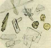
♦9_1
cystine, fibrin, mucin, fats, fatty acids, cholesterol, hippuric acid, bilirubin, hematin, melanin, indigo.—III. Solubility in (strong) hydrochloric acid. Dissolve: all sediments soluble in acetic acid, calcium oxalate, gypsum (with difficulty), xanthine (with difficulty), leucine, tyrosine, cystine, fibrin (swells beforehand), mucin. Do not dissolve: uric acid, fats, fatty acids, cholesterol, hippuric acid, bilirubin, hematin, melanin, indigo.—IV. Solubility in caustic soda. Of the substances insoluble in hydrochloric acid, those dissolving in caustic soda are: uric acid, fatty acids, hippuric acid, bilirubin, hematin, melanin (sometimes with great difficulty).—Methods for detecting arsenic and lead in urine, see Arsenic, Lead. V. Clinical examination of urine. The significance of urine examination for clinical diagnosis and therapy is enormous and perhaps unsurpassed by any other method of recognizing diseases, neither in the variety of pathological processes reflected by changes in urine nor in the ease, accessibility, and objectivity of the data obtained through a comprehensive examination of urine. In connection with the development of microchemical research methods and the widespread use of blood microanalysis over the past decade, however, there is a fascination with and overestimation of the role of the extremely unstable biochemical composition of urine and insufficient attention to, and sometimes unfamiliarity with, well-established facts regarding the patho-physiological significance of certain urine changes. The clinic utilizes macro- and microscopy, as well as chemical and physicochemical studies, in the study of urine. In most cases, it is even difficult to decide which methods are most important for solving specific diagnostic tasks. Schematically, it can be said that qualitative changes in urine (the appearance of pathological elements in it of both organic and inorganic character) mostly indicate overt pathological changes in the urogenital apparatus or other organs, whereas quantitative shifts in urine composition depend on the impaired function of certain organs or systems. However, this division is conventional, because experience teaches that apparently simple quantitative changes in the normal composition of urine can often in themselves reflect profound processes occurring either in metabolism (e.g., hypochloruria and low specific gravity of urine in diabetes insipidus or hyperazoturia in severe intoxications) or indicate a severe impairment of the excretory function of the kidneys, e.g., hyposthenuria and a drop in the cryoscopic index of urine in renal failure. Therefore, modern clinic, when evaluating the results of urine examination, pays attention not only and not so much to the appearance of pathological elements in it, but to changes in its normal properties and constituents (see above). Change in urine color. Normal urine, as indicated above, has a straw-yellow color, and the intensity of this B. M. E. vol. XIX. coloration depends most often on the concentration of urine, i.e., its specific gravity. Urine with a low specific gravity, e.g., after abundant drinking, appears almost colorless and, conversely, urine with a high specific gravity, e.g., after sweating or marches, possesses a saturated color resembling strong tea. Photometric and spectrophotometric studies have established that in these cases the quality of urinary pigments remains unchanged, while their concentration in urine fluctuates mainly. Since the pigments excreted normally in urine (so-called chromogens) arise as a result of general, basal, and in particular protein metabolism in the body, and are by no means the end products of blood (hemoglobin) metabolism, the amount of pigment excreted per day with urine can serve as an indirect indicator of the intensity of the basal metabolic process. (See Metabolism.) Under pathological conditions, light straw-colored or almost colorless urine is observed in diabetes mellitus and diabetes insipidus (see separate table, figure 5, 2). In phosphaturia (see), urine is whitish, almost milky in color, covered with an opalescent film both on the surface and along the walls of the vessel. The admixture of blood pigment (methemoglobin, hemoglobin, hematoporphyrin) observed in certain diseases without the appearance of formed blood elements in it gives the urine a cherry-red, ruby-red color, and sometimes, with high intensity or upon standing in the light, the urine acquires an almost black shade [see Porphyrinuria and sep. tbl. (vol. VII, art. 187–188), fig. 4]. It is important to remember that in these cases urine mostly retains its transparency. In hematurias of various origins (nephritis, cystitis, tumors, and renal tuberculosis), urine acquires a red coloration of varying intensity depending on the amount of admixed blood and the duration of its standing, sometimes with a dirty brown shade ("meat slop" color—see sep. table, fig. 5, 4). (See above and the article Hematuria.) Among organized sediments in urine, cellular epithelial elements of the kidneys and urinary tract, as well as formed blood elements—erythrocytes and leukocytes—are found in various lesions of the urogenital system. Also excreted with urine in various nephropathies are so-called urinary casts, organic formations of various origins that arose in the renal tubules and are shaped like casts of individual segments of the tubules. To detect formed elements in the urine sediment, and especially casts, the presence of freshly voided urine is required, since upon prolonged standing the majority of formed elements undergo destruction under the influence of fermentation processes and the vital activity of microorganisms. Cells of the squamous epithelium of the lower sections of the urinary tract—the urethra and the urinary bladder—are also found in normal urine. They are relatively large cells of irregular polygonal shape with a relatively small nucleus. In female urine, Figure 1. Koch's tuberculosis bacilli in sputum (Ziehl-Neelsen staining): 1—solid form; 2—Spengler fragments; 3—beaded form; 4—branched form with black granules; 5—filamentous form; 6—tuberculosis bacillus located inside a polynuclear leukocyte. Figure 2. Above—bacterioscopic picture of putrid sputum (abundance of microorganisms of various species); below—thrush fungus (Monilia albicans): 1—mycelium; 2—spherical and cylindrical conidia. Figure 3. 1—Pfeiffer's influenza bacillus; 2—catarrhal micrococcus Seyfert-Pfeiffer; 3—Fraenkel's lancet-shaped capsular diplococcus; 4—Friedländer's diplobacillus; 5—streptococcus; 6—staphylococcus; 7—leukocyte. Figure 4. Characteristic druse in actinomycosis in sputum; right and below—polynuclear leukocytes (Gram staining). Fig. 5. Urine: 1—freshly voided urine of normal straw-yellow color, with sp. gr. of 1.016; urine is transparent; only occasionally small clouds—nubeculae—float in it; 2—barely colored, slightly yellowish, transparent urine of low sp. gr. (1.001–1.002) in diabetes insipidus; 3—saturated orange-brown transparent urine of high sp. gr. (1.026–1.030) in cardiac congestion; besides normal pigments, urine also contains a certain amount of urobilin; 4—bloody urine of the "meat slop" type, turbid with a dirty brown sediment, of high sp. gr. in acute glomerulonephritis; 5—dark brown, beer-colored urine in mechanical jaundice, containing a vast amount of bile pigments; foam formed after shaking the urine is also colored brown; 6—saturated urine in the post-critical stage of croupous pneumonia; more than a third of the urine volume is occupied by an abundant pink-red sediment of precipitated urates (sedimentum lateritium); 7—almost black turbid urine containing melanins—in melanosarcoma of the liver, freshly voided urine rapidly blackened upon standing in air; 8—milky-white, opalescent freshly voided urine in phosphaturia; at the bottom of the vessel and along its walls, an abundant loose white sediment quickly precipitates, consisting of amorphous (calcium) and crystalline (magnesium, ammonium-magnesium) phosphates. (To the illustr. of the artt. Sputum, Urine.)
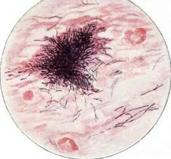
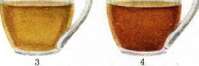

r


]
obtained without catheterization, larger cells of the squamous epithelium of the vaginal mucosa are also very frequently encountered. Epithelial cells are often observed under the microscope in groups closely adhering to each other [see separate table (p. 95 - 96), Fig. A], which indicates their shedding in entire patches from the surface of the epithelial lining of the aforementioned parts of the urogenital system. The number of such cells in normal urine is small and usually does not exceed 1-2 cells per field of view. The presence of significant amounts of desquamated squamous epithelium indicates a catarrhal, respectively inflammatory, process in the urinary excretion system, and along with pus cells, their detection serves as the main diagnostic symptom in these diseases. A reflection of pathological processes occurring in the urinary tracts is also the change in the structure of epithelial cells, their swelling, unclear outline of the nucleus, the appearance of fat droplets inside the protoplasm, and sometimes the nucleus. There is a generally accepted opinion that in inflammatory diseases of the renal pelves, peculiar cells appear in the urine in significant quantities, supposedly of the pelvical epithelial origin, having a pear-shaped or "tailed" form. However, there is indisputable proof that similar cells can also originate from the deep layers of the stratified epithelium of the lower sections of the urinary tract, which is why the aforementioned cells can be recognized only as having the value of a relative diagnostic sign of pyelitis (see). Renal epithelium cells are almost never found in normal urine and appear in it only when the tubular part of the kidneys is affected (nephroses of various origins, nephroso-nephritis, pyonephrosis, etc.). Most often they are observed under the microscope in the form of separately lying round or multifaceted well-contoured cells with a large, indistinctly outlined vesicular nucleus. In size, the cells of the renal epithelium are significantly smaller than the squamous epithelial cells described above (by 2-2.5 times) and somewhat larger than the leukocytes frequently observed in the urine. In necrotic and lipoid nephroses, the renal epithelium presents microscopically the phenomenon of the strongest lipoid infiltration ("fatty degeneration"), and in such cells it is not possible to discern either the clear outlines of the cellular contour or the nucleus. Often epithelial cells are observed in the form of groups, sometimes forming a cast of the tubule, in the form of so-called epithelial casts. The diagnostic significance of the renal epithelium for judging anatomical damage to the kidneys is undoubtedly great, especially in the presence of casts and protein in the urine at the same time. Leukocytes (pus cells) are found in an insignificant quantity (1-2 per field of view) also in the microscopic sediment of normal urine. Such a quantity of them, however, does not affect either the color or the transparency of the urine. In pathological conditions, the number of leukocytes in the sediment can increase significantly - from 15 - 20 to filling the entire field of view. In the latter case, they speak of pyuria (see). The urine appears cloudy and an abundant loose or mucous sediment precipitates at the bottom of the vessel, depending on the reaction of the urine. Under the microscope, leukocytes appear as small round cells with a sharply outlined and light-refracting nucleus (especially when moving the micrometer screw). They often stick together, forming clumps or groups of pus cells. A huge number of leukocytes covering the entire field of view in the microscope is observed in pelvic and bladder inflammatory processes (pyelitis, cystitis, urethritis), as well as in gynecological diseases (leukorrhea). Therefore, to resolve the question of the place of origin of the leukocytes found in the urine, it is necessary to examine, if possible, urine taken with a catheter, and also to compare the results of the study with other data: the reaction of the urine, the finding of other formed elements, as well as with clinical symptoms. To judge the place of origin of leukocytes excreted with urine, it was also proposed to determine the character of their nucleus. Senator claimed that urinary leukocytes of renal origin are usually mononuclear, in contrast to polynuclears, which originate in the vast majority from the urinary tract. This assertion found relative confirmation only for acute renal diseases - nephroso-nephrites and especially nephritis. In degenerative diseases of the kidneys, leukocytes, just like the renal epithelium, often exhibit phenomena of lipoid infiltration (steatophages), manifested by the appearance of birefringent substances in them. In nephroses, urolithiasis, renal tuberculosis, leukocytes are found in the urine in small quantities, but they always exceed the norm. Hematuria - the appearance of erythrocytes in the urine - is diverse both in intensity (from microscopic hematuria invisible to the eye to the appearance of urine of the aforementioned "meat slops" color) and in origin (see Hematuria). Erythrocytes under the microscope appear as round or biscuitoform plates, differing upon careful examination from other formed elements not only by the absence of a nucleus, but also by a greenish-yellow coloration. Depending on the movement of the micrometer, the structure of erythrocytes under the microscope changes - a dark rim with a light center, and vice versa. Often erythrocytes stick together, forming small groups, clumps, and sometimes so-called blood casts; the latter as a rule indicate an acute inflammatory process in the renal glomeruli. In renal diseases, as well as in poorly preserved urine, it is often possible to detect a significant number of leached erythrocytes, the so-called erythrocyte "ghosts", resulting from their hemolysis and taking bizarre shapes of mulberries, etc. In doubtful cases, to distinguish erythrocytes from yeast cells similar to them, one drop of dilute acetic acid is added to the urine, while erythrocytes completely dissolve, whereas yeast cells remain unchanged. Cylindruria is one of the earliest and at the same time important signs of a pathological process in the renal parenchyma. However, regarding hyaline and even granular casts most frequently observed in the urine, it must be taken into account that their presence cannot by any means serve as a measure of the severity of the process or its duration. Thus, a certain amount of hyaline casts can be found in the urine in orthostatic albuminuria, as well as in subjects with sensitive kidneys after hikes or swimming. Significant cylindrurias arising within 2-3 hours and disappearing just as quickly have been repeatedly observed. Therefore, in each individual case of finding hyaline and granular casts, to assess their diagnostic value, repeated examination is necessary whenever possible, as well as comparison with other urine examination data. Hyaline casts [see separate table (p. 95-96), Fig. B] - indistinctly outlined, almost transparent sausage-like formations, most easily detected in a darkened field of view; most frequently encountered in urine, they still have not found a generally accepted explanation. The opinion that hyaline casts are precipitates of fibrin from the inflammatory exudate of the glomeruli raises a number of valid objections. Hyaline casts are not observed in fibrinurias, they do not give the Weigert reaction for fibrin, and often represent a cast of the widest part of the collecting tubules, i.e., they must have arisen there, and not in the initial part of the tubules. Finally, hyaline casts appear in the urine in a whole series of non-inflammatory renal lesions. Waxy casts [see separate table (p. 95-96), Fig. C] represent more coarsely outlined formations, wider compared to hyaline casts, of a pale yellow color with a dull luster. They originate apparently from other casts (hyaline, granular, or epithelial) during the prolonged stay of the latter in the tubules. Granular casts [see separate table (p. 95-96), figure C] are frequently encountered in urine along with hyaline and epithelial casts. According to their structure, they can be coarse- (coarsely) granular and finely granular. The granularity of casts can be caused by fat droplets covering them ("fatty casts") or by lipoid and protein particles (cellular detritus). Granular casts are apparently the product of the decay of the renal epithelium, since one sometimes has to observe in one and the same cast on one half the preserved cellular structure, and on the other - typical granularity. Hyaline, granular, epithelial, blood casts appear in the urine both in lesions of the kidneys proper (nephroses of various etiologies) and in cardiac congestions, jaundice, acute pancreatitis, various comatose states, in other words, in a whole series of diseases accompanied by so-called secondary changes in the renal parenchyma. Waxy casts are usually observed in profound changes in the tubules - in the so-called necrotic nephroses (sublimate, meat poisoning), acute yellow atrophy of the liver, and a number of other severe intoxications. This is why their detection has special diagnostic, and to a certain extent prognostic, significance.
In rare cases, hemoglobin casts (see Hemoglobinuric fever), uric acid casts (see Uric acid infarct), and finally leukocyte casts (see Pyuria) appear in the urine. Blood casts are usually observed in the urine in inflammatory hematurias, such as in acute glomerulonephritis, etc. The diagnostic value of these casts is small, because simultaneously with them, a greater or lesser number of erythrocytes is always detected in the urine. Epithelial casts are found in the urine together with renal epithelium cells in various tubular processes in the renal parenchyma. These casts usually appear in nephropathies in which the amount of desquamating renal epithelium is very large, as a result of which the lumen of the tubules becomes clogged with renal epithelium and conditions are created for its gluing together. Urinary casts (especially hyaline casts) can sometimes be mixed up with formations similar to them, the so-called cylindroids. The latter are long, shiny mucous threads, sometimes occupying the entire field of vision, occasionally impregnated with salts. Cylindroids differ from hyaline casts by greater length, longitudinal striation, and lesser relative thickness. Cylindroids have no diagnostic value—it is only important not to mix them up with true casts. - Among the cellular elements in the sediment of male urine, spermatozoa are also frequently found in large or small numbers; spermatozoa are observed after coitus, masturbation, or convulsions (with unconsciousness), as well as in diseases of the urogenital system. Organic urinary sediments also include bacteria of various kinds that are very frequently encountered in it (see Bacteriuria). Among animal parasites, echinococcus hooks and vesicles are sometimes found in the urine—upon the rupture of some echinococcus vesicle into the urinary tract. In rare cases, filariae (in tropical chyluria) and distoma eggs (see Trematodes) have also been observed in the urine. In addition, details on pathological constituents of urine can be found in the articles: Albuminuria, Albumosuria, Acetonuria, Bacteriuria, Typhoid fever, Galactosuria, Galacturia, Hematoporphyrinuria, Hematuria, Hemoglobinuria, Hemoglobinemia, Diuresis, Jaundice, Lactosuria, Nephritis, Nephrosis, Nephrosclerosis, Oxaluria, Pneumonia, Heart defects, Pyuria, Porphyrinuria, Kidneys (functional diagnostics), Phosphaturia, and Chyluria. - Regarding changes in urine in individual diseases, see the corresponding diseases. M. Vovsi.
VI. Urine in children. Urination in the majority of cases begins immediately after birth, and sometimes even during it. Only very rarely does the first urine begin to be excreted later than the 3rd day after birth. According to Kocharovsky, in a third of cases after the first urination, anuria sets on, which lasts about a day. Anuria is shorter the earlier the child is put to the breast (Jaschke); thus, the moment of the beginning of urine excretion in a newborn is closely connected apparently with the moment of introduction of food, as well as its quantity. Table 1. (After Reusing.) Day of life Amount of milk Amount of urine Urine in percentage relation to the amount of milk Artificial Breast Artificial Breast Artificial Breast feeding feeding feeding feeding feeding feeding 96.0 38.3 35.8 8.4 37.0 21.8 150.6 120.8 71.0 26.8 47.0 22.2 229.5 176.6 135.8 40.9 58.8 23.0 253.1 220.0 187.0 60.0 74.0 27.6 364.6 271.5 283.0 119.1 78.1 43.9 369.0 296.0 246.0 148.6 66.0 50.0 410.0 297.0 325.0 157.0 79.1 57.6 530.0 338.0 406.0 208.0 77.0 62.5 Thus, children on breast-feeding excrete absolutely and relatively less urine. Absolute amounts of urine in a newborn fluctuate within fairly wide limits; they average: on the 1st day 10-20 cm3, on the 2nd day 20-40 cm3, 3rd day 40-70 cm3, 6th day 100-200 cm3, etc. According to Gundobin, at 12-30 days of age, the average daily amount of urine is 304 cm3; at the age of 30 days - 3 months - 421 cm3 and 3-6 months - 589 cm3; from 6 months to 1 year - 604 cm3; from 1 to 2 years - 759 cm3, from 5 to 6 years - 1,071 cm3 and from 12 to 13 years - 1,911 cm3. The number of urinations per day first increases with age, and then decreases. Table 2. (After Shanyavsky.) Age Number of urinations Amount of urine per 1 time (in cm3) 14-30 days....... 1-2 years......... 13 14 20 16 12 7-8 24 31 31 44 60 146
Already a newborn child has the ability to concentrate urine. Dohrn found specific gravity = 1.0018-1.006 shortly after birth. Mayerhofer even 1.006-1.012, Martin and Ruge determined the specific gravity in the first appearing portion of urine in a newborn to be 1.012. Subsequently, the specific gravity drops and stays within the limits of 1.003-1.005; however, this does not mean a decrease in concentration capacity; thus, in 2 children dehydrated due to pylorospasm, the specific gravity of the urine turned out to be 1.027. - The viscosity of urine in healthy infants, according to Mayerhofer, is less than in adults. An infant excretes no more than 40-60% of food nitrogen with urine and mainly in the form of urea. - Ammonia in the newborn's urine is very small, but already by the end of the 1st week it rises sharply (Reuss), its content is especially high in disorders of nutrition and digestion (Keller, Bendix), and besides, with food rich in fat and protein (Morev). According to Pfaundler, an infant excretes more N in the form of amino acids than an adult, in approximately 3/4 of cases (Goebel). Their amount often rises in acute toxic diarrheas, in heart diseases, pneumonias, leukemia, etc. The reason apparently lies in the increase in the kidney's ability to pass residual nitrogen. - Creatinine stays at the level of 10-15 mg per 1 kg of the child's weight; it is especially high in poorly nourished children; during muscular work (convulsions) its amount is 3-4 times higher than normal. - Sugar. In 40% of cases in newborns between 2 and 5 days there is lactosuria, and it is still not clear whether this is a normal or pathological phenomenon. Lactose also appears with great regularity in acute toxic dyspepsias in infants (see Childhood intoxication). Regarding the appearance of protein in the urine, two types of albuminuria in childhood should be mentioned, the so-called albuminuria (see) of newborns and orthostatic albuminuria. In the first days of life, the urine contains a lot of urates, and a few days later, when the washing out of the uric acid infarct (see) from the kidney begins, casts incrusted with uric acid salts can be seen in the urine.
The amount of uric acid on a purine-free diet is 14-25 mg per 1 kg of weight; with rickets it is significantly more - 30-36 mg per 1 kg. As a feature of the child, one can note the appearance in the urine in jaundice of newborns (see Jaundice, jaundice of newborns) of an insoluble bile pigment (masses jaunes), wherein the urine does not give a reaction for bile pigments. - Urobilin is usually not yet present in newborns; in cases where it is detected in the urine, its transition from the maternal blood through the placenta and excretion through the liver takes place. A few hours after birth, bacterial formation of urobilin in the intestine already begins and it appears in the urine, true, in meager quantities (Winternitz); it increases in diseases of the gastrointestinal tract (Langstein). - Urinary enzymes in the first days stay at a fairly high level
Figure 2. (transition from the maternal organism), and then drop sharply on the 3-4th day - parallel to the physiological drop in weight - and stay at a low level from 4 to 7 days, and then slowly rise again (Pokrovskaya). Regarding the formed elements of the urine, mention should be made of hyaline and granular casts, which often appear not only in acute, but also in chronic nutritional disorders; here a disturbance of water metabolism and acidosis plays a role.
Figure 3.
By this last factor is explained the cylindruria upon giving the infant calcium chloride, hydrochloric acid milk, etc. Collection of urine. In older children, collecting urine presents no difficulties and is carried out in the same way as in adults. In young children, one has to resort to special measures. In boys, it is most convenient to use an ordinary glass test tube attached with rings of
adhesive plaster, as shown in Fig. 1. In girls, urine is collected using an Erlenmeyer flask attached in the same way with an opening of a suitable diameter (Fig. 2). At the same time, the urine should be protected from contamination with feces. One can also lay children on a rubber ring, into the opening of which a vessel of a corresponding diameter is inserted (Fig. 3). Usually urine can be obtained
within 1/2-1 hour. Figure 4.
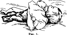


It is also necessary to know that in an infant, the act of urination usually occurs as soon as they are undressed, due to a spasm of the bladder under the influence of cooling. Taking advantage of this, one can very quickly collect the required amount of urine in infants of both sexes. The daily amount of urine in young children is more difficult to collect. In boys, as described above, a test tube is adapted, the end of which is drawn out into a tube; a rubber drainage tube is attached to the tube, lowering into a vessel for collecting urine standing on the floor near the bed. The entire apparatus for collecting urine looks as shown in Figure 4. Instead of a glass test tube, one can also use a rubber nipple with a hole cut in the top; the drainage tube is glued into this hole. In girls, collecting urine over a day is much more difficult, and sometimes this even has to be abandoned altogether.
Related articles
Cite this page
“Urine.” Soviet Medical Encyclopedia. English translation of Bolshaya Meditsinskaya Entsiklopediya, 1st ed. (Moscow, 1928–1936), ed. N. A. Semashko. https://sovietmedicalencyclopedia.pages.dev/article/urine/