Polarimetry
Historical document, translated for reference. It reflects medical knowledge of the 1920s–30s and is not medical advice.
Summary
Polarimetry is the determination of the direction and measurement of the angle of rotation of the plane of polarization using optical instruments called polarimeters. This method is primarily used in medical laboratories for the detection and quantitative determination of optically active substances, particularly glucose in urine.
Encyclopedia article (1928–1936)
POLARIMETRY, polarimeters. Polarimetry is the determination of the direction and measurement of the angle of rotation of the plane of polarization, performed with the aid of optical instruments—polarimeters. An essential part of the instrument (Fig. 1) consists of two Nicol prisms (see Nicol). The fixed Nicol (polarizer P), which decomposes ordinary light into two mutually perpendicular rays (polarized light) and transmits only one of them, and the rotating Nicol (analyzer A), which indicates the direction of the plane of polarization. If two Nicols are oriented so that their principal sections are mutually perpendicular, the instrument does not transmit light at all (zero point), whereas when the Nicols are in parallel position, light passes without attenuation. If a solution of an optically active substance is placed between two Nicols positioned in the dark position, the field of view brightens, and to restore the previous darkness, the analyzer must be rotated by an angle α, equal to the angle by which the plane of polarization of light was rotated under the influence of the optically active substance. The magnitude of angle α depends on the nature of the substance, solvent, concentration of the solution, length of the liquid layer through which the light passed, and the kind of light. A substance is called dextrorotatory if the analyzer needs to be rotated in the direction of the clock's hands to compensate, otherwise it is levorotatory. The angle by which the plane of polarization of light would be rotated under the influence of a solution containing 1 g of optically active substance in 1 cm³ with a layer length of 1 dm is called specific rotation and is denoted [α]. Under certain conditions, this value is characteristic of each optically active substance. The observation temperature (for example 20°) and the kind of light (homogeneous light of sodium flame—D or white light—j) are placed to the right, for example [α] D. Specific rotation can be calculated by the formula: [α] = α/l, where α is the found angle of rotation of the solution of the optically active substance, l is the length of the layer of this solution in decimeters, C is the concentration of the solution. Knowing the value [α] of a substance (in cases where this value changes little or does not change with concentration), its content can be determined by observing the angle of rotation and calculating by the formula—C = α/[α]l (polarimetric quantitative analysis). In application to urine, this method is most frequently used for the detection and quantitative determination of grape sugar. Instruments for determining the angle of rotation are divided into two groups: polarimeters (and polaristrobometers), which can be used for the investigation of all optically active substances, having a graduated circle and requiring illumination by homogeneous light, and saccharimeters with wedge compensation and a scale applied to a vertical ruler, illuminated by ordinary white light (electric or kerosene lamp). An instrument according to the scheme described above, consisting of a diaphragm, polarizer, place for a tube with liquid, analyzer, graduated circle, and a small telescope, is not very sensitive; the accuracy of adjustment is increased by including various optical devices. The Wild polaristrobometer, which has very limited application at present, includes a Savart polariscope consisting of two quartz plates cut at an angle of 45° to the optical axis and crossed. Sodium light passes through the system (Fig. 2): P—polarizer, A—analyzer, S—Savart plate; the field of view appears to be cut by a series of horizontal black bands, which disappear when the plane of polarization of the rays falling on plate S makes an angle of 45° with the principal section of both plates (zero point). In Wild's apparatus, the polarizer is rotated, accordingly with which the above-mentioned rule changes in relation to the recognition of dextro- and levorotatory substances.
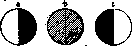
Half-shadow apparatuses. The field of view is divided into several parts, and one part is given a somewhat different direction of polarization than the others; in the Laurent apparatus (half-shadow apparatus of Mitscherlich), a quartz plate is placed in front of the diaphragm, and in better Lippich apparatuses (Fig. 3), a small Nicol (N) is included in front of the polarizer, which is rotated by a small angle δ with respect to the polarizer. If the analyzer is set perpendicular to the polarizer, the uncovered half appears dark, and a little light passes through the half covered by the small Nicol or quartz plate (Fig. 4a). If the analyzer is rotated by angle δ, then the other half of the field becomes dark, and the uncovered half transmits a little light (Fig. 4c). If, however, the analyzer is rotated only by γ, then completely equal in intensity light will pass through both halves of the field of view (half-shadow—zero point, Fig. 4b). The most perfect construction is represented by the Landolt-Lippich apparatus: the field of view is divided into three parts (two small Nicols are included), there is a device for maintaining a constant temperature in the tube with the test solution, angle δ can be varied depending on the strength of illumination and on the color and transparency of the solution. The eyepiece should be set so that the boundary line is sharply visible. Sodium flame is used for illumination. Saccharimeters. The Soleil apparatus (Fig. 5), little used at present, has between the polarizer P and the tube C a biquartz W. If the analyzer is set exactly parallel to the polarizer, both halves of the field of view will have the same grayish-violet tint; with the slightest rotation of the analyzer, the color in one half changes to red, in the other to blue. To determine the concentration of the solution, a wedge compensator is used instead of a graduated circle in degrees; it consists of a plate D of right-handed quartz and a plate L of the same thickness, composed of two wedges of left-handed quartz, which can slide one over the other. The system is used to compensate for the rotation of the test optically active substance. The movement of the prism is produced by a micrometer screw provided with a scale. The reading directly gives the percentage content of cane sugar in the saccharimeter or glucose in the glycosimeter. The most commonly used instrument for determining glucose in urine is the half-shadow apparatus with wedge compensation of Schmidt and Geschi.
Production of determination. The liquid to be examined must be transparent and as colorless as possible. The tube, completely dry or rinsed with the liquid to be examined, closed at one end with a glass disk by means of a sleeve, is filled with the liquid so that it stands somewhat higher than the edges of the tube; the other end of the tube is closed with a glass disk, cutting off the protruding part of the liquid to avoid the appearance of an air bubble, which could cover the field of view. The measurement is performed in a dark room. The light source is positioned so that its image is clearly obtained on the analyzer's diaphragm. The usual distance between the light source and the end of the apparatus is 22 cm. The eyepiece is set for the best visibility of the field of view, the zero point is determined, the tube with the liquid to be examined is inserted, the eyepiece is reset again, since the focus shifts at this point, and a new reading is taken from the instrument's scale. The difference between the second and first readings gives the magnitude of angle α.
Related articles
Mentioned in
Cite this page
“Polarimetry.” Soviet Medical Encyclopedia. English translation of Bolshaya Meditsinskaya Entsiklopediya, 1st ed. (Moscow, 1928–1936), ed. N. A. Semashko. https://sovietmedicalencyclopedia.pages.dev/article/polarimetry/