Polarity
Historical document, translated for reference. It reflects medical knowledge of the 1920s–30s and is not medical advice.
Summary
An overview of polarity as a biological phenomenon in organisms, organs, and cells, discussing cellular differentiation, embryonic axes, and Child's theory of physiological gradients.
Encyclopedia article (1928–1936)
POLARITY, the presence in an organism or a part of it of quantitatively or qualitatively different properties along some axis of the organism, organ, or cell. The phenomenon of polarity is discovered with greater or lesser distinctness everywhere, but even its formal statement has been accomplished far from completely, not to mention an exhaustive explanation of the origin and preservation of polarity. Cell polarity is especially distinct in epithelial and nervous tissue. In epithelial cells, as a rule, there is a clear differentiation into a basal or proximal region, i.e., the part of the cell that is connected with the underlying tissue or directed towards it, and a distal region directed towards the surface of the epithelial layer. The different morphological structure and physiological features of the proximal and distal regions of an epithelial cell are usually associated by histologists with different conditions of existence, since the distal ends of cells are exposed to the environment (epidermis) or the influence of processes occurring in various cavity organs (epithelium of the digestive tract, etc.), while the proximal ends of the same cells are in contact with underlying tissues and thereby experience completely different influences. The polarity of the arrangement of intracellular structural formations in epithelium is expressed primarily in the fact that the basal parts of cells are usually arranged more simply and are basically the same in various epithelia; the nucleus is usually located closer to the basal end. In the proximal region of the cell, the reticular Golgi apparatus is usually located, as well as the intracellular apparatus of ciliated epithelial cells; the granules of secretion of glandular cells also accumulate there. The distal surface of epithelial cells is provided with a resemblance of a dense darkly staining cuticle, sometimes with a so-called striated border; sometimes it is equipped with more or less long cilia originating from special bodies, the kinetic centers. Cells of nervous tissue also possess a clearly expressed polarity. From one of the poles of the cell departs a long process, the neurite; from the opposite side, the cell body gives off a large number of short, branching outgrowths, the dendrites. From the standpoint of the neuron theory, cells in nervous tissue are polarly differentiated in a dynamic sense, in the sense that only the dendrites and the cell body itself possess receptor properties, while the neurite exclusively conducts excitation. In most tissue cells, it is usually not possible to ascertain a clearly expressed polarity, especially in connective tissue cells; however, every cell capable of division periodically exhibits extremely clear polarity, specifically during mitotic division. The divided centrosome forms two centers, in relation to which all the constituent parts of the cell, especially the elements of the nucleus, are arranged symmetrically. The essence of indirect division consists precisely in this bipolarity of the dividing cell with the subsequent uniform distribution of nuclear chromatin substance (see Karyokinesis). The polarity of the ovum can often be ascertained before the start of cleavage and even before fertilization. Experiments with centrifugation of ova of various animals have shown that the displacement of strictly localized cytoplasmic inclusions in a number of cases does not disturb the arrangement of the axes of the developing embryo, and consequently the polarity of the ovum is not connected with these visible inclusions. Hypothetical submicroscopic structures upon which, according to various authors, the polarity of the ovum depends, have received various names (Lillie's "fundamental substance," Conklin's "spongioplasm," etc.). Morphological analysis at the present time is powerless to resolve the problem of the ovum's polarity, and in this area one must still be limited only to stating the fact of its presence. (See Promorphology.) Even more than in tissue cells, polarity is noticeable in so-called unicellular, protozoan organisms, where there is a differentiation of organoids of movement and digestion. In flagellates and especially ciliates, one can distinguish the arrangement of organoids along the long (anteroposterior) axis, and sometimes along the lateral and dorsoventral axes. Finally, the polarity of Metazoa is expressed both in the structure of the entire organism, especially in forms with bilateral symmetry, where the digestive organs, nervous system, skeleton, etc., are polarly arranged, as well as in the structure of individual organs, e.g., segmented appendages in crustaceans and insects and limbs in vertebrates. The so-called "physiological polarity" has been ascertained in various organisms, earliest and most distinctly in forms having an elongated body shape, specifically in hydroid polyps and worms. The totality of data on physiological polarity led C. M. Child to the theory of physiological gradients. The essence of this theory reduces to the assertion that there are regions of high activity in the organism that dominate over the body parts adjacent to them. This high activity reduces to enhanced metabolism, a more significant electrical charge, greater cell permeability, their strong sensitivity to toxic substances, etc. Between the region of high activity and the region of low activity, the corresponding properties drop gradually, forming a gradient, and the direction of the changing properties coincides with one or another morphological axis of the organism. The phenomena of the physiological gradient of various properties were studied by Child's school on a wide variety of objects. First of all, it is necessary to state the difference in the capacity for vegetative reproduction at various levels of the body of hydroid polyps. The closer to the oral end, the faster new hydranths develop and the greater the size of the latter. Thus, the gradient of capacity for vegetative reproduction drops from the apical end to the basal. In this same direction, i.e., from the oral to the aboral end, the sensitivity of various animals to various toxic substances decreases. The following substances and influences were tested for the differential susceptibility of various body parts: hydrocyanic acid and its salts, various anesthetics (ethyl alcohol, ethyl ether, chloroform, chloral hydrate, chloretone, ethylurethane), formaldehyde, strong and weak acids (a solution of CO2 in water, acetic acid, HCl, H2SO4), strong and weak bases (NaOH, NH4OH), various other electrolytes (LiCl, KMnO4, HgCl2, CuSO4), alkaloids (caffeine, strychnine, pilocarpine, atropine), dyes (Toluidinblau, Victoriablau, Krystallviolett, Methylenblau, Brilliantkresylblau, Janusgrün, Neutralrot), hypo- and hypertonic seawater (for marine forms), ultraviolet rays, the action of high and low temperature, oxygen deprivation. Permeability to alkalis drops in the direction from the oral end in Paramaecium (Child, Deviney; 1925), and from the oral end to the aboral in various hydroid polyps (Child, 1926). Preliminary staining with Neutralrot was performed, and the rate of penetration of ammonium hydroxide was monitored by the yellowing of the introduced dye. A respiratory gradient, expressed in the varying intensity of tissue respiration (oxygen consumption and CO2 production), was also discovered in various objects. As a rule, the intensity of respiration drops from the head end to the basal, i.e., parallel to the gradient of sensitivity to poisons and parallel to the permeability gradient. However, there are data on the presence in the posterior (aboral) part of the body as well of a center of high metabolic activity. Parallel to the properties described above, the capacity for oxidation-reduction reactions changes. The drop in this capacity along the body axis, starting from the oral end, was ascertained by Child and Galigner in protozoa, ova and embryos, and hydroid polyps. The indicator was the rate of appearance of a brownish or blackish coloration after the introduction of potassium permanganate. In the hydropolyp Corymorpha palma, Child (1926) showed with the help of the nitroprusside reaction that the amount of glutathione also drops from the apical to the basal end. A large amount of data exists on the question of the difference in electrical potentials between various parts of the organism. Mathews (1903) found that the distal ends of some hydroids are electronegative in relation to the middle, proximal region. The same was established by Morgan and Dimon (1904) for the earthworm. Mathews' data were confirmed by Hyman (1920) on the hydroid Tubularia and by Hyman and Child (1922) on Corymorpha. Hyman and Bellamy (1922) found the potential difference between individual body parts in various forms of sponges, hydroid polyps, jellyfish, ctenophores, flatworms and annelids, and frog tadpoles. These authors noted that regions of high metabolic activity are usually electronegative in relation to other parts of the body. This fact agrees with the fact that in an electric field the earthworm takes the shape of the letter U and is directed by its anterior and posterior ends toward the cathode (Moore, Kellog; 1916). Hyman and Bellamy confirmed the observation of Moore and Kellogg and noted that this characteristic galvanotaxis is in correlation with the gradient of metabolism and electrical potential.
A further deepening of ideas regarding the connection of polarity with electrical phenomena is found in the works of Lund on the hydropolyp Obelia comissuralis, in which the aforementioned author influenced the course of the regeneration process with an electric current. In his first experiments, Lund passed an electric current from accumulators or dry batteries through seawater along a lying polyp with cut ends. The current density varied from 60 to 100 <5. In control experiments, heads formed at both ends—apical and basal. The difference lay only in the fact that the apical heads began to develop earlier than the basal ones. In the experiment with passing a current of the specified density, heads formed only at those ends that were directed toward the anode, regardless of whether this end was previously apical or basal. Thus, morphological polarity can be altered under the influence of an electric current; the latter acts in an inhibitory manner on the formation of the polyp's heads. For this inhibition on the cathode side, the minimum current density is 66 d. To stop the regeneration of heads on the anode side as well, the current density must be increased to 132 d. The closer to the apical end a piece is cut from the polyp's body, the less sensitive it is to current density (in the sense of cessation of regeneration). Lund and Spek, who cites his data, explain these phenomena by the assumption that «bioelectrical phenomena, regular potential differences, or an electric current, which, arising as a result of cell activity, goes from one morphological pole to another, are an integrating component of morphological polarity.» The presence of a potential difference between the apical and basal ends of the polyp was established using a potentiometer with a sensitivity of 13,000 megohms. The apical end turned out to be electropositive relative to the basal one. In addition, the outer surface of the coenosarc is electropositive relative to the inner surface of the endoderm (Fig. 1 A). The reverse
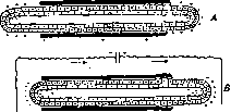
Figure 1. A—Potential difference between the coenosarc surface and the inner surface of the intestine in the hydropolyp Obelia comissuralis; B—Influence of electric current on the intrinsic charge in Obelia. (From Lund.)
relations, i.e., the electropositivity of the inner layers of plasma relative to the outer ones, were established by Osterhout and others in individual cells of the algae Valonia and Nitella. An electric current at the cathode reverses the latter relation, while at the anode it makes it even more distinct (Fig. 1B). As a result, in the opinion of Lund, shared by Spek, the phenomena described above arise with the inhibition of regeneration at the cathode. One of the expressions of polarity during regeneration is the migration of certain cell elements along the axis of the regenerate, as was established by Spek for the formation of winter buds in the ascidian Clavelina lepadiformis, a phenomenon very close to regeneration. According to Spek, the differentiation of the bud proceeds at the expense of young cells provided with drops of protein inclusions susceptible to vital staining. At the beginning of bud formation, a polar distribution of these cells takes place.
Isolated amoeboid cells with protein drops migrate upward (in the oral direction), while cell aggregates and granular cells migrate downward (in the aboral direction) (Figure 2). Regarding the influences from the outside on the process of polar differentiation, there are many data. The cut piece of the trunk of the polyp Antennularia antennina forms a head at the end which, when vertically suspended, ends up at the top (J. Loeb, 1892). This species of Antennularia is rather an exception than the rule regarding the influence of gravity on polarity, since in the closely related form Antennularia racemosa, regardless of position, the regeneration of hydranths proceeds from both ends if the piece is cut from the middle; pieces cut closer to the apex retain their original polarity, i.e., in the apical direction the oral end regenerates, and in the basal—the aboral. In many polyps, for example in Tubularia mesembryanthemum, the basal cut end gives rise to the stem part only upon contact with the substrate, otherwise a head regenerates from it. The apical end gives rise to a head. In the polyps Serturarella polyzonias, according to Driesch's data, and in Eudendrium racemosum, heads are formed only under illumination, and with side light only from the illuminated side. The same has been established for many plants, where gravity also plays a major role in determining polarity. A very effective experiment illustrating the influence of temperature on the emergence of polarity was set up by Gilchrist (1928). By exposing the dividing egg of the amphibian Triturus torosus to heating on one side and cooling on the other, Gilchrist obtained the formation of additional medullary plates on the warm side, i.e., foci of increased activity, which, according to Child, are sources of polarity and consequently of the physiological gradient. Hence, in particular, stems Child's assertion that areas of high activity correspond to the «organizational centers» or «organizers» of Spemann (see Developmental mechanics, Organ, organogenesis). Of particular importance is the question of polarity and its inversion during regeneration in vertebrates. Kurz (O. Kurz, 1912, 1922) showed that the knee joint of a newt with parts of the thigh and lower leg, being implanted by its distal end into the skin sleeve of a limb, produces the regeneration of a paw at the proximal end. Thus, an inversion of polarity occurs. The same results were obtained by Graper, Milošević, and others. Recently, Efimov obtained the regeneration of a hand on both sides of a limb grafted by its middle part into the tissues of an axolotl's tail. Weiss, expounding these data, does not consider it possible to treat them as an inversion of polarity, but his argumentation is unconvincing. The study of physiological processes accompanying the phenomena of morphological polarity undoubtedly sheds light on this poorly studied field. It would, however, be erroneous to believe that the discovery of certain physiological or even physical relations, like the Lund data cited above, resolves this problem. Child in one of his summaries on the issue of physiological gradients makes, in particular, a conclusion that can lead to deeply erroneous conclusions. «First, polarity and symmetry are more related to physiological conditions in the protoplasm than to hereditary structural features; second, the presence of physiological axes in most cases, if not always, is determined rather by quantitative than qualitative differences at different levels; third, conditions that destroy physiological axes are precisely those conditions that reduce quantitative differences between different regions; fourth, if the axis disappears, the corresponding gradient no longer exists, and conversely, the presence of a new axis is associated with the appearance of a new gradient. In short, gradients are an adequate physiological basis of the phenomena of polarity and symmetry, which leads to the rejection of the hypothesis of a stereochemical or other structural basis of these phenomena» (1928). A one-sidedly physiological, as well as a one-sidedly morphological interpretation of the phenomena of polarity is unable to penetrate the essence of these phenomena. Further research must undoubtedly proceed along the line of combining morphological and physiological methods, along the line of studying the dynamics of structural and functional features in their interrelation and interdependence.
Related articles
Mentioned in
Cite this page
“Polarity.” Soviet Medical Encyclopedia. English translation of Bolshaya Meditsinskaya Entsiklopediya, 1st ed. (Moscow, 1928–1936), ed. N. A. Semashko. https://sovietmedicalencyclopedia.pages.dev/article/polarity/