Mechanics of Development
Historical document, translated for reference. It reflects medical knowledge of the 1920s–30s and is not medical advice.
Summary
Mechanics of Development is a field of biology that studies the laws of structure formation in the individual development of organisms. It was theoretically and experimentally established by Wilhelm Rou, who used a causal-analytical method to study morphological problems.
Encyclopedia article (1928–1936)
MECHANICS OF DEVELOPMENT. Contents: History......................18 Materials and methods of research........20 Problem of determination.............22 Two main types of morphogenesis......26 M. r. and regeneration................30 Practical significance of M. r............31 Mechanics of development, a field of biology that studies the laws of formation of structures in the individual development of organisms. Theoretically and experimentally, M. r. was substantiated by Wilhelm Rou. The main feature of the new field, according to V. Rou, was the causal-analytical method of studying morphological problems. The name proposed by him, as well as the understanding of the tasks of the new field, caused a number of objections (O. Hertwig, H. Wolff and others). First of all, a wrong idea arose that the new field should explain the phenomena of development in biology by the laws of mechanics. Rou pointed out, for example, that the word "mechanics" is used by him not in the narrow sense of physics, but in the broad sense in which Kant refers to mechanics every phenomenon subject to causality. In the programmatic introduction to the first volume of the Archive of Developmental Mechanics (Archiv fur Entwicklungsmechanik der Organismen) founded by him, Rou writes (1895): "Mechanics of development, or causal morphology of organisms... is the doctrine of the causes of the origin, preservation and reverse development of structures," and further: "every subordination of causality of certain phenomena according to the assessment of the mechanism given by Kant and Spinoza should be designated as a mechanical phenomenon. Thus, the doctrine of the causes of becoming can be called mechanics of development." The narrowing of research tasks by analyzing developmental phenomena without taking into account the need for synthesis and the one-sided understanding of M. r. as the causal morphology of organisms characterizes the ideological connection of Rou with mechanistic materialism and with the methodology of Kant, since the latter appeared as a mechanist. However, Rou spontaneously transcended the mechanistic understanding of developmental phenomena, and in his methodological views, the concepts of a materialist-dialectician appear. A number of authors proposed other names for the new discipline, for example, developmental physiology (H. Wolff and H. Driesch). Jacques Loeb called physiological morphology the field that essentially coincided with the mechanics of development of V. Rou. Indifferent terms are also used: experimental embryology, experimental morphology. History. The beginning of experimental research on morphogenesis dates back to the 18th century - the famous experiments of Trembley on the regeneration of hydra (1744), experiments of Bonnet, Spallanzani and others. This field received a solid foundation with the victory of the evolutionary idea in the second half of the 19th century, when descriptive-morphological research on development was followed by a second stage - the study of the process of morphogenesis. The beginning was laid almost simultaneously - V. Rou, who began his experimental embryological work in 1883, and in 1888 published his historical work ("On the artificial production of half-embryos after the destruction of one of the first two cleavage spheres and on the subsequent development of the missing parts of the body"), and L. Chabry in France (1887), who published a remarkable study on experimental embryology of ascidians. The Naples Zoological Station played a prominent role in the development of this field, where at the same time students of V. Rou - Herbst, Driesch, and through them the Americans E. Wilson and T. Morgan working at the station, the new field moved to America, where Jacques Loeb was already working independently. The beginning of the 20th century is characterized primarily by the complication and enrichment of research techniques: the development by Spemann of the method of constriction, the introduction by him of micro-operations with glass instruments, the method of vital marking of parts of the embryo by V. Focht, and in the most recent time - the combination of tissue culture methods with microsurgery (Strangeways laboratory in Cambridge). At the same time, this period brought essentially new things in the theoretical field. First of all, one should note - the doctrine of organizers by H. Spemann. In the history of embryology, the ideas of preformation and epigenesis played a major role. Rou stopped at the point of view of preformationism, but, not losing sight of the importance of the epigenetic moment in development, he proposed to speak of two types of organ formation - about development through self-differentiation and about the dependence of differentiation. In the first case, the moment of preformation is revealed, in the second - of epigenesis. A special role in the history of mechanics of development was played by H. Driesch. Having created for himself a certain model of materialism (extremely vulgar), in the struggle with his opponent he slid to the recognition of the absolute autonomy of life phenomena and the presence of original purposefulness (entelechy), becoming one of the most reactionary figures in biology of recent decades. The basis of Driesch's theoretical views is the so-called equipotential system. From the factual side, the matter comes down to the fact that at early stages, part of the embryo (or part of the body of an animal during regeneration) often gives a whole organism - for example, one of the blastomeres of the 4-cell stage in a sea urchin gives a whole larva. Driesch draws from this the conclusion that the egg is isotropic, uniform in all directions, and from this - the further consequence that the forces building the embryo are of a non-material order. This position is developed by him in the so-called two proofs of vitalism. Their essence comes down to the fact that if living beings were three-dimensional machines, then when a part is taken from such a machine, the remaining part could not restore a whole structure. However, since such restoration occurs (in experiments with regulation of embryos and in regeneration of adult organisms), this, in Driesch's opinion, indicates the impossibility of explaining phenomena in a living organism on the basis of physics and chemistry. The vicious circle in which Driesch is located is as follows: either living organisms are three-dimensional machines, all processes in which are completely reduced to physico-chemical phenomena, or they are controlled by non-material forces. Starting from such a basis, Driesch created a series of his unrealistic concepts. Only in the most recent time, after all the artificiality and confusion of concepts with which he shone for 40 years, Driesch came to the following conclusions: the machine theory is not the opposite of vitalism; the opposite of vitalism would be the concept "automatic"; the machine theory is itself a vitalistic theory, since a machine is unthinkable without the one who operates it; in living organisms, such a managing moment is entelechy. Tasks and scope of M. r. The practical task, the goal of M. r. - is the possibility of controlling the phenomena of the emergence of traits in the process of ontogenesis. Obviously, any phenomenon of morphogenesis belongs to M. r., regardless of age (from the egg cell to the death of the organism). But it should be emphasized that we are dealing with M. r. only when it comes to studying the laws of morphogenesis, and not to establishing the fact of the transition of one completed state to another. In particular, in the field of endocrinology, one should distinguish the emergence of a new structure from the detection with the help of various stimuli of already existing structures. The doctrine of tumors, which currently belongs to pathology, can be thought to enter the field of M. r. Of other disciplines, genetics is especially close to M. r., studying the regularity of inheritance of traits. Hecker proposed to merge M. r. with phenogenetics, i.e. to consider M. r. as a field that studies the implementation of a trait. Such a narrowing of tasks cannot be considered successful - M. r. studies the laws of morphogenesis and regardless of the phenogenetic implementation of the trait (regeneration, asexual reproduction). Thus, at present, the core of M. r. is the field of experimental research on embryonic development, regeneration and asexual reproduction, as well as in a number of cases the study of morphogenesis in the adult state of higher vertebrates. M. r. already now plays a huge role for the practical doctor, engaged in the causal study of teratogenesis. Materials and methods of research. The most favorite material for research on embryonic development continues to be the eggs of amphibians, frogs and other tailless amphibians, and even more suitable for early stages is the striped triton, the special qualities of which were shown by Spemann and allowed him to discover a new stage in M. r. Another valuable material is the eggs of sea urchins - since the early works of the Hertwig brothers, it has been the most frequent and most suitable object for the study of M. r. in invertebrates. For questions of regeneration, favorite objects are hydrozoan polyps (in particular, freshwater hydra) and flatworms (planarians), among vertebrates - the triton. Recently, the synthesis of tissue culture methods and M. r. has begun to be implemented, and it has become possible to use the embryos of warm-blooded birds; in the most recent time, operations on mammalian embryos have begun in the Strangeways laboratory. This opens the way to the study of regeneration in higher vertebrates (see below). The methods of research in M. r. are very diverse - various devices for studying the action of gravity, salt solutions, centrifugation, selective destruction of certain parts of the embryo with ultraviolet rays (Chakhotin apparatus), etc.
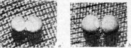
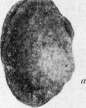
Microsurgery (or so-called microsurgical technique) acquired particular importance in research on embryonic transplantations. The method of embryonic transplantations was introduced in the 1890s by Born. Born, as did a number of later researchers, worked with late embryos and used metal instruments—thin needles, needles shaped like knives, fine scissors, etc. A major contribution of Spemann was the introduction of glass instruments for operating on early embryos. The most important types of Spemann's instruments are visible in the accompanying figure (Fig. 1). First of all, these are glass needles used for cutting tissues; the hair loop, the application of which at present is extremely diverse, represents a bent piece of a child's hair inserted into the finely drawn-out end of a pipette. Depending on the size of the loops, they can not only orient embryos but also perform all the finest operations up to the so-called exchange of small pieces {see below). Spemann also improved the method of re-ligation for the Triton egg. He used fine hairs, making a loop with a reef knot from them (Fig. 2). Re-ligations make it possible to study the time of determination, various questions of the causal genesis of double malformations (deviation of one of the twins, frequency of situs inversus viscerum etc.), the boundary of the so-called organizational field, the process of gastrulation. In the very recent past, G. A. Schmidt, working in Spemann's laboratory, succeeded in developing a method of re-ligation for eggs of tailless amphibians. [See photomicrographs on a separate plate (Fig. 1)—re-ligation of eggs of a tailless amphibian—Bombina pachypus: A—stage of early gastrulae (embryi connected by a narrow bridge); B—late gastrulae; C—stage of neural ridges; D—dorso-ventral re-ligation; right embryo—the so-called ventral piece.] An important role in research on M. r. is played by the so-called method of vital marking. First applied by the American Goodale, it was developed in recent times (1922-25) by V. Vogt. In its essential features, the method consists in that pieces of agar-agar are soaked in a solution of some vital dye (most often Nile blue sulfate, sometimes also Neutralrot) and then applied with the help of special devices to the surface of the embryo in one or another areas. This method makes it possible to determine the rate of multiplication of different parts of the embryo, which allowed Vogt to make a detailed analysis of the process of gastrulation and to draw a map of the presumptive rudiments in the eggs of tailed and tailless amphibians. A number of authors have used this method for research
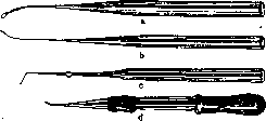
Figure 1. Main glass instruments
for microsurgery of early embryos: a—hair loop; b and c—glass needles; d—micropipette. questions of the location of rudiments on early stages (for example, rudiments of the eye cup on the stage of the neural plate). Finally, the method of tissue culture will obviously play a very prominent role in M. development. With its help, Waddington succeeded in 1930 in obtaining induction of the neural plate in embryos of chicken and duck. This method also makes it possible to cultivate parts of embryos and bring them to later stages. At the anatomical congress in Amsterdam (1930), Fell, head of the Strangeways laboratory, demonstrated rudiments of bones of chicken embryos, which she had brought to complete formation and differentiation of hist.
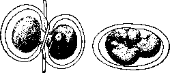
Figure 2. Re-ligation of the Triton egg with a loop
from a child's hair: a—before the beginning of cleavage; b—after removal of the loop (inside the shell there are two monozygotic twins). structure. Previously, for cultivating parts of embryos, methods of Murphy-Danchakova were used. The study of factors enhancing and inhibiting regeneration processes constitutes one of the nearest practical tasks of modern M. development. The problem of determination. The basic problem of morphogenesis in relation to the cell was posed by A. Weismann and developed in his teaching on the germ plasm. Then V. Rou transferred to the mechanics of development the Weismannian concept of the unequal value of nuclear divisions and of the correspondence of nuclear divisions to the structure of the organism being formed. The first work by Rou on M. r. "On the time of determination of the main axes of the frog embryo" appeared in 1883, the first research with puncturing blastomeres—in 1885, the work of Shabry—in 1887. Shabry devised a number of ingenious devices and instruments with which he selectively destroyed various cells of the cleaving egg and observed the subsequent stages of development. The formulation of the problem by Rou was as follows: when is the fate of parts of the embryo determined? He began with the two-cell stage. It was necessary to decide whether the first two cells of the cleaving egg are equipotential or determined to correspond to certain parts of the organism. First of all, he established that when all interfering circumstances are eliminated, the first plane of cleavage coincides with the median plane of the embryo. To this assertion, 20 years later, Brachet joined. On the basis of research on re-ligation of Triton eggs, Spemann came to the conclusion that in 3/8/8/« cases the median plane coincides with the 2nd furrow, in x/4-Vs—with the 1st furrow. With the help of the vital marking method, Vogt could note an even greater variation in the relationship between the 1st furrow and the median plane of the embryo. It should also be noted that in a number of other animals there is an even rarer coincidence of cleavage planes with planes of symmetry. Julin and van Beneden established in ascidians the coincidence of the 1st cleavage plane with the plane of symmetry. In Ctenophora, the 1st cleavage plane coincides with the gastric plane, and the 2nd—with the plane of tentacles. In a number of cases, the plane of symmetry of the future embryo is revealed in the structure of the egg even before fertilization, which led Conklin (1929) to note,
2 that

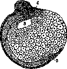
Figure 3. Rou's experiments: a-Hemiembryo dexter; b-Hemiembryo sinister; 1 and c-neural ridge; 3-in the unclosed part of the blastopore, b—the primitive gut; 4—notochord; 5-undifferentiated cells. In b the punctured part has completely turned into a cellular mass—the beginning of postgeneration.
that 'ontogeny begins before fertilization'. Roux's experiments consisted of destroying one of the blastomeres of a two-cell stage with a red-hot needle and studying the fate of the remaining living half of the egg. He found that as a rule the remaining living half of the egg gives half an embryo. Doing the same with 4, 8 cells and so on, he found that the parts of the egg are determined in relation to the future organism (figure 3). O. Hertwig objected, showing that if the living half is freed from the membranes, it gives a whole embryo, and therefore the half-embryo develops only in the presence of the killed half. T. Morgan also showed that if, without removing the killed half, the egg is turned over, a whole embryo develops from the remaining half. Morgan made use of the idea of Schulze's experiment, which shows the inadequacy of Roux-Weismann's concepts. Schulze's experiment consisted in the fact that a frog's egg, clamped between glass plates, was turned over shortly before the appearance of the first furrow of division. In this position it remained until the blastula stage (see), i.e., until a relatively late stage of development. As a result, in a large percentage of cases, double formations developed. Recently, Schulze's experiment was repeated by Schleip on a larger material with somewhat improved technique. These authors simplified Schulze's method: the frog's egg is clamped between two slides, between which strips of plasticine are inserted at the ends; the thickness of the latter is approximately equal to the diameter of the egg. This eliminates the need to secure the slides with special rubber rings, as Schulze did. These experiments showed that double formations are obtained in a certain percentage of cases not only when the egg is turned over at the beginning of cleavage, but also during early stages up to and including eight cells. In short, a number of facts showed that in frogs it is impossible to speak of a correspondence between nuclear divisions and the structure of the embryo. W. Roux observed that in many cases half-embryos turned into whole ones, and called this process supplementary development, or postgeneration. From this he derived his extremely important concepts of independent differentiation, or self-differentiation, and dependent differentiation. The first is, according to W. Roux, a typical case, the second an atypical one, caused by special experimental conditions. Almost simultaneously with Schulze's experiments, the Italian scientist Herlitzka succeeded in tying off a triton's egg with a thin silk thread and showing that whole embryos developed from both halves. The method of ligation had to play a prominent role in the study of the Roux-Weismann hypothesis of hereditarily unequal divisions of the nucleus of the fertilized egg. Previously, it should be pointed out to another experiment, historically produced much earlier, which also had a decisive significance. This concerns Jacques Loeb's experiment: sea urchin eggs shortly after fertilization were transferred to a hypotonic solution (sea water + distilled water 1:1). A sharp swelling and rupture of the shell occurred. Part of the contents came out into the water, taking a round shape (so-called extra-ovate) (figure 4). In some cases, the nucleus was initially in one half of the egg (inside the shell) and here it performed a certain number of divisions. Then at a certain stage one of the daughter nuclei passed into the extra-ovate, which thus received the product of the 2nd, 3rd or 4th division, i.e. 1/2, 1/4, 1/8, 1/16, 1/32 part of the nucleus of the fertilized egg. Nevertheless, a normal embryo developed from the extra-ovate. In recent times, the verification of the Roux-Weismann rule was made by Spemann (figure 5). From his experiments it followed that the first furrow divided the egg into dorsal and ventral parts. These studies finally showed that the determination of parts of the embryo has a different basis than W. Roux had assumed. In particular, in the mechanism of embryo formation, an area of the plasma, so-called gray crescent, plays an important role, already noticeable in amphibians before the beginning of cleavage. Spemann showed that only the part of the egg that received the gray crescent can develop the nervous system, axial skeleton and musculature; the part without it represents the so-called ventral piece. The important role of plasma in the process of morphogenesis has been established for a number of animals. The best-known example is the ascidian
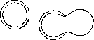
Figure 4. Schematic representation of Jacques Loeb's experiment: a-egg before being placed in hypotonic solution; b-egg in hypotonic solution - the shell has ruptured, part of the egg's contents has come out.
K? L'
» »
* *%*
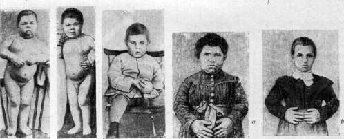
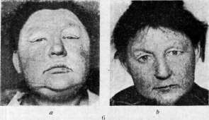
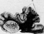
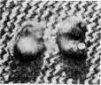
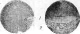
Figure 1-3. Mechanics of development (explanation in the text). Figure 4. Sporadic cretinism (10 years): a - before treatment; b - after 2 weeks of treatment with thyroidin; c - after 4 months of treatment with thyroidin. Figure 5. Sporadic cretinism (21 years): a and b - before and after treatment with thyroidin. Figure 6. Myxedema: a and b - before and after treatment. Figure 7. Atrophy and shriveling (a) of one convolution of the brain in infantile paralysis.
To the article Mechanics of development, Microgiria, Myxedema, Styela partita, in which Conklin in the egg before the beginning of cleavage distinguishes 5 differently colored plasmas [see separate table, fig. 2 - egg of ascidian Styela (Cynthia) partita: A - from the side; B - from behind; C - two-cell stage from the side; 1 - light plasma (mesenchyme); 2 - yellow crescent (mesoderm); 3 - directing bodies; 4 - material for the notochord and neural tube]. These plasmas then pass into the parts of the embryo corresponding to different organs. This case is also interesting in that the final distribution of plasmas is connected with the process of egg maturation. F. Lillie succeeded by affecting certain
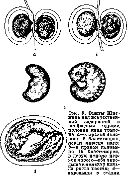
larvae with developed gills. The larvae were kept for 140 days, and at the time of fixation no difference could be noticed between them, whereas the left one developed from the half of the fertilized egg that had 1/16 of the nucleus, and the right one 1/32.
to induce differentiation in the egg of the polychaete worm Chaetopterus without cleavage, to obtain a single-celled blastula, gastrula and trochophora, i.e. 'formation of a ciliated cover corresponding to the normal cover'. Peterfi succeeded in removing the nucleus from the egg of an axolotl with the help of a micromanipulator and still obtain development up to a late stage of cleavage. Wilson in his famous experiment on the eggs of the mollusk Dentalium showed the definite significance of the plasma, so-called polar lobe, in the determination of the apical organ and the plume (figure 6). Fischel showed the determining significance of plasma in the development of ctenophores. In recent times, the organ-forming plasma of ctenophore eggs was investigated by Speck with the help of the dark field method, which made it possible to show its movements at the beginning of cleavage in an extraordinarily demonstrative way. Organ-forming substances are known for a number of worms. These facts show that plasma plays an important role in the mechanism of embryo development - in the process of determination of its various organs. The fact of the important role of the plasma component of the cell in the process of determination does not
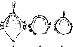
Figure 6. Wilson's experiments on Dentalium: a- normal trochophora; b- larva after removal of the primary yolk lobe; c- larva after removal of the secondary yolk lobe; in stage b the plume did not develop at all.
contradict the chromosome theory of heredity, since it is a matter of a completely different category of phenomena - not about the mechanism of transmission of hereditary traits, but about the mechanism of morphogenesis - development of the trait. Two main types of morphogenesis and the problem of their relationship. The doctrine of organizers, concepts of labile determination and double assurance. Experiments on the ligation of triton eggs were begun by Spemann in 1898 and continued until 1904. Subsequently, Spemann took up the question of the determination of the eye (figure 7). As is known, in the process of development of the nervous system, eye vesicles, which are outgrowths of the intermediate brain, grow to the epithelium, and
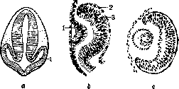
Figure 7. Development of the eye in vertebrates: a- stage of eye vesicles (2); b- lens pit (1) (2 and 3- two layers of the resulting eye cup); c- later stage- the lens rudiment has separated from the connection with the ectoderm.
at the point of contact, a cup-shaped depression of the epithelium forms—the rudiment of the lens. At the same time, the outer and distal walls of the eye vesicle begin to invaginate—the eye vesicle turns into a cup. One might have thought that the influence of the forming lens should explain the transition of the eye vesicle into the eye cup. It turned out to be exactly the opposite—in a number of amphibians, the invagination of the lens does not occur if the rudiment of the eye vesicle is excised (the area corresponding to the eye vesicle is excised at the stage of the neural plate). In some amphibians (the edible frog), the lens nevertheless develops. In other cases, the eye cup was transplanted under the skin of another part of the head or body. In the tree frog (Hyla arborea), a lens forms in any part of the body. In the gray frog, a lens forms in any part of the head. In the edible frog, not every area of the head skin is capable of transforming into a lens, but at the same time the eye cup has the ability to induce the lens, as shown by Filatov's experiment, in which he replaced the skin over the eye vesicle in the edible frog with a piece of toad skin taken from any part of the head—lens formation occurred. These experiments, carried out by a number of authors (Spemann, Lewis, Ekman, Filatov), lead to the concept that the development of one organ occurs under the influence of another organ, and therefore V. Rou's principle of dependent differentiation can be understood in the sense of a direct connection, in the sense of one organ being built by another. In his works on eye determination, Spemann was also able to make an observation in a new direction. By exchanging pieces over the area of the eye cup with the help of a special instrument, the so-called micropipette, Spemann tried to perform such exchanges between pieces of different areas of ectoderm in two embryos. He established that at the gastrula stage there is no stable determination, that pieces from the area of the presumptive neural plate and from the area of abdominal skin continue to develop in accordance with their location after exchange (Fig. 8). Further, Mangold
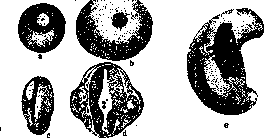
Figure 8. Spemann's experiments on exchanging pieces of presumptive neural plate and abdominal epithelium between embryos Tr. taeniatus (striped newt) and Tr. cristatus (crested newt): a-embryo Tr. taeniatus with transplanted (white) piece of presumptive abdominal epithelium of Tr. cristatus; b-the same at the neurula stage; d-section through the same embryo at the stage of primary eye vesicles; c-embryo Tr. cristatus shortly after the operation with a piece of presumptive neural plate; e-the same at the stage of primary eye vesicles and tail kidney-the dark implant has turned into body epidermis. (Mangold) showed the absence of stable determination at these stages and also with respect to the various germ layers: a piece of ectoderm transplanted into mesoderm develops in accordance with its location. Schotte showed the same with respect to mesoderm—its ability to transform into ectoderm. Right at the beginning of his work, Spemann made the observation that sometimes pieces influence the tissues surrounding them. It turned out that pieces of ectoderm
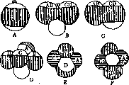
Figure 9. A-two gastrulas of the striped newt, connected in such a way that their median planes diverge forward; B-a larva that arose by the fusion of these halves-duplicitas anterior (anterior doubling); C-anterior doubling in a calf; D-two embryos of the striped newt are connected in such a way that their median planes converge forward; E-a larva that arose from two embryos (D) with two posterior ends-duplicitas posterior (posterior doubling); F-duplicitas posterior in a calf.
pieces taken from the dorsal lip of the blastopore, when transplanted into the area of the ventral ectoderm, induce in it the neural plate. Connecting this fact with experiments on constriction, which indicated the presence in the area of the gray field of some special zone determining the construction of the axial complex, Spemann came to the concept that in the area of the dorsal lip of the blastopore there is the so-called organizational field. A piece of it, transplanted into an indifferent area, causes in it the formation of highly differentiated organs. Two more important series of experiments strengthened him in the concept of the organizational field: he performed a series of experiments on the fusion of halves of gastrulae. The results of experiments on the fusion of dissimilar halves are presented in Figure 9. If similar halves of gastrulae were fused, then the material from the cut-off dorsal lip on each side is supplemented with ordinary ectoderm. As a result, an embryo with two neural plates and two axial complexes arises. The second series consisted in the fact that if the roof of the gastrula was cut off and turned 90° or 180°, no change in the position of the neural tube occurred. Subsequent works of Spemann and his students clarified a number of other interesting phenomena—the area of the organizational field passes into the roof of the archenteron, and a piece of the roof of the archenteron induces the neural plate (Marx) [see separate table, Figure 3: A and B—microphotographs of an embryo with an induced secondary neural plate (A—its side; B—from the ventral side, on which the secondary plate is located); C—section of the same embryo; 1—medullary plate; on both sides axial complexes are visible—neural plate, notochord, mesoderm]. Indifferent tissue, placed in the organizational field, acquires inducing abilities. Spemann discovered that a piece of the organizational field causes in the ectoderm phenomena similar to the initial stages of normal induction. Bautzmann clarified the area of distribution of the organizational field and showed that the notochord is also capable of inducing the neural plate in the ectoderm. Mangold and Spemann discovered the remarkable phenomenon of equivalent induction—a piece of the neural tube of an older embryo forms a neural plate in the ectoderm of a younger one. Hamburger, studying the differentiation of the nervous system of the limb, established 4 stages: 1) the ingrowth of nerves into the limb bud (their attraction by the bud), 2) the differentiation of plexuses, 3) the differentiation of large nerve trunks under the influence of blood vessels, 4) the differentiation of terminal branches under the influence of muscles or skin. From all these facts, Spemann derived two extremely important theoretical principles: 1) the principle of labile determination—at the gastrula stage, all ectoderm is labilely determined, as shown by experiments with exchange of areas; 2) determination occurs according to the principle of double or multiple provision; in other words, an organ is determined from at least two sources, for example, the neural plate is determined from the gray field and from the roof of the archenteron. Research on the structure of the organizing center gave only the most general indications of its lateral and anteroposterior segmentation. The basic but most obscure question is about the nature of the inductive action. Recently, attempts have been made to clarify the properties of organizing centers. Thus, Anikin (1927) showed that the lips of the blastopore are a source of mitogenetic rays (see). The same author managed to establish the radiation of the medullary groove, ceasing after its closure. Blyakher and Zamarayev established (1930) the presence of mitogenetic radiation in the hypostome and budding zone in hydra. These areas of the hydra's body have the properties of organizing centers (see below). The data presented do not fully explain the mechanism of the formative action of organizing centers. In any case, they can serve as material for judging this mechanism from the point of view of the causes of cell multiplication in the sphere of action of the organizational field. Thus, the works of Spemann and his school showed the presence of two principles in determination—these are, as it were, two types of Rou's principle (principle of self-differentiation and dependent differentiation), only applied with much greater clarity and consistency. In the development of any organ, there is an element of mosaicism—an element of predetermination and an element of possibility of change, i.e., regulation. Focht's research by the method of defects showed that both moments can be present simultaneously. The problem of the relationship between mosaic development and regulation—or labile determination and the action of an organizer of one kind or another—is the next problem of developmental mechanics. Mechanics of development and regeneration. Research by V. Isaev showed the existence of zones of high morphogenetic activity in the adult organism. In hydra, the object of his research, such a zone is the hypostome, the area from which tentacles surrounding the mouth grow, the budding zone, and the sole. Gotsch confirmed Isaev's observations on hydra. He showed the existence of zones of high activity in planarians during regeneration and asexual reproduction. In them, this zone is located in the head region. The latter observation agrees well with the works of the American embryologist Child, who came to the concept of morphogenetic nonequivalence of the adult organism by another path. Studying the rate of metabolism, Child found that it varies in different tissues. In a number of animals, the area of enhanced metabolism is at the anterior end of the body. In bilaterally symmetrical animals, the progressive decrease in the intensity of metabolic processes coincides with the animal's axis. The intensity of oxidative processes was determined by Child with potassium permanganate, reduced in the protoplasm with the formation of a brown color of the dioxide compound of manganese. Poisoning and dying processes also coincide with the animal's body axis. The electrical potential also changes along the axis. Ultraviolet rays kill tissues in areas of high activity first. This was shown by experiments with hydra, in which the zone of highest activity, as indicated above, is located in the hypostome. Zones of high physiological activity have been established in a number of animals. In addition to the main axis, there are secondary axes directed to the sides of the body. The morphogenetic difference of different body areas manifests itself in the process of regeneration and asexual reproduction. If a piece is cut from the body of a flatworm so that the long axis of the segment coincides with the long axis of the body, then the head end regenerates in the direction of this axis. If the piece is cut so that its apex touches the long axis, then this apex is the place of greatest physiological activity. During head regeneration, there must be a certain difference in the rate of reaction between the anterior cut surface and the end of the piece. All these facts led Child to the so-called theory of axial gradients, according to which the intensity of morphological and physiological processes decreases along the animal's body axis; in the objects he studied—in the direction from front to back. The conclusions that Child draws are as follows: a mature organism does not represent a predetermined harmony of independent parts. There are mutual influences between different organs. The basis of the organism's integrity is physiological dominance—an area dominating in the organism's system. Physiological dominance varies in its direction; during this, isolation of parts of the organism can occur, which under certain conditions become new individuals. Recently, Child has drawn parallels between the theory of axial gradients and the doctrine of organizing centers in amphibian development. One of Child's students showed the presence of a zone of high physiological activity in the area of the organizing center. Child believes that the organizing center does not fundamentally differ from other dominant areas of the body. At the anatomical congress in Amsterdam (1930), Focht made a report on the relationship between regulation and regeneration and showed that regeneration is a specific property that develops only at late stages. For higher vertebrates, the problem of the origin of regeneration or the solution of the question of why regenerative ability is not realized is one of the greatest tasks of modern developmental mechanics. In the plane of research of these specific regularities of the regeneration process, the works of the school of Guillot are of particular interest. Schotte showed that the processes of regeneration in the triton depend on the nervous system—regeneration does not occur if the limb is deprived of innervation and, as he established in a series of interesting experiments, specifically in the absence of sympathetic innervation. Limbs that are immobile and deprived of sensitivity (with motor and sensory nerves cut) but retaining sympathetic innervation regenerate; limbs that retained sensitivity and motor function but were deprived of sympathetic innervation do not regenerate. Other series of experiments established that the sympathetic nerve has no independent significance; this nerve only transmits impulses coming from the endocrine glands.
In adult tritons and salamander larvae, after destruction of the hypophysis, the limbs do not regenerate with unchanged innervation. This direction of research does not concern the factors of differentiation. The study of the latter in the aforementioned school led to the concept of so-called territories. In limb regeneration, such a territory is the soft tissue surrounding the skeleton. Upon their removal, regeneration does not occur. The works of Schotte on the influence of endocrine glands and the sympathetic nerve on the regeneration process speak of a central component of the process. In this regard, this is one of the first indications of the role of endocrine glands in the processes of morphogenesis during regeneration. Practical significance of M. r. I. M. r. and teratology. The works of Spemann had a great influence on the study of teratogenesis. In one of his first studies, Spemann describes a case of cyclopic malformation and analyzes it. The genesis of cyclopia became the subject of a number of studies by other authors. A whole series of studies, both from Spemann's laboratory and from other institutes, is devoted to various questions of the genesis of double malformations. With the help of the constriction method, Spemann and Falkenberg were able to approach such a subtle question as the origin of asymmetry phenomena. The main types of double malformations (anterior, posterior, and cruciform) receive a perfect explanation in connection with the principle of the organizational field—the divergence of the axes of the gastrula gives the anterior, convergence gives the posterior, and direction toward each other gives the cruciform double malformation (Figure 9). For more details on works on M. r. in connection with teratogenesis, see Teratology. II. M. r., surgery and pathology. The relationship of M. r. to surgery follows from everything said regarding regeneration. The method of embryo culture in a nutrient medium opens a new era here. Questions of regeneration and the conditions in which it occurs require further study. The surgery of the future must use the data of M. r. to master the processes of morphogenesis. In the field of pathology, oncology can expect to be illuminated by the data of M. r., when such problems as the phenomena of integrity, the relationship of parts in the organism, the essence of the differentiation process, and the phenomena of physiological isolation are deciphered and understood. All these problems await research and solution and will form the basis for understanding the tumor process. So far, only the first steps have been taken on the path to the study of these most difficult problems.
Related articles
Mentioned in
Cite this page
“Mechanics of Development.” Soviet Medical Encyclopedia. English translation of Bolshaya Meditsinskaya Entsiklopediya, 1st ed. (Moscow, 1928–1936), ed. N. A. Semashko. https://sovietmedicalencyclopedia.pages.dev/article/mechanics-of-development/