Organ
Historical document, translated for reference. It reflects medical knowledge of the 1920s–30s and is not medical advice.
Summary
The article defines an organ as a specific collection of tissues with a particular function, distinguishing between morphological and physiological definitions. It discusses organ classification, development, temporary and permanent organs, regressive and progressive organs, and various specialized organ systems.
Encyclopedia article (1928–1936)
ORGAN (from Greek organon - tool), a specific collection of several tissues possessing a special function. The cells of an animal organism, grouping together, form anatomical units of a higher order - tissues. The latter, in turn, joining together, give anatomical units of an even higher order - O. This is the morphological definition of an organ of a multicellular organism. In a physiological sense, however, any part of the organism that performs a specific function may be called an organ. From this point of view, sometimes even an individual cell (for example, unicellular glands in crustaceans) and even a part of a cell (for example, cilia in infusorians) may be called an organ. In the latter case, in contrast to the organs of multicellular animals, the term organoids or organelles is also used. The skeleton and organs of active movement are classified as organs of animal life or animal organs, while all others are classified as organs of plant life or vegetative organs. In lower animals, the same part of the body performs diverse functions, for which higher animals have special, distinct organs. Thus, for example, the gastrovascular system of coelenterates replaces four organ systems: digestive, circulatory, respiratory, and excretory, while in unicellular organisms all functions are concentrated even in a single cell. As the organization becomes more complex, the process of organ isolation, or differentiation (see), also proceeds, and simultaneously with morphological differentiation, physiological differentiation also occurs. But along with such a process, which is essentially analytical, there also occurs a synthetic, integral process, in the sense of combining simple anatomical units into more complex ones, i.e., from cell to organ. In turn, organs, joining together and participating jointly in any complex function, form a system. Such are the excretory, or urogenital system, the system of respiratory, digestive organs, etc. Organs participating in one common function, in some cases, such as the urinary organs of mammals, are anatomically closely connected to each other, as if passing one into another (kidneys, ureters, urinary bladder), while in other cases such a close anatomical connection does not exist, and only a functional connection exists. Such are the hematopoietic and endocrine organs. The latter are united into one system on the basis of internal secretion (see), although their functions are complex and far from uniform in all respects, and often directly antagonistic. The physiological connection between them is carried out both directly between them (purely humoral pathways or through the autonomic nervous system) and (which may be of even greater importance) peripherally (see Internal secretion). The reticulo-endothelial and chromaffin systems are also mentioned, since in both cases we are dealing with scattered formations in the body, united by a unity of function. Organs are distinguished as permanent and temporary, regressive and progressive. Permanent, or definitive, organs are those that, once appearing, do not disappear until the end of the individual's life, progressing in their development up to a certain age of the latter, and then begin to regress. Temporary, or provisional, organs are those that appear only for a certain period, and then disappear, as for example the abdominal legs of butterfly caterpillars. In this case, it often happens that during the development of the animal, one temporary organ is replaced by another carrying the same function, and such a replacement may occur more than once. Thus, for example, in the development of higher vertebrates, three kidney systems are successively replaced: pronephros, primary kidney, or Wolffian body, and secondary, or final kidney. This phenomenon is called substitution. The fate of the replaced organ varies: it either disappears completely, or passes into a rudimentary state, or finally changes its function; thus, for example, the excretory canal of the primary kidney in male individuals transforms into the excretory canal of the seminiferous gland. Temporary organs can also include the so-called embryonic organs. They serve partly as coverings to protect the delicate body of the embryo, partly for respiration and for receiving food. These organs either undergo reverse development at the end of embryonic life or are discarded at birth. Such are the yolk sac, amnion, chorion, allantois, and placenta. Some confusion in terminology is evident in relation to such organs as the notochord, pronephros and primary kidney, pharyngeal arches and pharyngeal clefts: some are ready to classify them also as embryonic organs, while others consider them rudiments of permanent organs. But the first definition, according to Bonnet, cannot be considered correct because in Anamniota the first three organs function outside of embryonic life, and from the pharyngeal pouches and pharyngeal arches in Amniota, for example, the thyroid gland, parathyroid glands, larynx, etc. develop. In view of this, regarding the last two embryonic formations, the second definition should be considered quite successful specifically for Amniota, although along with the formation of permanent organs from them, there also occurs a process of reverse development to a considerable degree. Bonne therefore proposes to call the germ layers primary organs of the first order, and the above-mentioned and similar ones primary organs of the second order. The term "primary" seems appropriate to Bonne in that it says nothing about the function of the organ or its further differentiation or reverse development, but simply indicates that from these simplest organs, more complex organs can later be formed. There is also no unity and sufficient definiteness regarding what should be understood under the names of regressive and rudimentary organs. Under the name of regressive organs, some authors mean "those that, having reached a certain degree of development, do not go further and remain little suitable or entirely unsuitable for use or even undergo reverse development (reduction), i.e., gradually decrease and disappear." Along with such organs as the gill arches of higher vertebrates and many other temporary organs, the thyroid gland is also included here. Regression is considered here from the standpoint of ontogeny. But one can also approach the question of regressive organs from a phylogenetic point of view. A similar divergence in understanding, but to an even greater extent, is also noted in relation to the term rudimentary. Most understand rudimentary organs as those that have reached such degrees of regression that they are anatomically preserved only in the form of a remnant, and physiologically are incapable of function; such are, for example, the lateral hooves of some ruminants, the reduced eyes of cave animals, the unpaired parietal eye of some lizards, the remains of limbs in some snakes, the right ovary of birds, the remains of the bony skeleton in cetaceans, the left lung of snakes, the vermiform appendix and wisdom teeth in humans, etc. Bonne, however, based on the word derivation (Latin rudimentum - beginning, first experience), on the contrary, applies this term to organs that are far from fully formed, even in the adult individual, and are only on the way to complete development in the process of evolution. Such are, for example, the horns of fossil deer, the cochlea in the ears of reptiles and birds, the tongue of fish, one ampulla instead of seminal vesicles in canids, the flying organ in flying fish (compared to birds), etc. Bonne therefore proposes to distinguish between cataplastic-regressive and anaplastic-progressive organs. He in turn subdivides the former into 1) transient embryonic, 2) abortive, 3) reduced, and 4) variable, and the latter into 1) rudimentary and 2) excessive formations (for example, excessive branching of deer antlers). Abortive, like embryonic, occur only in embryonic (or larval) life, but differ from the latter in that they generally do not develop to the ability to function independently and disappear early. (Such are the abortive rudiments of the upper incisors in ruminants, the excessive embryonic rudiments of mammary glands in pinnipeds that do not reach hypertrophy and later disappear, the rudiments of the 1st and 5th toes in ostriches, which subsequently also undergo reverse development; the abortive rudiments of the sensory ganglia of the posterior roots of the hypoglossal nerve in humans, etc.). Reduced organs differ from abortive ones in their preservation after the birth of the individual despite their uselessness in a functional sense (see the examples listed under the heading of rudimentary organs, to which much else could be added, for example, mm. palmaris longus and brevis, mm. plantaris, piriformis abdominis, as well as the muscles of the auricle in humans, etc.). Reduced organs, however, differ in their ability to vary: they disappear in some individuals in certain generations, only to reappear in subsequent ones; they also vary in form, as well as in size (wisdom tooth, m. palmaris, cervical ribs in humans), and as a result, they may sometimes again acquire some importance with a change in function. Bonne therefore calls them variable, or changeable organs (Wechselorgane). Such is the functional transformation of the cloacal glands in female tailed amphibians into a seminal receptacle.
Similarly, in the python, in the region of the cloaca, there are two bony formations, the remains of a reduced pelvis along with the remains of reduced limbs, which, according to current opinion, play the role of a copulatory organ (when introduced into the cloaca, they hold the heavy snake bodies in a state of connection during copulation). In this case, such a change in function can protect the Organ from gradual disappearance over several generations and, as Bonnet thinks, under especially favorable conditions, even promote its transition to an anaplastic state. As for the process of development of abortive and reduced Organs, Menert notes two peculiarities: first, slowness, and second, shortening (retardation and abbreviation). The latter Menert understands as premature arrest in development, but apparently this aforementioned peculiarity is not always realized in such a simple way, and in a number of cases, as some think, the Organ in ontogenesis first passes through all stages of development, but the later stages then disappear depending on the partial reverse development of the Organ, as a result of which the impression is created that the Organ never reached the final stages of morphogenesis. As an example, they point to the shortening of the tail, which in humans and many animals is laid down longer and then atrophies even in embryonic life. Progressive, or anaplastic Organs (according to Bonnet's terminology) are those which, being absent or less developed in lower related forms, show a tendency to greater and greater development with the evolution of the latter. Such are, for example, some muscles of the human body (m. flexor pollicis longus, which is absent as a separate structure in monkeys; facial muscles, in contrast to what the ear and skull roof muscles are; a finer differentiation of the laryngeal muscles in connection with articulate speech), the brain. Also included here is the improvement of the entire lower limb in adaptation to the vertical position of the body in humans, the development of the iliac region in women in connection with the widening of the pelvic inlet, etc. Anaplastic Organs, according to Bonnet, can thus pass through 3 stages: 1) of a rudimentary Organ, 2) of a complete Organ and 3) in some cases-excessive formation. Kataplastic Organs, in turn, also have 3 stages: 1) of a reduced Organ, 2) of an abortive one and 3) of complete agenesis. As for regressive Organs, the most interesting question is about the causes of their reduction, sometimes even to their disappearance. Since Darwin's time, a satisfactory answer has been considered to be the indication on non-use of the Organ, which has become unnecessary, and the weakening influence of such non-use, transmitted from generation to generation in increasing degrees. As can be seen, with such a point of view, the question is connected with the assumption of the possibility of inheritance of acquired characteristics. Weismann, who denied the latter, sought an explanation for the phenomenon in the cessation of the influence of natural selection-in panmixia (general crossing), and he saw proof of the correctness of this view, among other things, in the preservation of non-functioning Organs in their former state in parthenogenetic species, in contrast to species that reproduce sexually. Selection, according to his explanation, not only creates new forms but also preserves existing ones, since the maintenance of the status quo of any part of the body in a genus is possible only on the condition that individuals not making proper use of it will be eliminated from reproduction, i.e., will perish in the struggle for existence. However, many disagree with this point of view. Doubt is expressed as to whether panmixia by itself can lead to the disappearance of any Organ: natural selection can cause strengthening of the Organ through the extinction of individuals in which it is insufficiently developed, and weakening of it through the extinction of those in which it is developed beyond a certain measure. But it is not easy to prove how panmixia, or the cessation of the influence of natural selection, can affect in any direction. In any case, regression and disappearance of Organs that have become unnecessary in the evolution of organisms prove to be just as necessary as progress. Moreover, underdevelopment of unnecessary parts is a necessary condition for further development, and if organisms inherited everything from their ancestors literally, then, in the words of the same Weismann, the result would have to be a monstrous animal, unfit for life. Organs of adults originate from the germ layers, and are complex structures built from two, and sometimes three, embryonic layers. Thus, for example, a muscle develops from cells of the middle layer and mesenchyme; the intestinal canal with its glands-from elements of three layers: the inner, middle, and mesenchyme, etc. The laying down of permanent Organs occurs in such a way that in a certain place of the primary Organs of the 2nd order (see above), as a result of processes sometimes of pressure, but more often of protrusion, with simultaneous multiplication of cells here, an anlage of the Organ is formed. Thus, for example, the thyroid gland originates from the entoderm of the pharyngeal intestine, and at the place of its formation a tubercle appears, which grows in depth and turns into a tube opening in the middle of the tubercle. The unpaired tubular anlage of the thyroid gland soon gives rise to two lateral lobes, consisting of branching cellular outgrowths resembling the anlage of grape-like glands, for example, the salivary gland. The parathyroid glands develop in the human embryo from the so-called 3rd and 4th pharyngeal pouches by the ingrowth of epithelium into the depth of the tissues, etc. All formed, mature Organs in multicellular animals have more or less a common structural plan. Organs have a basis, which serves as a kind of skeleton of the Organ and consists mostly of connective tissue (in the brain, of neuroglia); in this basis or stroma, in one form or another, is located the functional part of the Organ or its parenchyma (epithelial, secreting cells in glandular Organs, muscle fibers in muscular Organs, etc.). In the stroma of the Organ pass blood vessels, lymphatic pathways, nerves, excretory ducts. On the outside, Organs have a covering of fibrous connective tissue with an admixture of elastic fibers. This covering has a whitish or white appearance (tunica albuginea) and is often nothing other than a serous membrane, for example, the peritoneum, pleura. Outward from the proper covering, Organs often have other coverings, sometimes of the nature of a fatty capsule.
G. Sakharov. Organogenesis, the process of organ formation during the course of development (see also Morphogenesis). Corresponding to the difference between individual development-ontogenesis and the developmental history of the species-phylogenesis, one should also distinguish between ontogenetic organogenesis and phylogenetic. The study of the latter constitutes the task of comparative anatomy, the study of the former-the task of embryology. In addition to the most profound analysis and description of the course of the organogenesis processes themselves, the tasks of these disciplines also include the causal explanation of these processes-in phylogenesis and ontogenesis (mechanics or physiology of development). Evolutionary organogenesis considers questions about the emergence of new Organs and their transformation, division, about the progressive development of Organs and their reduction, about the processes of rudimentation, etc. The consideration of the form of Organs in an inseparable connection with their function as an indivisible whole has led to some major generalizations, which outline for us the basic regularities of phylogenetic organogenesis. These are especially the principle of differentiation, associated with the laws of physiological division of labor (Milne Edwards) and integration (H. Spencer), and the principle of change of function as a guiding principle in phylogenetic transformation of Organs (Dohrn). Since ontogenetic organogenesis to a certain degree repeats phylogenetic (see Biogenetic law), it also finds its expression in the process of sequential differentiation of Organs. The task of causal study of ontogenetic organogenesis is however much more accessible to exact study, especially thanks to the availability of the experimental method of research in this area, which cannot be said for phylogenetic organogenesis. Thanks to experiment it is possible first of all for a much more profound analysis of differentiation and secondly for the study of dependencies in the development of individual parts. Purely descriptive study of embryogenesis already allowed K. Baer (Baer) to distinguish certain characteristic phases of it. During the first phase, the period of cleavage, the egg divides into ever smaller units, without noticeably changing either its size or shape. During the second period, the formation of primary organs-germ layers-occurs. During the third phase, the formation of Organs takes place and finally embryogenesis is completed by histological differentiation. We are interested here only in the general process of Organ formation. The study of the mechanism of these processes has long made it possible to speak of certain elementary principles of morphogenesis. Thus, W. His revealed the importance of the principle of unequal growth. Due to the unequal growth of epithelial plates, their bending-formation of folds, tubes, bulging outward or inward-occurs. Usually the rudiments of epithelial Organs are in the form of such local protrusions or indentations of cellular layers. An example of such formation of rudiments can serve in vertebrate animals the processes of formation of the neural groove and then of the tubular central nervous system from a simple thickening of the ectoderm-the neural plate, the formation of eye vesicles by protrusion of the lateral walls of the anterior brain vesicle, the formation of the labyrinth rudiment-the auditory vesicle-from an invagination of the ectodermal epithelium, the formation of the lung rudiment in the form of paired protrusions of the epithelium of the pharyngeal region, the formation of digestive glands from the endoderm of the intestine, etc. Later the importance of another factor-active cell movement-was also revealed. This phenomenon was taken into account especially by D. Filatov in the study of the development of skeletal rudiments of the vertebrate skull, which are formed by the active contraction of mesenchymal cells to certain places (to the notochord, to the nervous system, to the auditory vesicle). The fact of such formation of skeletal rudiments by active cell movement was then experimentally proven by the same Filatov (formation of the auditory capsule), Stone (L. Stone) (formation of the visceral skeleton), and others. It is natural however that a whole series of other questions immediately arise and first of all-what determines the movement of cells in a certain direction. Here one can think of some pre-existing paths, which purely mechanically make cell movement possible, and experiments with fibroblast cultures indeed show the importance of the mechanical factor. And in
the growth of epithelial rudiments the mechanical conditions of the environment undoubtedly to a large extent determine morphogenesis, this is especially evident in the development of organs of the abdominal cavity (e.g. formation of the liver, even at late stages). Thanks to the "descriptive" (Roux) experiment it was also revealed that active movement of cellular material plays a major role not only in the formation of mesenchymal rudiments but also in the formation of epithelial rudiments. The method developed by V. Foth of vital staining (application of markers) made it possible to trace the movement of entire cell masses, as if flowing in continuous layers. Thus, among other things, the rudiment of the nervous system (neural plate) is formed by the contraction of ectodermal cells of a certain area toward the middle of the dorsal part of the embryo. And in many other cases the formation of epithelial rudiments is not so much a consequence of unequal growth as a result of the movement of cell
tular material, not losing at the same time its mutual connection.
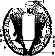
Figure 1. Embryo of the earthworm, according to Wilson: 1-mouth; 2-mesoblasts of the ring musculature of the skin with developing strips from them; 3-mesodermal teloblasts; 4-mesodermal strips; 5-neuroblasts and developing from them strips-rudiments of the nervous system; 6-endoderm; 7-ectoderm. It is clear however that all these factors, however important they may be, reveal only the gross mechanism of morphogenesis and do not answer the question about the essence of the processes themselves. If we see that some parts of the embryo grow at a certain time faster than others, that at some moment the movement of certain cells begins in a certain direction, then questions immediately arise-why do these particular parts grow faster and why do those particular cells move and in such a direction. It is clear that to the apparent process of formation of the Organ rudiment there already precedes some differentiation, which determines the material for this rudiment, and obviously the process of formation of the latter is determined not only by environmental conditions. There are already some internal factors in the material of the rudiment itself, which predetermine its fate. In some cases it is possible to show that the material going to the formation of the rudiment of a certain part or Organ is already outlined extremely early and then consists of a very small number of cells. Sometimes the material of the future Organ rudiment is even in the form of only one cell, which by successive division gives rise to all the cells of this rudiment. Such cells are called teloblasts. The phenomenon of teloblastic development is especially widespread in higher worms (earthworms, leeches), in which most Organ systems develop from special teloblasts (fig. 1). This shows that the fate of some cells is determined very early and sometimes it can be traced back to the first stages of cleavage. In some animals the first products of egg cleavage, the first blastomeres, already have a perfectly definite fate and are as it were rudiments of certain parts of the embryo. Such "determined" cleavage is observed in roundworms, in annelid worms, in mollusks, in ascidians (fig. 2). Individual blastomeres differ in this case from each other""by position, size, and sometimes by the apparent composition of their protoplasm, containinggnot only different amounts of nutritive substances (yolk), but also various other inclusions.
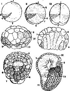
Figure 2. Distribution of organ-forming substances in the egg and at early stages of development in ascidians Cynthia partita according to Conklin: I-undivided egg; II-stage of two blastomeres; III-stage of four blastomeres; IV-beginning of gastrulation; V-later gastrula-view from below; VI-embryo with organ rudiments (all figures except V-view from the left); 1-animal pole; 2-vegetative pole; 3-area of light plasma; 4-"yellow crescent"; 5-future epidermis; 6 and 9-material of the future nervous system and notochord; 7-future endoderm; 8-future mesoderm; 10-future notochord.
Such inclusions are arranged in a specific way in the protoplasm of an undivided or even unfertilized egg and later become part of certain blastomeres, and then also of the rudiments of certain Organs. In other cases of determined cleavage, one can postulate the existence of such 'organ-forming substances,' invisible with current research methodsg, localized in a specific way in the egg and marking out special 'organ-forming areas' (W. His). In any case, with determined cleavage, the egg's protoplasm is always already differentiated ('mosaic' eggs), even if this differentiation is not expressed in visiblet differences between individual parts. With other types of cleavage (in eggs with 'regulation'), the egg has a simpler organization, but in any case it has at least a 'polar' structure from the very beginning, which is already indicated during egg formation in the ovary. Noting a certain differentiation already between blastomeres, we connect the further fate of these blastomeres with this differentiation. Indeed, the removal or displacement of individual blastomeres or areas of the protoplasm of an undivided egg, or sometimes even the displacement of egg substances by centrifugation, in the case of determined cleavage leads to underdevelopment or abnormal development of certain parts. We call the plasma differentiation preceding the formation of a known rudiment and obviously determining the fate of a known part of the embryo determination. A certain part (blastomere) may be determined from the very beginning by the substances it received along with the area of egg plasma as a result of its cleavage. In other cases, however, determination is the result of later processes obviously occurring within the protoplasm. Determination may initially be unstable, 'labile,' i.e., allowing 'regulation' within certain, gradually narrowing limits in case of deviations from the normal course of development. Thus, one speaks of determination as a process. The process of determination is studied by the method of transplanting embryonic rudiments at different stages. A firmly determined part (and even more so a already formed rudiment of an O.) develops in any environment (with any transplants) according to its original purpose through 'self-differentiation' (W. Roux). However, an undetermined part develops (H. Spemann and his school) depending on the neighboring parts with which this part comes into contact through 'dependent differentiation.' Thus, it turns out that, for example, in amphibians, the material from the posterior half of the animal hemisphere of the embryo at the blastula stage, which under normal development gives rise to the central nervous system, when transplanted into the area of future epidermis, gives rise to epidermis; when transplanted into the mesoderm area, it itself becomes part of the mesoderm. The same material, however, at a later gastrula stage is already finally determined, and no matter what area of the embryo it is transplanted into, it will always develop only into a specific part of the central nervous system. The same applies to most other Organs—one can establish certain stages of development during which the fate of the known parts of the embryo is finally determined (determined) as the material of the rudiment of a known Organ. Summarizing, it can be said that the fate of a certain part depends, first, on the properties of the cellular material, which sometimes contains certain specific substances received from the egg's protoplasm, and second, on the surrounding parts that interact with this material. Experimental research reveals in any organogenesis both elements of preformed structure and elements of dependent development. If the preformed structure is more stable, experimental intervention cannot significantly change the fate of individual parts ('mosaic' development), if this structure is labile, then disruption of relationships can lead to its change and to the restoration of more or less normal relationships through 'regulation.' A more or less labile preformed structure of the egg determines the course of at least the first processes of morphogenesis (formation of embryonic layers). The interaction of parts arising from these processes determines 'epigenetically' the formation of rudiments of individual Organs, their time, and especially their localization. The determination of Organs turns out to be a process of dependent differentiation, however, the further development of their rudiments proceeds, as numerous experiments of embryonic transplantation show, through self-differentiation, most complete in the first, 'prefunctional' period of development. The interaction of parts, which thus becomes a factor determining their further differentiation, is often carried out through direct contact. It is interesting to note that in this case the influence of parts already differentiated or at least determined on neighboring still indifferent parts is especially evident—a differentiated part already acts in the role of an 'organizer' for undifferentiated parts of the embryo. The earliest determined part of the amphibian embryo—the roof of the primitive gut (containing the substance of the 'gray crescent' of the egg)—thus acts in the role of a 'first-order organizer,' determining through direct contact the formation of the neural plate and consequently the brain from the initially indifferent ectoderm. The neural plate itself and its parts then act in the role of 'second-order organizers,' determining the formation of other parts (lens of the eye, auditory vesicle), etc. Transplanted to an abnormal location, organizer tissue causes the formation of corresponding Organs even at this abnormal location (see Mechanics of Development). Thus, the study of organogenesis processes at early stages of development is closely connected with a number of problems of more general significance. The mere fact of the mutual conditioning of the development of individual parts, of course, does not exhaust the significance of experimental research—there is a whole series of other questions concerning the process of formation of the harmonious whole structure that is the animal organism. This is achieved not only as a result of the implementation of such particular structures (organs), but also through the establishment of quite typical relationships between them. The interaction of known parts determining their final fate, i.e., the time and place of their laying down, is a necessary consequence of preceding, strictly regular processes occurring both in the whole and in individual parts, and any particular process turns out to depend both on internal factors contained in the material and on external factors for this area of the 'whole.' In determining the time of laying down, besides the characteristic interaction of parts, a determining factor is undoubtedly the maturity of the material—obviously the result of certain age changes of the protoplasm itself. In any experimental 'induction,' its result is always manifested only at a precisely defined stage of development. In determining the place of laying down, the 'double assurance' of this is always manifested—through labile pre-determination of the material itself and through typical interaction of parts (place of contact). Only a specific material is an adequate stimulus (inducer, organizer) in this case, and only a specific material is capable of giving, at a certain degree of maturity, a reactive response in the form of a typical (for this material) formative process. Labile pre-determination of parts gives some freedom in determining the place of laying down, and this also determines the greater or lesser regulatory ability of the developing embryo. The amount of material with labile determination is always greater than what is needed for the construction of a rudiment, and the influence of a neighboring part (inducer), specifying the place of future laying down, selects from this mass a more limited area. With minor violations in the topography of parts, it is thus still possible for a normal reaction of a neighboring area, i.e., replacement of the normal material by at least a similar nearby material is possible within certain limits. These phenomena of the existence of an excess amount of material capable of reacting to the irritation coming from the inducer thus also underlie a certain regulatory ability of the embryonic organism (D. Filatov calls such excess of material 'multiple laying' of organs). And the material with its specific properties and the environment or surroundings into which it falls turn out to be equally important in determining the time and place of formation of an early rudiment of an Organ. The revelation of this complex conditioning of formative processes does not allow us now to reduce embryogenesis to the former simple formulas of 'preformation' or 'epigenesis.' Undoubtedly, in any embryogenesis there are always elements of both.
Undoubtedly, the egg already has a certain structure, and all the educational material of later primordia already possesses its own specific properties (expressed at least in purely quantitative differences), but it is also undoubtedly true that this initial differentiation of the egg plasma has nothing to do with later differentiation—it only determines the direction of the first formative processes, and all the diversity of later structures is the result of the complex interaction of parts that epigenetically determine differentiation, and consequently the properties of the material and further formative processes. The interaction of the stimulus (inducer) and the substrate leads to the formation of a new primordium, i.e., a new quality not as such inherent in the material of the preceding stage. In this case, both factors (external and internal) are united in their inseparable connection and are unthinkable without one another. Embryogenesis thus consists not only of the transformation of an invisible multitude into a visible one, but also of true neogenesis. The determined primordia of Organs differentiate further in a specific manner characteristic of a given animal species. This species specificity of later structure is undoubtedly inherent from the very beginning in the material itself. In this respect, the following experiment performed by Spemann is particularly demonstrative. At the early gastrula stage, an exchange transplantation was performed of the ectoderm from the area of the future neural plate and from the area of the future epidermis between embryos of two species of newt (crested and striped), which differ in pigmentation, allowing for precise tracking of the fate of the transplanted material. The material of the future epidermis of the crested newt, transplanted into the area of the future neural plate of the striped newt, under the influence of contact with the roof of the primitive gut of the latter, gives rise to the corresponding part of the neural plate, which fits harmoniously into the composition of the central nervous system of the striped newt; however, the internal structure of this part (developed under the influence of the organizer of the striped newt) completely reveals all the specific features of the nervous tissue of the crested newt, from which the indifferent material was taken. Thus, the specific reaction always depends on the peculiarities of the material that responds to the formative stimulus in a precisely determined manner. The material always possesses its specific "norm of reaction," characteristic of a given type of organism. This specificity is determined by the hereditary substance of the nucleus, its genetic structure. During individual development, there occurs only the realization of the inherited structure through the process of increasingly complex interaction between the hereditary substance of the nucleus and the gradually differentiating protoplasm. This interaction determines the selective realization of one or another specific structure in one or another part of the embryo. During organogenesis, it would seem that the effects of the external environment should have some significance. However, it must be noted that during the embryonic period of development, the significance of the latter is exhausted mainly by the significance of conditions that limit the possibility of typical formative formation within certain limits of some average norm (temperature, humidity, nutrition, gas exchange, reaction and osmotic pressure of the medium, salt equilibrium, etc.). In the subsequent post-embryonic period of organogenesis, one can point, for example, to the often observed dependence of pigmentation development on light, temperature, etc. However, during this period, the external environment exerts to a much greater extent only an indirect influence through function, which in many cases determines the fine structure and mutual adaptation of parts. An example can be the structure of the spongy substance of bones, the dependence of skeletal parts on muscles, of the nervous system on peripheral Organs, and other facts of correlative development. Finally, one cannot fail to note the significance of the internal environment of the organism itself on the later development of Organs. In this sense, the hormones of vertebrates have particular significance, which in some cases definitely act as formative stimuli. Thus, for example, the hormone of the thyroid gland is a stimulus that in amphibians affects metamorphosis, i.e., an entire chain of very complex formative processes. The significance of the sex hormone, associated with the development and existence of a whole series of secondary sexual characteristics in vertebrate animals, is also very substantial. All these facts in part indicate the lability of the structure even of the adult organism, which is in a continuous process of internal reorganization.
Related articles
Mentioned in
Cite this page
“Organ.” Soviet Medical Encyclopedia. English translation of Bolshaya Meditsinskaya Entsiklopediya, 1st ed. (Moscow, 1928–1936), ed. N. A. Semashko. https://sovietmedicalencyclopedia.pages.dev/article/organ/