Vegetative Nervous System
Historical document, translated for reference. It reflects medical knowledge of the 1920s–30s and is not medical advice.
Summary
This article from the 1928–1936 Soviet Great Medical Encyclopedia provides a historical overview of the development, anatomy, and connections of the vegetative (autonomic) nervous system. It details the embryological formation of sympathetic ganglia, the structure of the sympathetic trunk, and the organization of the sympathetic centers within the spinal cord.
Encyclopedia article (1928–1936)
VEGETATIVE NERVOUS SYSTEM
VEGETATIVE NERVOUS SYSTEM III
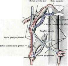
Figure 1. Diagram of the fibers of the sympathetic system (variant according to Toldt and Moller).
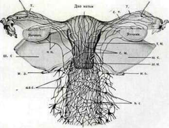
Figure 12. Diagram of the nerves of the uterus and vagina of a human (preparation and method of A. N. Zhuravlev): a. t. - tube; n. o. - ovarian nerve; g. c. - cervical ganglion; a. u. - uterine artery; p. p. - paracervical plexus; v. - vagina; p. v. - vaginal plexus; r. l. - round ligament; p. m. - plexus on the uterus; c. t. - plexus on the tube. VEGETATIVE NERVOUS SYSTEM IV
The formation of cell columns into sympathetic ones, their condensation in places corresponding to individual segments of the spinal cord into ganglia, and the simultaneous appearance of connections between the ganglia, which occurs through the formation of internodal branches (rami internodiales); the main constituent part of these branches are axons passing through rows of ganglia, and axons passing from one ganglion to another. The sections of the developing spinal cord from which the sympathetic cells migrated themselves turn into a column of gray matter and are the sympathetic spinal cord center, in which Jakobsohn (1908) distinguishes three divisions. The sympathetic center (tractus intermediolateralis) extends from the I thoracic segment to the II-IV lumbar segments (see figure 2), lies in the lateral horn and in the area lying between it and the posterior horn, and is connected for life with the boundary trunk and peripheral ganglia. This connection is extremely important, as its presence to a significant extent determines the coordination and transmission of processes from the periphery to the brain and back. The connection is carried out, on the one hand, by medullated centrifugal axons of the sympathetic spinal cord centers, which pass through the anterior root and, having formed a white communicating branch connecting the root with the ganglion of the boundary trunk, are directed to the cells of the trunk ganglia or to the peripheral ganglia; on the other hand, by non-medullated, centripetal axons of the cells of these ganglia; these fibers, having passed through the ganglia of the boundary trunk and forming a gray communicating branch between the trunk ganglion and the spinal nerve, penetrate into the posterior root and then into the posterior horn of the spinal cord (see figure 11). In both communicating branches, as in the peripheral sympathetic branches in an adult, the presence of both medullated and non-medullated fibers is often confirmed. The cells of the sympathetic centers, the axons of which go to the cells of the ganglia of the boundary trunk or peripheral ganglia, are called prevertebral neurons, and the axons themselves—preganglionic fibers; the cells of the ganglia of the boundary trunk, the processes of which are directed to the spinal cord or to the periphery, and their fibers—postganglionic; the ganglia of the boundary trunk, by their location at the vertebral column—vertebral (see figure 11).
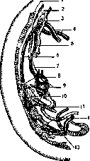
Figure 1a. Sympathetic boundary trunk and abdominal ganglia of a human embryo 10.5 mm in length (Brüning, after W. His jun.): 1-truncus sympathicus; 2-pharynx; 3-respiratory tube; 4-aortic arch; 5-n. vagus; 6-thoracic aorta; 7-esophagus; 8-nn. splanchnici; 9-stomach and plexus of the vagus nerve on the stomach; 10-plexus coeliacus; 11-a. omphalo-mesenterica; 12-pelvic part of the sympathetic trunk; 13-arteriae umbilicales. In the same way, i.e., by way of cell migration, the cervical part of the sympathetic trunk develops, which consists of a varying number of ganglia (v. d. Broeck, 1907-08), in humans 1-2-3, connected to each other by long internodal branches (rami internodiales) and connected to the spinal cord by means of communicating branches (rami communicantes). The development and connections of the cervical ganglia have been studied less, just as have the sacral ganglia; a greater number of works is devoted to the study of the question of the development of the ganglia of the head. It is believed that the sympathetic ganglia of the head develop from the walls of the neural tube and from the head ganglia (Kuntz). The ganglionic cells migrate from the cell mass designated for the formation of the semilunar ganglion of Gasser (ganglion Gasseri), and, exiting the cranial capsule, occupy specific places and group into the following 4 ganglia (Keibel, 1911): 1) ciliary ganglion (gangl. ciliare) lies in the orbit, on the outer surface of the optic nerve; 2) pterygopalatine ganglion (gangl. sphenopalatinum) in the pterygopalatine fossa; 3) otic ganglion (gangl. oticum) under the oval foramen of the sphenoid bone, on the inner surface of the mandibular nerve; and 4) submandibular ganglion (gangl. submandibulare).
Figure 2. Diagram of the location of the sympathetic center in the spinal cord (after Villiger): cervical segment; Th. I - first thoracic segment; Th. XII - twelfth thoracic segment; L. I - first lumbar segment; L. III - third lumbar segment.
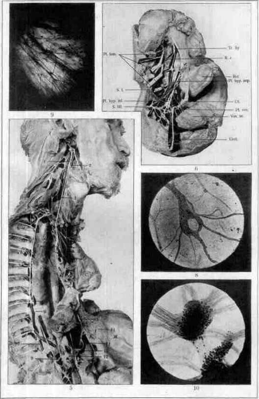
[...] ciliary [ganglion]—above the submandibular gland. In the early stages of development, in a 6-week embryo, one can observe long and short branches connecting the ganglia that migrated earlier with the starting point of their emigration—the semilunar ganglion (Huber, 1897). Fibers for the ciliary ganglion run along the ophthalmic branch of the trigeminal nerve, for the sphenopalatine ganglion—along the maxillary branch of the trigeminal nerve, fibers for the otic and submandibular ganglia—along the mandibular branch of the trigeminal nerve. The ganglia of the head receive, in addition, supplementary sympathetic cells migrating along other paths. Thus, the sphenopalatine and submandibular ganglia, which are in connection with the geniculate ganglion (gangl. geniculi), receive supplementary cells, the former—along the greater superficial petrosal nerve (n. petrosus superf. major), the latter—along the chorda tympani. Branches for the otic ganglion run along the ramus tympanicus from the glossopharyngeal nerve (according to Müller—from the petrous ganglion through the lesser superficial petrosal nerve). An exception is the ciliary ganglion in humans, all cells of which migrate from the semilunar ganglion (according to Kuntz and from the n. oculomotorius). The boundary trunks in an adult human represent long cords interrupted by ganglia, which, lying, in general, along the sides of the vertebral column, extend to the coccyx (see figure 5). The cervical part of the trunk includes 2-3 ganglia: superior, middle, and inferior. The superior ganglion, usually of an oval-oblong shape, lies at the level of the transverse processes of the II-IV cervical vertebrae, on the rectus capitis muscle, in front of the prevertebral fascia and is located behind the internal carotid artery and the sheath of the vagus and hypoglossal nerves; sometimes the ganglion is closely connected with the gangl. nodosum of the vagus nerve and is difficult to separate from it (according to Fick—4 times in 28 cases). The segment of the trunk departing from the ganglion lies on the prevertebral fascia, the longus capitis and longus colli muscles, and enters the middle cervical ganglion, which lies at the level of the VI cervical vertebra next to the inferior thyroid artery and is distinguished by extreme variation in form; sometimes the ganglion is completely absent. The second segment of the trunk begins from the lower pole of the middle cervical ganglion; having formed a loop around the subclavian artery (ansa subclavia Vieussenii), the trunk enters the inferior cervical ganglion, lying in front of the transverse process of the VI cervical vertebra at the site of the origin of the vertebral artery (in the absence of the middle ganglion, the first segment passes directly into the inferior cervical ganglion). The latter almost always fuses with the first thoracic into a flattened, curved plate, often designated as gangl. stellatum. The thoracic part of the sympathetic trunk includes 10, 11, 12 ganglia, is covered along its entire path by the diverging leaves of the posterior mediastinal pleura and lies in the upper section—in front of the rib heads and intercostal arteries, in the lower section—more medially. Its ganglia lie in front of the rib heads, they are triangular or square, flattened; the first thoracic is often fused with the second. The lumbar part with its 4-5 ganglia is shifted toward the midline, so that the ganglia lie directly on the vertebral bodies, medial to the m. psoas major, being covered on the right side by the v. cava inf., and on the left—by the abdominal aorta (see figure 6). The ganglia of this part are small, the connecting branches between them (as in the thoracic part) are often bifurcated. The smallest part of the trunk is the sacral section, located medially from the sacral foramina: being interrupted by 3-4 small ganglia, it ends in a small unpaired coccygeal ganglion. In the head, the presence of a boundary sympathetic trunk is also noted (see figure 7). Its first segment is the trunk of the internal carotid nerve from the superior cervical ganglion. Having entered the external opening of the carotid canal, the trunk usually divides into lateral-anterior and medial-posterior branches, the anastomoses of which form the second segment of the trunk—the carotid and cavernous plexuses. The internal carotid plexus usually includes a small flat or star-shaped gangl. caroticum, lying on the second bend of the carotid artery. The twigs of the plexuses to the sympathetic ganglia of the head constitute the third segment of the boundary trunk. The ganglia of the head section are considered to be the above-mentioned gangl. ciliare, gangl. spheno-palatinum, and ganglion oticum. The peripheral sympathetic system is divided into the system of the soma (body) and the visceral system, intended for the internal organs. The somatic system is composed of the sum of axons of cells whose bodies lie in the ganglia of the boundary trunk. Upon exiting the ganglion, the axons are assembled into small trunks, join the trunks of the spinal nerves, and terminate in peripheral vessels, lymph glands, in the glands and smooth musculature of the skin, as well as in some skeletal muscles. Thus, the somatic part of the peripheral sympathetic system consists of the aggregate of the first postganglionic neurons. The visceral part of the sympathetic system is characterized by the fact that it innervates the internal organs (viscera) and consists more often of two, sometimes three neurons. The first neurons of the visceral sympathetic system are also located in the vertebral ganglia; however, their axons do not follow directly to any organ, but terminate in peripheral ganglia that have migrated from the boundary trunk, the cells of which send axons to the destination; these cells are the second postganglionic neurons, and the peripheral ganglia bear the name prevertebral. The small trunks are for the most part gray in color, transparent, their course is often tortuous. The shape of the peripheral, prevertebral sympathetic ganglia varies from round to star-shaped and often depends on topography; thus, in the plexuses of the intestines the ganglia are flattened, in the urinary bladder they are round, the ganglia of the heart are round, but on the anterior surface of the atria there are always 1-2 flat ganglia (see figure 8, 9, 10). The ganglia, by their position, can interrupt the course of the trunk or, by being located inside, push apart its fibers; furthermore, they often lie adjacent to the trunk or are included in the network of its anastomoses. In the latter case, if there are many ganglia, extensive nodal plexuses are formed, lying outside or inside the walls of organs (e.g., in the intestines), or small nodal fields (on the heart). The size of the ganglia varies from the limits of visibility under a microscope (in the case, for example, when the ganglion consists of 2-3 cells) to the size of the os pisiforme. Smaller ganglia are found inside the trunks (they are always located in the vagus nerve), medium and large ones lie adjacent to the trunks of large vessels or are located near organs and within the organs themselves. The trunks often form plexuses, which are also found outside or in the walls and inside the organs. Plexuses are divided into large and small; small ones are formed independently or are part of large ones. The question of the nature of the plexuses in a number of cases is controversial, because parasympathetic cerebro-spinal nerves are often mixed with the sympathetic trunks. Sympathetic nerves for the head, neck, and chest depart from the cervical and thoracic sympathetic ganglia, partly from their internodal branches, and are divided into an ascending group, heading to the head, into a group of branches descending to the viscera of the chest, and a group for the organs of the neck, heading to them almost directly from the site of departure. Nerves for the head (see figure 7) depart from the superior and inferior cervical ganglia and are divided into a group penetrating into the cranial cavity, and a group approaching from the periphery. The first group consists of a plexus located along the course of the internal carotid artery, which departs from the superior cervical ganglion, and a plexus from the inferior cervical ganglion, following the course of the vertebral artery; both plexuses anastomose inside the cranium, give branches to the vessels of the brain, meninges, pituitary gland, sympathetic ganglia, trunks of the III, IV, V, VI pairs of cranial nerves and the tympanic nerve. The second group is composed of two branches of the superior cervical ganglion, which, having formed plexuses around the external carotid artery, accompany its ramifications on the head. A part of the twigs of the plexus penetrates inside the cranium along the arteries of the dura mater and gives a small trunk to the otic ganglion; the external maxillary plexus—plex. maxillaris ext.—gives a root to the submandibular [ganglion].
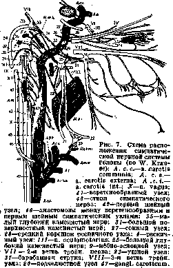
node. The organs of the neck receive nerves from three cervical ganglia; part of the nerves originates from the cervical part of the sympathetic trunk, and part from the plexuses of the carotid arteries formed by the branches of all the cervical ganglia. The twigs from the plexuses follow the course of the branches of the carotid arteries and approach the organs along with them, due to which the number of individual sympathetic plexuses is equal to the number of arterial branches. Among the trunks originating from the cervical part of the sympathetic trunk, the laryngopharyngeal branches from the superior cervical ganglion—rami laryngo-pharyngei—are noted, which partly go with the superior laryngeal nerve to the larynx, and partly descend to the lateral wall of the pharynx, where, together with branches of the glossopharyngeal, vagus, and superior laryngeal nerves, they form the pharyngeal plexus—plexus pharyngeus. Independent twigs also depart from the superior and middle cardiac nerves and accompany the superior laryngeal and recurrent nerves. The plexuses for the organs of the thoracic cavity are composed of branches of the cervical and thoracic ganglia, to which trunks from the vagus nerve system are added; the plexuses can be divided into three groups. The trunks of the upper group depart from the superior and middle cervical ganglia, from various segments of the interganglionic connecting branches, and anastomose with branches from the trunks and ramifications of the vagus nerve. The trunks of the middle group depart from the inferior cervical ganglion and receive twigs from the vagus nerve. Both groups follow, in general, the course of the vessels, the upper one the common carotid, the lower one the subclavian artery, and are directed toward the trachea and large vessels; in this segment, the plexus again receives branches of the vagus and recurrent nerves and trunks from the lower group, composed of twigs from a number of thoracic ganglia. The plexuses are divided into sections, although their nomenclature is not precisely established, which depends on the difficulty of classification. The entire plexus of the thoracic cavity can be called the plexus pectoralis, with its subdivision into the plex. praetrachealis, plexus cardiacus, plexus pulmonalis, plexus trachealis, and plexus oesophageus. The plexuses for the organs of the abdomen, by the nature of the branches composing them, are primarily sympathetic. The approaching pathways are composed of the vagus nerves descending from the esophagus, the lesser and greater splanchnic nerves, and a large number of branches departing throughout the entire length of the sympathetic boundary trunks of the abdominal cavity. The branches of the vagus nerves are well traceable to the stomach. Some physiologists and morphologists indicate that the vagus nerves take part in the innervation of the intestine; they note branches going from their trunks into the celiac plexus and the liver. The nn. splanchnici major and minor begin from the VI to IX and from the X to XII sympathetic thoracic ganglia, enter the largest unpaired celiac plexus (plexus coeliacus), lying on the anterior semi-circumference of the abdominal aorta, behind the pancreas, and surround the initial parts of the celiac and superior mesenteric arteries. The plexus occupies the area between the renal arteries, the adrenal glands, and the aortic opening and always includes a varying celiac ganglion (gangl. coeliacum) and sometimes the superior mesenteric ganglion (gangl. mesenteric. super.), lying under the root of the latter. From the plexus, a number of smaller plexuses depart to the diaphragm, adrenal glands, kidneys, and the spermatic plexuses, following the course of the internal spermatic artery. Furthermore, the plexus gives rise to a number of smaller unpaired plexuses directed to individual organs, and the superior mesenteric plexus, intended for the pancreas, small and large intestines, up to half the length of the transverse colon. The second main source of nerves for the organs of the abdominal cavity is the plexus on the aorta, composed of two trunks from the celiac plexus and twigs from the sympathetic lumbar ganglia. From the aortic plexus departs the inferior mesenteric plexus for the transverse and descending parts of the colon, the sigmoid flexure, and the upper sections of the rectum. The aortic plexus continues initially into the unpaired superior hypogastric plexus, which at the promontory bifurcates and passes into the pelvic plexus. The terminal sections of the aortic plexus are considered to be the parts located along the course of the iliac, femoral, and popliteal arteries. All pelvic organs—the urinary bladder with the prostate gland, the vagina, the uterus with the ovaries (partially), the rectum, etc.—receive nerves from the inferior hypogastric plexus (see figure 6). The plexus is composed of branches of the I-III sacral spinal nerves, 1-3 branches of the sacral sympathetic ganglia, a number of trunks from the inferior mesenteric plexus, and forms a plate extending from the sacrum to the urinary bladder (the nodal plate of older authors). Careful study allows one to distinguish: 1) the anteroinferior section of the plate, the lower part of which is directed to the prostate gland, to the seminal vesicles, to the vas deferens, and to the cavernous bodies, the upper one to the urinary bladder; 2) in women, the middle section, the lower part of which gives branches to the vagina, the cavernous bodies of the clitoris, the upper one to the uterus and ovaries; and 3) the posterior section, intended for the rectum. In the plexus, a number of small nodules are embedded, connected by anastomoses with the nodal plexuses lying in the organs. The assumption about the existence of large ganglia that are centers for the innervation of the uterus must be refuted. The parasympathetic system consists of: 1) centers lying in various sections of the brain and spinal cord, 2) fibers that pass inside individual cranial and sacral nerves and are directed to peripheral ganglia, and 3) peripheral ganglia lying outside or inside the organs of the chest, abdomen, and pelvis. The cells of the centers are the preganglionic neurons of the system, the cells of the ganglia are the postganglionic neurons. Müller considers it possible to attribute to the system hypothetical fibers which, in his opinion, should follow to the periphery along the posterior roots of the thoracic and lumbar nerves. The system was isolated by Langley on the basis of the limited territory of distribution of certain cranial and sacral nerves, their antagonism to the sympathetic nervous system, and also on the basis of the peculiarity (not recognized by everyone) of the structure of the cells of the central nuclei and peripheral ganglia of a certain group of nerves. In general, the morphology of the system is poorly developed. The system is divided into central and peripheral sections. The central part is subdivided into cranial and sacral, with the cranial part, in turn, divided into centers embedded in the midbrain and medulla oblongata (see figure 13). The peripheral part passes inside the trunks of the III, VII, IX, and X pairs of cranial nerves and in the trunks of the II-IV sacral nerves. The fibers of the system receive their name from the nerves inside which they pass; they follow from the cells of the centers directly to the peripheral ganglia and do not form anastomoses. The parasympathetic center, the fibers of which pass inside the 3rd pair (n. oculomotorius), lies, according to Bernheimer, medially from the nucleus of the oculomotor nerve, under the anterior section of the superior colliculus, and consists of a group of small cells. The fibers are directed to the ciliary ganglion, the cells of which send axons to the muscles—the sphincter of the pupil and the ciliary muscle. The location of the three nuclei embedded in the bulbar part is also not fully determined. The anterior nucleus lies in the form of a small and non-isolated (like all parasympathetic nuclei) cluster of cells, dorsomedially from the nucleus of the facial nerve, bears the name of the superior salivatory nucleus (nucl. salivatorius sup.), and sends part of the conductors, which go together with the facial nerve (VII pair), as the n. intermedius. From the geniculate ganglion of the facial nerve, the conductors go along the n. petrosus superficialis major into the pterygopalatine ganglion, the cells of which send fibers to the glands and vessels of the mucous membranes of the nose, the upper part of the pharynx, the oral cavity, to the upper lip, Waldeyer's ring, and the pulp of the upper teeth. The latter fibers are postganglionic, according to Müller, non-medullated, whereas the fibers of the n. petrosus superf. major are preganglionic and medullated. Another part of the parasympathetic fibers lying in the facial nerve follows through the chorda tympani and the lingual nerve into the submandibular ganglion, the cells of which send postganglionic fibers into the submandibular gland. The second parasympathetic nucleus, the inferior salivatory nucleus of the bulbar part, nucl. salivatorius inf. (Kohnstamm), lies in front of the frontal part of the nuclei ambigui and sends fibers along the glossopharyngeal nerve (IX pair). The fibers pass into the petrous ganglion and from there, in the form of the tympanic nerve or the superior lesser petrosal nerve, into the otic ganglion, the cells of which send fibers into the auriculotemporal nerve, into the parotid gland (according to Langley, to the mucous membrane of the lower lip, part of the cheek, the dental pulp, and the orbital glands). The third nucleus of the bulbar part is considered to be part of the dorsal nuclei of the vagus nerve. Parasympathetic fibers, exiting from the brain, follow along the vagus nerve and its ramifications and are considered preganglionic in relation to the ganglia embedded in the heart, stomach, and intestine, which are considered as peripheral parasympathetic ganglia (plexus parasympathicus postganglionaris—Dresel, Gaskell).
It is believed that parasympathetic fibers follow the branches of the vagus nerve; the function of those of its branches which are directed to the heart and intestines has been most studied. According to Müller, sympathetic cells are also included in the plexuses of the intestine and stomach, which migrate here somewhat later than the migration of cells following the path of the vagus nerve. In the vagus nerve itself, the presence of sympathetic trunks entering it from the superior cervical sympathetic ganglion has been established (W. Krause, R. Fick). According to Keibel, although the facial, glossopharyngeal, and vagus nerves are not directly connected to the sympathetic system, sympathetic cells are included in their stem ganglia, central nuclei, or roots.-Sacral nuclei of the parasympathetic nervous system ^Midbrain Ciliary Sphincter of the pupil,
Ciliary ganglion, ciliary muscle Lacrimal gland Parotid
>*L>
Cranial part
Medulla oblongata 511 Sacral Hypogastric
^ S 6'III part plexus
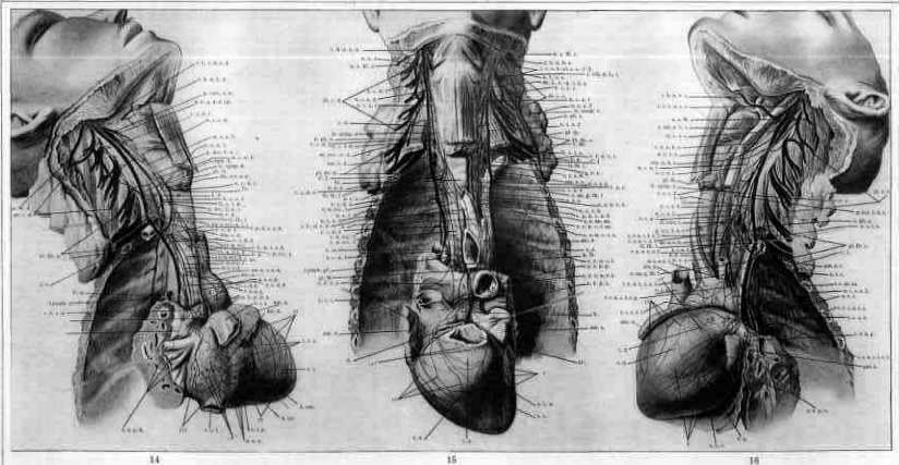
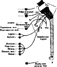
Rectum; Urinary bladder; Genital organs. Figure 13. Diagram of the location of parasympathetic centers in the brain and spinal cord and the course of parasympathetic fibers in the III, VII, IX, and X pairs (according to Villiger): III—n. oculomotorius; VII—n. facialis; IX—n. glosso-pharyngeus; X—n. vagus. They lie in the region of the lower segments of the lumbar part of the spinal cord. Preganglionic fibers follow the course of the I-III sacral nerves and, emerging from there, give rise to a series of branches passing through the sympathetic plexus of the pelvis and heading to the ganglia located inside the pelvic organs. The cells of these ganglia are the second neurons. Vegetative nervous system of individual organs. Morphological data on the vegetative nervous system of individual organs are poorly developed. The participation of the sympathetic nervous system in the innervation of individual organs is proven by tracing the course of individual sympathetic branches with the eye. The inability to understand intrastruncal topography and to recognize the axons of various nerves, the inaccuracy of the method of determining trunks by the caliber and number of fibers contained in them (measurements by Gaskell, Billingsley, Hanson)—all this complicates the research process. It is even more difficult to morphologically prove the fact of the participation in the innervation of organs of those sympathetic axons which are classified as belonging to the parasympathetic nervous system. It is clear that the cardinal question for all of biology regarding the participation of various nerves in the formation of mixed trunks and plexuses will be resolved simultaneously with the finding of a method for studying intrastruncal topography. The data provided below concern those organs whose innervation is clearer. Nerves of the heart. The nervous system of the heart consists of: 1) incoming trunks, 2) plexuses in the heart itself, and 3) nodal fields connected to the plexuses. The incoming nerve pathways depart from the cervical and thoracic parts of the vagus nerve and its branches and from the three cervical ganglia of the sympathetic trunk and, exchanging branches, are organized into two groups—superficial and deep. The first group adheres to the vessels: in the upper segment—to the carotid and subclavian arteries, in the lower—to the aorta and pulmonary artery; the second, composed of part of the branches of the superficial plexus and branches of the vagus and recurrent nerves, lies on the anterior surface of the lower third of the trachea. In both groups, the number of branches, the place of their origin, the number of anastomoses and ganglia included in the trunks, and the topography and diameter of individual nerves vary greatly. Most often, 4-5 branches are encountered from the left and 6-7 from the right sympathetic trunk and its ganglia, and 5-6 branches from each of the vagus nerve trunks; in addition, branches from the superior laryngeal and recurrent nerves go to the heart. All cardiac branches from the sympathetic trunk are called 'nervi', and from the vagus—'rami'. Usually, the following sympathetic branches are present: superior, middle, and inferior cardiac nerves. The superior (see figures 17 and 18, 1c) departs from the lower pole of the superior ganglion, runs medial to the sympathetic trunk and the common carotid artery, lies in front of the prevertebral fascia, crosses the inferior thyroid artery, and, following the left side of the common carotid, and on the right—the brachiocephalic, passes to the aorta and pulmonary artery. The nerve anastomoses with a branch from the superior laryngeal nerve, with the superior cardiac from the vagus, and with the recurrent nerve. The middle cardiac (see figure 17, 2c), departing from the middle ganglion or directly from the trunk (the nerve is often absent, as in fig. 18), lies on the posterior surface of the common carotid artery and enters the cardiac plexuses, adhering on the left to its posterior-lateral surface, and on the right—to the same surface of the brachiocephalic artery. The inferior cardiac nerve departs (usually by a series of roots) from the inferior cervical ganglion, or the stellate ganglion, lies on the left—behind the subclavian, on the right—behind the subclavian and brachiocephalic arteries, and in this way reaches the heart. Among the cardiac branches of the vagus nerve, branches from the trunk itself and small trunks from its branches are noted. The cervical part of the trunk gives 1-2 small branches, with the place of their origin being inconstant. The thoracic part of the trunk sends 3-4 branches, of which the lowest always separates somewhat above the bifurcation of the trachea, lies between the superior vena cava, the trachea, and the lymph node adjacent to the trachea, and gives branches to the third cardiac plexus (see below) and to the lungs. It is of interest to physicians that an enlargement of the node (pulmonary tuberculosis) can cause compression of the nerve and a change in the heart rhythm. The branch of the superior laryngeal nerve, which always anastomoses with a branch from the superior cervical ganglion (superior cardiac nerve), and the branches from the recurrent nerve are distinguished by greater constancy; the latter on the left side depart from the place where the recurrent nerve bends under the arch of the aorta and immediately, penetrating through the pericardium, enter into the composition of the first and fourth plexuses (see below), and on the right, departing from the place where the nerve bends under the subclavian artery, they immediately anastomose with the branches of other nerves lying behind and outside the brachiocephalic artery. In general, the cervical part of the vagus nerve and the superior and middle sympathetic ganglia send fewer branches than the thoracic part of the vagus nerve and the inferior cervical and stellate ganglia. On the heart itself, the nerves are organized into six clearly distinguishable separate plexuses, the presence of which is confirmed by a series of comparative anatomical studies; they occupy a precisely defined position and, in the region of the atria and on the arterial cone, are connected with extensive nodal fields that transition into one another. Two anterior plexuses, two posterior, one plexus for the anterior surface of the atria, and one in the region of the sinus of Haller are distinguished. The anterior plexuses—left and right (first and second)—descend from the pulmonary artery and the aorta onto the ventricles and, lying initially on both sides of the arterial cone, innervate the corresponding part of the anterior wall of the ventricles, giving small trunks to the muscle, vessels, pericardium, and sending branches to the anterior sections of the septa of the ventricles, atria, and to the endocardium (see figures 15 and 16). The posterior plexuses—right and left (third and fourth)—are located more complexly (see figure 14). The third plexus lies along the upper boundary between the atria and, adhering to it, passes onto the posterior wall of the right ventricle, innervating the outer and posterior walls of the right atrium and the posterior wall of the right ventricle. The fourth plexus spirally winds around the outer wall of the left atrium, crosses the coronary sinus of the heart, passes onto the posterior surface of the left ventricle, and innervates the outer and posterior walls of the left atrium and the posterior wall of the left ventricle. Both plexuses send branches to the musculature of the atria and the posterior walls of the ventricles, to their vessels, to the corresponding parts of the endocardium, and also small trunks for the superior and posterior sections of the atrial septum, the ventricular septum, and to the His-Tawara bundle (see figure 21). The fifth plexus is located on the anterior wall of both atria and, innervating it, gives branches to the anterior part of the wall of the septa of the atria and ventricles. The plexus of the sinus of Haller—the sixth plexus—lies in the upper section of the posterior wall of the left atrium and innervates the parts adjacent to it (see figure 22). All plexuses are accompanied by nodal fields, occupying, like the plexuses, a definite territory, although the number of ganglia constituting them, their size, and their interrelationship often vary. The field of the first and second plexuses is located in the region of the arterial cone and represents a group of ganglia connected to each other in a chain, analogous to the anterior chain of the heart of birds. The field of the third plexus occupies a section of the right atrium between the superior and inferior venae cavae and, being limited on the outside by the terminal sulcus (sulcus terminalis), continues onto the posterior surface of the right atrium, where it reaches the coronary sinus and connects with the field of the fourth plexus. Small trunks of the third plexus and thin branches of the plexus of the sinus of Haller (see below) enter the field and connect with the small branches of a series of ganglia. The lateral ganglia of the field themselves give off small branches, which organize into 1-2 larger trunks and a series of small ones, which, after giving off branches in the direction of the lateral surface of the inferior vena cava, lie on the outer wall of the right atrium, where they bend anteriorly and anastomose at the right surface of the coronary sulcus with the branches of the second plexus. The branch from the vagus nerve described above, which lies between the trachea and the superior vena cava, enters this field. The field accompanying the fourth plexus begins at the place where the trunks of the fourth plexus transition onto the left atrium, in the region of the plicae nervinae, and extends predominantly in the region of the posterior wall of the atrium, between the posterior pulmonary veins and the coronary sinus. The field of the fifth plexus is small and consists of several ganglia, which occupy the middle of the extent of the entire anterior wall of the atrium. The field of the 6th plexus is also small, the region of distribution of which is limited to the posterior wall of the left atrium in the space between the anterior trunks of the pulmonary veins. The position of the fields, as well as the number and size of the ganglia, change with age, as well as by classes of animals. In newborns, the fields are located closer to the vessels, they are less developed, and include ganglia of smaller size.
In birds (Anser domesticus, Columba vulgaris, Gallus domesticus, Corvus cornix, etc.), the fields also encompass the ventricles, and on the atria, in the region of the posterior part of the sulcus interatriosus, a long spindle-shaped node is always noted, sharply standing out among a number of other small ones (Sinelnikov). It has been noted that the ventricular nodes of marine birds are smaller than those of domestic birds (Kondratyev). The nodes of the fields are very small in cats (Anufriev) and rabbits (Zhuravlev), the nodes in dogs are somewhat larger (Zhuravlev), and the nodes in cattle are extremely small in relation to the mass of the heart (Volynsky). Nodal fields reach their greatest development in humans (see Heart, nerve cells, terminal apparatus). Nerves of the trachea. The main trunks innervating the trachea are the inferior laryngeal branches of the vagus nerve, the trunks of the latter, and the twigs of the superior laryngeal nerve, which directly anastomose with the inferior laryngeal (recurrent) nerve. Sympathetic small trunks depart from the cardiac branches, from the stellate and superior cervical ganglia, and mix with the branches from the vagus nerves. Lying in the space between the trachea and the esophagus (cf. nerves of the esophagus), the recurrent nerves ascend upward, the left for a greater distance, the right for a lesser one, and send twigs into the posterior segments of the annular ligaments and into the lateral parts of the membranous wall. In the upper and lower parts of the trachea, twigs depart from the anastomotic branch between the superior laryngeal and recurrent nerves, and on the right side, from the right trunk of the vagus nerve. The approaching twigs enter from the lateral sides of the trachea into its annular ligaments and into the lateral segments of the membranous wall, with the first twigs directed toward the anterior surface of the A9 annular ligaments, and the posterior ones entering directly into the processes of numerous nodes embedded in the membranous wall. The nodes are embedded throughout the entire extent of the membranous wall; they are all flattened and send out 4-6 long, thick, anastomosing processes; some of them are perforated in the middle; in general, these nodes represent a formation sui generis, not encountered in other organs. Nerves of the lung. The approaching nerves depart from a series of twigs of the vagus nerve, twigs from the stellate and 3-4 underlying ganglia, and form 4 anastomosing plexuses, which lie on the anterior and posterior surfaces of the bronchi and lung vessels at their hilum. The right anterior plexus lies on the anterior surface of the right pulmonary artery; it includes branches from the cardiac small trunk, which travels from the vagus nerve into the III cardiac plexus, and 2-3 twigs from the tracheal plexus, which lies on the anterior surface of the trachea. Branches for the left anterior plexus depart from the trunk of the vagus nerve, immediately below the point where the recurrent nerve departs from it, and from the initial part of the recurrent nerve itself, and lie on the anterior surface of the left branch of the pulmonary artery. Among these branches, the number of which can be 2-3, there is always a twig descending along the anterior surface of the left branch of the pulmonary artery into the IV cardiac plexus (see above—nerves of the heart). Surgeons believe that when the root of the left lung is pulled backward during an operation, the tension of the twig can cause a change in heart function (Taft). The anterior branches of both sides follow the course of the bronchial and vascular branches inside the lungs; in the upper lobes and in the middle lobe of the right lung, they are located along its anterior and medial surfaces. The posterior approaching branches are thicker and more numerous than the anterior ones; departing partly from the trunks of the vagus nerves in the segment at the hilum of the lungs and from twigs from the sympathetic ganglia, they lie against the posterior surfaces of the bronchi, with the twigs of the left plexus forming a series of parallel small trunks, and the twigs of the right forming three small trunks, which adhere to the upper, middle, and lower surfaces of the right bronchus (Taft). Penetrating into the lung, both posterior plexuses innervate the posterior and lateral surfaces of the bronchi and vessels and form a series of branches which, descending obliquely downward, pass to the anterior and medial surfaces of the vessels and bronchi of the lower lobes of both lungs (see figure 20). Along the course inside the lung, the nerves are accompanied by a series of separately scattered small nodes, the size of which decreases toward the periphery (Remak). Nerves of the esophagus. The nerves of the esophagus are formed by branches of the vagus nerve and small trunks of the sympathetic nerve, which depart at various levels from the boundary trunk or its branches. The main branches of the vagus nerves depart from the recurrent nerves and the main trunks of the vagus nerves. Both recurrent nerves, the right higher, the left lower, lie in the gap between the trachea and the esophagus and give off a series of anterior twigs for the trachea and posterior ones for the esophagus. Both vagus nerves, after giving off the recurrent nerves, approach the esophagus, with the left trunk lying on the anterior surface of the esophagus, the right on the posterior, and they give the esophagus a large number of branches. Thus, the place where the recurrent nerves depart is the boundary of the supply to the esophagus by small trunks from the trunks of the vagus nerves and small trunks from its branches. The origin of the departure of the sympathetic nerves of the esophagus is not precisely determined. Branches from the stellate ganglion are indicated, the departure of twigs from the laryngopharyngeal branches is probable, and the participation of twigs from a series of thoracic ganglia is undoubted. The twigs of the approaching nerves penetrate through the thickness of the outer muscles and immediately turn into a wide-meshed network, in the intersection of the loops of which large, rounded nodes are embedded, sending twigs into the submucosal plexus. The loops of the network are elongated along the longitudinal axis and sharply change character at the cardia, passing into the myenteric plexus of the stomach. Nerves of the stomach. The nervous system of the stomach is constructed very complexly. Branches of the sympathetic nerve, following the course of all the arteries supplying the stomach, approach together with them to the greater and lesser curvatures and form here a group of clearly distinguishable nodes, which pass onto the stomach and are so connected with the system of the vagus nerves that the very recognition of their belonging to one or the other system is very difficult. The branches of the vagus nerves are the terminal segments of the trunks themselves, which, as indicated, after giving off branches to the esophagus below the level of the lung hilum, descend to the stomach, adhering: the right to the posterior-right surface, the left first to the anterior-left and then to the anterior-right surface of the esophagus. Subsequently, both trunks pass onto the stomach, lie on the lesser curvature, the left closer to the anterior surface, the right to the posterior, and follow to the pylorus. Along the way, both trunks give 6-8 branches, which immediately submerge under the serous membrane and, together with the sympathetic nerves, take part in the formation of three very complexly constructed plexuses. The first superficial plexus lies under the peritoneum, is called "subserous," and represents a wide-meshed network of branches, at the intersection points of which nodes are embedded (Vorobyov, 1910). The network is located throughout the entire extent of the anterior and posterior surfaces of the stomach and is connected at the greater and lesser curvatures with the sympathetic small trunks of the approaching arteries. The largest nodes lie at both curvatures; the fundus and body of the stomach are occupied by small nodes. The shape, size of the nodes, and width of the loops vary depending on the class to which the animal being studied belongs: they are well expressed in dogs, weakly in cats, and comparatively well in humans. The plexus is connected with the deeper myenteric plexus by a series of twigs, which both plexuses exchange. The second plexus—myenteric—was described by Auerbach (1862-1864), represents a strongly developed network, and is formed, mainly, by branches of the vagus nerve (see figure 4). Descending from the lesser curvature, 6-8 twigs of the vagus nerve enter between the circular and longitudinal musculature and, forming a bend directed toward the fundus of the stomach, go to the greater curvature and give off a series of twigs along the way, in the intersection of which cells are embedded. Thus, an extensive nodal plexus is formed, embedded throughout the entire extent of the stomach and passing directly onto the duodenum. In it, the following three parts are distinguished: fundus, body, and pylorus. The loops of the fundus plexus are comparatively wide, the fibers constituting the walls are thin, and there are few cellular elements. In the part of the body, the cellular elements increase, and the walls of the loops become thicker. In the part of the pylorus, the walls of the loops are thick, and cells are embedded inside and around the nodal points, which makes the plexus similar to the Auerbach plexus of the small intestines. Thus, the pyloric part of the stomach, by the shape and structure of its myenteric plexus, differs sharply from the fundus and, partly, the body, and can be compared with the adjacent segments of the duodenum. The third plexus—submucosal—was described by Meissner (1857) and consists of a dense network of fibers and a series of well-formed nodes embedded in the network. The nodes of the plexus in cats lie above the network, and their processes connect the third plexus with the second (Shabadash). The degree of participation of each plexus in the innervation of the parts of the stomach wall has not yet been fully established; it must be assumed that the subserous plexus innervates the peritoneum and subperitoneal vessels and that its nodes do not carry a motor function, as is assumed by Openchowski, Knaut, and Dobert and is set forth by some physiologists. The second plexus, besides branches to the muscles of the stomach, sends small trunks to the first and third plexuses, and the third to the mucous membrane (see
(Stomach—subserous, intermuscular, and submucosal plexuses). Nerves of the intestine. The system of intestinal nerves is less developed than others. The afferent pathways are the solar plexus, the superior and inferior mesenteric plexuses, as well as branches of the hypogastric plexus, which participate in the innervation of the rectum, and thin branches of the vagus nerve, which pass to the initial segment of the duodenum. Furthermore, as physiologists point out and morphologists are unable to establish, the trunks of the vagus nerve, which enter the solar plexus, participate in the plexus. All afferent nerves, following the course of the vessels, are directed toward the intestinal tube and participate in the formation of the subserous, intermuscular, and submucosal plexuses, which, being, in general, similar to the plexuses of the stomach, differ in structural and partly in topographical respects (see Figure 25). In human material, under conditions of successful macro-microscopic staining, the following is always established: 1) there are no ganglia in the subserous plexus of the intestine, 2) the thickness of the loops and the number of cells in Auerbach's plexus of the small intestine decrease in the direction of the large intestine, 3) Auerbach's plexus of the large intestine, where longitudinal muscle bundles (taeniae) are absent, lies not “intermuscularly” (between muscle layers), but within the circular musculature. Greater details are known about the structure of the plexuses in animals. Auerbach's indication that the structure of his plexus differs in different animals, and his assumption about the complication of the plexus as the organization increases, is confirmed. Very recently, the fact of the peculiarity of the structure of Auerbach's plexus in different sections of the small intestine (cat, monkey, see Figure 24) has been confirmed, details of the arrangement of the subserous plexus throughout the entire length of the small and large intestines are described, and the topography of the ganglia of the submucosal plexus is established, which is different from their position in the stomach: they lie not only between Auerbach's and Meissner's plexuses, but especially within Meissner's plexus, forming 2 layers: superficial and deep; furthermore, the presence of long trunks in the subserous plexus of the rectum has been noted (Lavrentyev). However, the structure of all intestinal plexuses requires further development. Nerves of the urinary bladder. The afferent pathways are composed of parts of the anterior-superior sections of the inferior hypogastric plexus. Branches from the plexus are composed of 2-4 small trunks, which, embracing the ureter from the front and back at 1-1.5 cm from the place of its entry, enter the ganglia lying along the lateral surface of the bladder and adjacent to it. Having emerged from the ganglia, the trunks are directed upward, give off a series of short branches to the ganglia of the anterior and posterior surfaces of the body of the bladder, and themselves, being interrupted by ganglia, are traced most often to half the extent of the lateral surfaces. The presence of ganglia in both the trunks and the large and small branches causes the entire nervous system of the bladder to represent an extensive ganglionic network, the arrangement of which, due to the irregular course of the trunks connecting the ganglia, cannot be precisely determined. The entire plexus, the superficial parts of which are located under the peritoneum (where it is absent, on the muscle bundles of the bladder), is connected with ganglia lying between the muscle bundles and ganglia of the submucosal membrane (see Figure 23). In general, larger ganglia lie at the place of entry of the ureters and in the lower sections of the lateral surfaces of the bladder; smaller ones are scattered along the anterior and posterior surfaces of the body and the apex and are especially concentrated at the base, in the region of the bladder triangle (trigonum Lieutaudi). Nerves of the vagina. Nerves for the vagina depart from the medial and lower segments of the inferior hypogastric plexus, immediately include rows of ganglia, and themselves turn into a ganglionic plexus, which, being located at the lateral surface of the vagina, passes to its anterior and posterior walls, where it connects with the plexus of the other side (see Figure 12). Thus, the vagina is enclosed as if in a network, at the intersection points of which are included ganglia connected with ganglia lying in the thickness of its muscular wall. Large ganglia are located at the lateral surfaces of the lower and, partly, upper sections of the vagina, smaller ones, often consisting of 2-3 cells, in the thickness of the anterior and posterior walls. It is interesting to note the absence on the v. n. s.
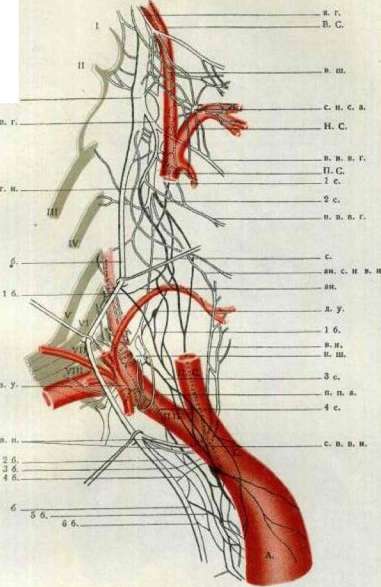
Figure 17. Scheme of the branching of the sympathetic and vagus nerves of the right side (schematic, preparation by L. A. Orbeli): 6. - vagus nerve; g. - sympathetic nerve; t 6. - 6 6, - branches to the heart from the vagus nerve; 7 s. - 4 s. - branches to the heart from the sympathetic nerve; s. sh. - superior cervical ganglion; i. sh. - inferior cervical ganglion; st. g. - stellate ganglion; I-VIII - anterior branches of the spinal nerves; s. g. b. - superior cervical ganglion of the vagus nerve; s. l. - superior laryngeal nerve; r. n. - recurrent nerve; i. r. a. l. - internal branch of the superior laryngeal nerve; e. r. a. l. - external branch of the superior laryngeal nerve; c. r. s. l. n. - cardiac branches of the superior laryngeal nerve; c. r. r. n. - cardiac branches of the recurrent nerve; c. p. e. c. a. - plexus of the external carotid artery; p. s. a. - plexus of the subclavian artery; s. g. - stellate ganglion; v. p. n. - vagus-pulmonary trunk; A. - aorta; b. a. - brachiocephalic artery; R. C. - right common carotid artery; R. S. - right subclavian artery; E. C. - external carotid artery; I. C. - internal carotid artery; a. g. - accessory ganglion; c. s. n. a. r. n. - connection of the sympathetic nerve and the recurrent nerve. Vegetative Nervous System VI
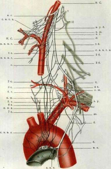
Fig. 18. Cardiac branches of the sympathetic and vagus nerves of the left side (semi-schematic, after L. A. Shinkarev). v. - vagus nerve; s. - sympathetic nerve; r. v. - branches to the heart from the vagus nerve; r. s. - branches to the heart from the sympathetic nerve; v. c. - superior cervical ganglion (indicated by a dotted line); m. c. - middle cervical ganglion; i. c. - inferior cervical ganglion; s. g. - stellate ganglion (indicated by a dotted line); I-VIII - anterior roots of the cervical nerves; v. g. - fusiform ganglion of the vagus nerve; n. l. - superior laryngeal nerve; l. n. - laryngeal nerve; i. r. l. n. - internal branch of the superior laryngeal nerve; e. r. l. n. - external branch of the superior laryngeal nerve; c. b. s. l. n. - cardiac branch of the superior laryngeal nerve; c. b. i. l. n. - cardiac branch of the inferior laryngeal nerve; p. c. a. - plexus of the common carotid artery; l. s. a. - loop of the subclavian artery; n. h. - hypoglossal nerve; a. c. - carotid artery; A. - aorta; A. P. - pulmonary artery; S. - subclavian artery; L. C. - left common carotid artery; L. S. - left subclavian artery; E. C. - external carotid artery; I. C. - internal carotid artery; v. s. - connection of the pulmonary and subclavian loops and the inferior cervical ganglion; r. r. n. - branches of the recurrent nerve. Fig. 19. Nerve plexus of the heart (after A. A. Shinkarev). a. - aorta; p. a. - pulmonary artery; v. c. s. - superior vena cava; v. c. i. - inferior vena cava; H. B. - His-Tawara bundle; n. g. - cardiac nerve ganglion (after A. A. Shinkarev); c. - cardiac plexus, located in the thickness of the wall of the heart, between the atria and the ventricles; r. - cardiac plexus in the wall of the heart, between the atria and the ventricles; s. p. - sympathetic plexus; v. - vagus nerve; r. c. - cardiac branches; l. c. - left cardiac nerve; r. c. - right cardiac nerve; s. c. - superior cardiac nerve; i. c. - inferior cardiac nerve; p. c. - pulmonary plexus; a. p. - aortic plexus; c. p. - coronary plexus; r. c. a. - right coronary artery; l. c. a. - left coronary artery; v. c. - cardiac veins; n. - nerves of the heart; s. n. - sympathetic nerves; v. n. - vagus nerves. Fig. 20. Nerve plexus of the bladder (after A. A. Shinkarev). v. - bladder; u. - ureter; n. v. - nerves of the bladder; n. p. - pelvic nerves; p. p. - pelvic plexus; s. n. - sympathetic nerves; g. - ganglion. Fig. 21. Nerve plexus of the rectum (after A. A. Shinkarev). r. - rectum; n. r. - nerves of the rectum; p. p. - pelvic plexus. Fig. 22. Nerve plexus of the uterus (after A. A. Shinkarev). u. - uterus; n. u. - nerves of the uterus; p. p. - pelvic plexus. The sheath of individual long trunks; they are composed of branches of the ganglia only in the region of its fornices and immediately ascend to the uterus. Nerves of the uterus. The plexus for the uterus also departs from the medial section of the inferior hypogastric plexus (see figure 6) and, being part of the upper segment of the vaginal plexus, lies between the lateral surface of the fornices and part of the uterine artery at the place of its approach to the uterus. The plexus is composed of several large ganglia and a number of small ones, the boundary of which does not extend beyond the boundaries of the fornices. The ganglia located in the noted area (see figure 12) send out small trunks, which lie on the sides of the body of the uterus and can be divided into a lateral group, from which one or several branches go to the ovary (these branches lie in the distal segment of the broad ligaments) and branches for the tubes, lying at the tubes themselves, in the thickness of the mesovarium, and a medial group—a group of branches intended for the uterus itself. The latter group consists of a series of branches that ascend along the lateral, anterior, and posterior surfaces of the body of the uterus and anastomose with the already noted branches departing from the ganglia of the vaginal fornices (see figure 3; Zhuravlev). Just as the nervous system of the vagina is composed of a network of ganglia, so the nervous system of the uterus represents, thanks to the anastomoses, a network of trunks located along its longitudinal axis, which lie on the surface of the body of the uterus and also penetrate its walls in the direction from bottom to top (see fig. 12).
Vegetative Nervous System.
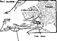
Vorobyov. Anatomy of vegetative centers. Central vegetative apparatuses consist of complexes of ganglionic cells and fibers embedded in the corpus striatum and the diencephalon, midbrain, medulla oblongata, and spinal cord. 1. Corpus striatum. Based on the research of Dresel and Lewy, the highest vegetative center, from which an impulse is conducted to the vegetative nuclei of the diencephalon, is the corpus striatum. According to comparative anatomical data (Kappers), the globus pallidus, which is part of the corpus striatum, is its phylogenetically oldest part (palaeostriatum), while the nucleus caudatus and putamen are newer (neostriatum). The neostriatum, according to Blumenau, develops from a thickening of the lower wall of the lateral vesicle, i.e., genetically it appears to be a modified part of the cerebral cortex and matures by the 6th month of life. The palaeostriatum is fully formed at birth and, based on phylogenetic data, is close to the substantia nigra (Sano). Under a microscope, the neostriatum contains, besides neuroglia, many ganglionic cells resembling cortical cells with short, branched axons, and a smaller number of round multipolar cells with long neurites that transition into bundles running through the putamen and heading toward the globus pallidus [see Vol. II (art. 639-640), Table II]. In the pallidum, there are relatively few ganglionic cells; they are significantly rounder, with long processes; there are many nerve fibers entering these nuclei. Spatz discovered in the globus pallidus: iron in the cells, lipoids in the neuroglia, and lime in the arteries and around the capillaries. This nucleus is very labile with respect to certain poisons (hydrocyanic acid, illuminating gas, carbon monoxide, and manganese). All axons of the neostriatum cells terminate in the globus pallidus. Almost all centrifugal fibers of the palaeostriatal system, which connects it with other nuclei of the nervous apparatus, depart from the globus pallidus. Grinshtein, while destroying the nuclei of the nucleus caudatus, discovered that all pathways originating in this nucleus do not go further than the globus pallidus. Furthermore, the majority of fibers starting in the putamen also terminate there. Apparently, the neostriatum is an organ of a higher order, communicating with lower centers (including vegetative ones) only through the pallidum and regulating, and sometimes inhibiting, the function of the pallidum. Connection of the pallido-striatal system with other centers of the nervous system. From the inner side of the corpus striatum, a few fibers depart to the thalamus—strio-thalamic fibers. Conversely, powerful bundles pass from the thalamus to the corpus striatum through the anterior peduncle of the thalamus in a central direction. A large part of the central pathways from the apex and lower side of the pallidum to the subthalamic region are the so-called hypothalamic fibers. The latter include: 1) nerve bundles that are part of Forel's lenticular fasciculi and run under the name of strio-rubral fibers to the red nucleus and further to the posterior commissure and its nuclei (O. and C. Vogt); 2) Greving's pallido-infundibular tract, connecting the globus pallidus with the mamillo-infundibular nucleus (Malone et al.). Ramon y Cajal calls the latter the "nucleus of Forel's field," and Lewy calls it the "periventricular nucleus"; 3) Dejerine's strio-Luysian fibers, heading to the body of Luys through the ansa lenticularis; 4) strio-nigral fibers to the substantia nigra; 5) fibers forming the ansa lenticularis and reaching the lower section of the thalamus (Blumenau); 6) the fronto-supraoptic tract to the supraoptic nucleus. Of other centrifugal pathways, nerve bundles to the cerebellum through the inferior olive and to the superior colliculus through the posterior commissure are currently known. Regarding the connection of the pallido-striatal system with the cortex, there is disagreement. Most authors deny this connection. Dejerine and Flechsig assert that the cortex is connected by nerve pathways directly to the globus pallidus. Ramon y Cajal discovered collaterals to the neostriatum from descending cortical fibers but did not find a direct connection of the latter with subcortical ganglia and vegetative nuclei. On the other hand, daily observations register the dependence of the vegetative nervous system on the psyche ("blushing," "bear disease" [diarrhea due to nervousness], hyperhidrosis during emotional experiences, "banker's sugar," etc.). Smirnov, based on physiological experiments, comes to the conclusion that the cortex influences vegetative centers in the medulla oblongata. Morphine, as well as extirpation of the cortex, destroys this influence. 2. Diencephalon. The diencephalon develops from the anterior cerebral vesicle. The cavity of the latter turns into the third ventricle, the walls of which develop strongly and form the main mass of the diencephalon with its nuclei. On the inner side, the sulcus of Monro is marked, which divides the diencephalon into the upper section—the thalamus, the region of the geniculate bodies (metathalamus), and the lower section (hypothalamus). The latter is located at the base of the brain and is bounded in front by the optic tract and anterior commissure, and behind by the cerebral peduncles. It includes the anterior perforated substance, the tuber cinereum with the infundibulum, and the mamillary bodies, which are located freely on the basal part of the brain. In addition, the hypothalamic region contains the hypothalamic body (body of Luys) lying dorsally to the latter. These regions form earlier than others and are phylogenetically very old. In lower vertebrates, they play the role of higher regulatory centers. Based on phylogenetic, experimental, and morphological data, it can be asserted that in the diencephalon there is a central nervous apparatus for the regulation of vegetative functions. I. Cytoarchitectonics and morphology of cellular forms.—A. Region of the optic chiasm. In the region of the optic nerve decussation, there is a cell group—the supraoptic nucleus. It lies dorsally in relation to the optic tract, accompanies it for a considerable distance, and closely adjoins its lateral side. Some cells surround the optic tract from the medial and ventral sides, and are also scattered along the tuber cinereum. According to Röthig, this nucleus appears phylogenetically very old. The cellular elements of the supraoptic nucleus are quite large, massive, pine-cone shaped, with two or three thick and long processes, and they impregnate poorly by the Bielschowsky method. Throughout the tuber cinereum, small cells are distributed, sometimes as single specimens, sometimes in whole groups: the central substantia nigra. A large number of them are observed in the oral part of the tuber cinereum. These cells have two or three processes, their nuclei are very small, surrounded by a narrow rim of protoplasm, and their shape is round, oval, oblong, or rarely polygonal. Their morphological similarity to the cells of the vegetative nucleus of the medulla oblongata and to the cells of Jacobson's nuclei in the spinal cord, according to Greving, serves as proof that they carry out vegetative functions. Among the central gray substance, there are several more nuclei. The anterior convexity of the tuber cinereum is formed by the tuber nuclei. On frontal sections, they represent three cell groups that increase in the lateral direction, with the lateral group extending in the caudal direction to the mamillary bodies. The cellular elements of this nucleus are small, with 3-4 processes, a large nucleus, and a narrow belt of protoplasm. The tuber nuclei are phylogenetically young (Spiegel, Zweig) and therefore it is unlikely that they are connected with vegetative functions. In various places of the tuber cinereum, throughout the area to the mamillary bodies and the bundle of Vicq d'Azyr, around the tuber nuclei, encircling the column of the fornix, large multipolar cells with long, richly branched processes appear in groups. Malone designates them as the mamillo-infundibular nucleus. Based on the degeneration of this cell group upon destruction of the vegetative nucleus of the medulla oblongata, it can be considered, apparently, as a direct center of the sympathetic and parasympathetic system. Greving describes two more nuclei in the tuber cinereum: the pallido-infundibular nucleus and the interfornicate nucleus. The pallido-infundibular nucleus (named so because it is connected by nerve bundles of the pallido-infundibular tract with the globus pallidus) occupies the space between the optic tract, the tuber nucleus, and the mamillo-infundibular nucleus. Morphologically, these nuclei are undoubtedly independent units. Their cells appear extremely characteristic, unlike the ganglionic cells of other parts of the hypothalamus. Their body is massive, oval, or oblong in shape. They give off powerful processes that impregnate poorly by the Bielschowsky method. Since these cells are similar to those in the globus pallidus, Greving raises the question of the functional connection of the pallido-infundibular nuclei with the globus pallidus. Between the middle and inferior tracts of the fornix, in the caudal part of the tuber cinereum, there is a group of bipolar cells that impregnate well with silver, with long, thin processes: the interfornicate nucleus. In the region of the mamillary bodies, the medial ganglion and lateral ganglion are distinguished. Greving distinguishes two nuclei in the medial ganglion—the magnocellular nucleus and the parvocellular nucleus.
He calls the lateral ganglion the nucl. mamillaris cinereus. The n. magnocellularis is located in the ventro-medial part of the corp. mamillaris. It consists of densely packed large multipolar cells. Dorso-laterally from this group is located the nucleus parvocellularis, represented by sparsely arranged small cells with short processes. Laterally from these cell groups lies the nucleus mamillaris cinereus, consisting of small, sparse, pinecone-shaped elements similar to the cells of the substantia grisea centralis. Closely adjoining the latter, on the side, is the nucl. intercalatus Malone—very small nuclei consisting of delicate multipolar cells.
B. The region located under the tubercles. In the middle between the commissura anterior and the floor of the diencephalon, in the wall of the third ventricle, near its ependyma, there is the nucl. paraventricularis. The cells of this nucleus, lying in a densely intertwined network of fibers, are very similar to the ganglion cells of the nuclei supraoptici. In the medial plane, the nucl. paraventricularis extends up to the region of the chiasma. Its bundles at the bottom, together with the tr. supraopticus inferior, enter the tuber cinereum. In the middle of their extent is an additional group of cells. Greving calls the latter the nucleus paraventricularis accessorius, and the nerve bundles—the tractus paraventricularis cinereus. In the wall of the third ventricle there are two more nuclei, named by Malone the nucl. reuniens and the nucl. paramedianus. The nucl. reuniens consists of large multipolar cells, is placed in the commiss. intermedia, and protrudes into both sides of the thalami. The nucl. paramedianus lies dorsally and medially from the nucl. reuniens. Its unipolar and bipolar cells are small, arranged closely, have a pinecone shape, and are very similar to the elements of the central gray substance.
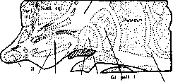
In the caudal part of the subthalamic region, laterally from the bundle of Vicq d'Azyr, outside the substantia nigra, there is a cluster of gray matter—the corpus Luysii (corpus subthalamicum)—densely enveloped and permeated by fibers, which impregnates well with silver; the multipolar cells of this formation have several processes each. Based on phylogenetic data and the histological picture, Greving considers the following cell groups of the diencephalon to be vegetative nuclei (see figures 26 and 27): 1) Substantia grisea centralis; 2) Nucleus mamillaris cinereus; 3) Nucleus interfornicatus; 4) Nucleus paramedianus; 5) Nucleus supraopticus; 6) Nucleus paraventricularis; 7) Corpus Luysii.
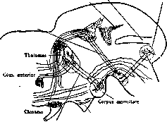
II. System of pathways. A. System of pathways of the corpora mamillaria (see figure 28). From the bulbus olfactorius to the corpus mamillare passes a system of thin fibers—the basal olfactory bundle of Edinger and Wallenberg; some of these fibers reach the gangl. interpedunculare.* In the corpus mamillare ends the anterior column of the fornix, originating in the Ammon's horn. A part of the fibers of the columna fornicis forms a decussation with the opposite column and extends to the tegmentum. From the gangl. medullare (nucl. magnocellulare) emerges the bundle of Vicq d'Azyr, which soon divides into two bundles (Ramón y Cajal and Kölliker): 1) tr. mamillo-thalamicus, directed toward the nucl. anterior thalami, and 2) tr. mamillo-tegmentalis (Gudden's tegmental bundle), reaching the dorsal ganglion of the tegmentum behind the corpora quadrigemina. From the gangl. laterale (nucl. parvocellulare) emerges the pedunculus corpor. mamillaris. In it, Wallenberg, along with efferent fibers to the gangl. tegmenti profundum, proved the existence of afferent fibers from the medial lemniscus. * The termination of the olfactory bundle in the corpus mamillare indicates the connection of the latter with olfactory functions (Greving).
B. System of pathways of the floor of the diencephalon (see figure 29). In the floor of the diencephalon, near the chiasma, are located: 1) Gudden's commissure, according to Edinger—decussatio supraoptica ventralis, 2) decussatio supraoptica dorsalis, 3) Meynert's commissure. The significance of the first two bundles has not yet been finally clarified; as for Meynert's commissure, Economo and Karplus found (in the monkey) that its fibers emerge in front of the red nucleus from the region of the lemniscus, curve laterally around the cerebral peduncle, run under the optic tract before and after the decussation, and end in the nucl. commissurae Meynerti, lying at the outer edge of the end of the peduncle. Wagner, however, classifies the comm. Meynerti, based on morphological data, to the globus pallidus.*
Through the dorsal region, crossing the nucl. supraopticus and ending in the lower part of the thalami, passes a bundle named by Greving the tract. supraopticus thalamicus. Pathways of the nuclei supraoptici: 1) tr. supraopticus superior; its fibers, going in a dorsal direction to the thalamus, take part in the formation of its lower part; 2) tr. supraopticus hypophyseus or tr. supraopticus inferior of Greving, also known as the fasciculus hypophyseus of Pines. This bundle from the region of the nuclei supraoptici runs through the hypophyseal part of the tuber cinereum to the posterior part of the hypophysis, where it forms a dense plexus (Greving, Pines).* The nucl. supraopticus receives, in addition, fibers (as Greving asserts) from the ansa peduncularis. He calls this bundle the tr. fronto-supra-opticus.
Region of the tuber cinereum. From the ansa peduncularis emerge fibers of a bundle named by Greving the tr. fronto-tuberalis. This bundle extends dorsally past the nucl. supraopticus and encompasses the dorsal and ventral convexities of the nucl. tuberis. In addition, the nucl. tuberis are connected by nerve bundles with the lower part of the thalami, and in the caudal direction from them departs the tr. tuberis, adjoining the dorsal longitudinal bundle; in the medial region from the cells of the central gray matter. * According to Marie and Cajal, there is a connection between the commissure and the nucleus supraopticus.
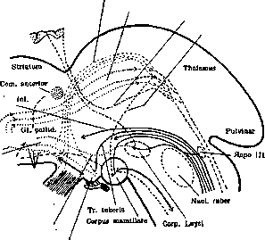
Frontal tract, supraoptic tract, fronto-tuberal tract, ansa peduncularis, supraoptic tract, superior supraoptic tract to the hypophysis, ansa lenticularis, tuber cinereum. Figure 29. Substances extend in the diencephalic floor, in a caudal direction, dorsally under the corpora mamillaria; fibers of the tr. substantiae griseae infundibularis (Greving). From the nucl. paraventricularis, together with the tr. supraopticus inferior, the tr. paraventricularis cinereus enters the tuber cinereum. The globus pallidus is connected to the infundibulum by means of the tr. pallido-infundibularis. Both Luys' bodies are connected by the Forel commissure, or commissura hypothalamica posterior, located behind the corpora mamillaria. By means of the fibrae perforantes, the corpus Luysi is connected to the globus pallidus. Furthermore, the ansa lenticularis passes through this nucleus, connecting with the globus pallidus and the nucl. ruber. Medial region. Within the subst. reticularis hypothalamicae, fibers of the tr. reticularis hypothalamici of Greving are formed. They adjoin the tr. mamillo-tegmentalis, bypass the dorso-medial surface of the nuclei rubri, and head toward the midbrain. Lateral region. According to the studies of Edinger and Riese, fibers from the striatum to the substantia nigra—the tr. strio-peduncularis—pass through the lateral region of the regio subthalamica. The structure of the thalami optici is extremely complex and has not yet been precisely clarified. It is sufficient to point out that 40 nuclei are described in the monkey brain. Undoubtedly, the true anatomo-physiological significance of the thalamus will be established upon studying the connection of these nuclei with other parts of the brain. 3. Midbrain. On the border between the base and the tegmentum of the midbrain lies a layer of substance—the substantia nigra Soemmeringi. The cells of the latter are of diverse shape and contain black pigment only in humans. According to Bauer, they are arranged in the following three groups: outer, intermediate, and inner. Their axons, according to various authors, go in two directions: into the cerebral peduncle and into the tegmental region. Fibers from the striatum terminate in the substantia nigra. Experimental studies have proven its connection with the cortex, the lemniscus, and Luys' body. Economo experimentally localized a general coordinating center for the entire act of eating in the inner sections of the upper half of the substantiae nigrae. On both sides of the raphe is the nucleus ruber, from which the Monakow tracts (fasc. rubro-spinalis) depart, forming a decussation immediately upon exiting the nucleus. Behind and somewhat lateral to the red nucleus passes the dorsal longitudinal fasciculus (fasc. longitudinalis dorsalis). Central impulses from vegetative centers to the motor cells of peripheral neurons pass along the Monakow and dorsal longitudinal fasciculi. Directly behind the fasc. longitudinalis dorsalis, in the region of the anterior colliculus, ventral to the Sylvian aqueduct (in the oculomotor zone), there is a cluster of two types of cells: 1) large multipolar ganglion cells with long processes, similar to the cells of the anterior horns of the spinal cord, and 2) small, pine-cone-shaped cells with a narrow strip of protoplasm and bipolarly arranged processes. They are similar to the cells of the nuclei supraoptici. The main nuclei of the oculomotor nerve consist of cells of the first type. The anterior cells penetrate into the thickness and up to the anterior surface of the fasc. longitudinalis dorsalis. From this nucleus, forming a decussation between the nuclei rubri, the bundles of the n. oculomotorii pass through them. Closer to the oral part of the main nucleus are smaller cellular groups. Inside and behind it are located the paired Edinger-Westphal nuclei. They consist of cells of the second type, among a dense network of nerve fibers, and are divided into two parts—inner and lateral. Between both main nuclei lies the nucleus medianus (central nucleus of Perlia). It consists of cells of the first type. The Edinger-Westphal nuclei innervate the smooth musculature of the eye, the mm. ciliaris and sphincter iridis, through the gangl. ciliare; consequently, both morphologically and functionally, they belong to vegetative formations. A portion of the fibers of the third pair, contained in the ganglion ciliare, belongs to the cranial parasympathetic system. 4. Medulla oblongata. On the floor of the fourth ventricle, lateral to the nucleus of the hypoglossal nerve, macroscopically identifiable as the fovea vagi, is located a group of cells known as the dorsal nucleus of the vagus nerve or (according to Marinesco) the sympathetic nucleus of the X pair. Through experimental and physiological studies, Molhant proved that retrograde degeneration is observed in this nucleus upon resection of various visceral organs (stomach, lungs, liver, etc.). Further research by the Kraus school showed that all organs with smooth musculature, visceral organs, and endocrine glands are projected in this nucleus, and they are located in reverse order compared to their localization in the body, i.e., from top to bottom. The cells of this nucleus differ morphologically sharply from other nuclei of the vagus nerve. Among them are many bipolar, pear-shaped, or pine-cone-shaped cells; in size, they are significantly smaller than the cells of other nuclei of the medulla oblongata. In its structure, the nucleus resembles the sympathetic nuclei of the spinal cord. Levi's experiments showed that after removal of the first cervical ganglion, a portion of the small ganglion cells in the anterior part of the dorsal nucleus (“small cells” of Ramón y Cajal) dies, and they proved the relationship of this nucleus to the sympathetic system. The small ganglion cells that do not change in this experiment, according to Dresel, are the initial cells of the sympathetic fibers, which extend through the cervical spinal cord, pass into the sympathetic trunk, and finally reach the peripheral sympathetic system. Physiological and morphological data (the presence of sympathetic and parasympathetic cells) enabled the Kraus school to call this nucleus the “vegetative nucleus of the medulla oblongata.” Brugsch, Dresel, and Levi definitively established the vegetative significance of this nucleus. By destroying it, they obtained degeneration in the nuclei of the dorsal region of the III ventricle (including the nucl. periventricularis—Levi). Apparently, this degeneration occurred in a retrograde order due to the destruction of fibers coming from the regio subthalamica, likely as part of a conglomerate of association pathways known as the fasc. longitudin. dorsalis (according to Kohnstamm). Transection of the vagus nerve in various places showed that the vegetative nucleus also contains cells belonging to the vagus nerve. Furthermore, the n. vagus has another motor nucleus—the nucl. ambiguus, located dorsal to the olive, between the subst. reticularis and the subst. gelatinosa. The large multipolar cells of this nucleus are very similar to the cells of the anterior horns of the spinal cord. Fibers innervating visceral organs originate from the vegetative nucleus, with the exception of the striated musculature of the pharynx and larynx, which receive branches from the nucl. ambiguus. The sensory fibers of the n. vagi (fasc. solitarius) terminate in the nucl. vagi sensibilis (in the nucl. tr. solitarii—descending branch, and in the nucl. alae cinereae—ascending branch). In addition to the dorsal nucleus, there are other cellular clusters in the medulla oblongata that have vegetative significance. According to data confirmed by Brugsch, Dresel, and Levi, an increase in the secretion of saliva and water occurs upon damage to cells located in the substantia reticularis. Here, one can detect large multipolar cells connected to the corpora mamillaria (Dresel) and lower nuclei, named by Kohnstamm the nucleus salivatorius superior et inferior. The substantia reticularis consists of association fibers going up to the brain and fibers descending to the upper part of the spinal cord. According to Edinger, pathways pass through the subst. reticularis that associate the activity of the nuclei of the facial, vagus, and phrenic nerves, as well as fibers coordinating cardiac and respiratory activity. The substantia reticularis is located at the medial edge of the substantiae gelatinosae trigemini, medio-ventral to the corpus restiforme, and dorsal to the nuclei of the n. facialis and the nucleus of the lateral columns. This region of the medulla oblongata constitutes the bulbar part of the parasympathetic system. From the nucl. salivatorius superior, lying dorso-medial to the nucl. facialis, emerge vasodilator and salivary-secreting fibers. As part of the n. intermedii and then the chordae tympani and n. lingualis, as preganglionic branches, they head toward the gangl. submaxillare. The postganglionic fibers of this ganglion terminate in the gland. submaxillaris and ganglion sublinguale. Small ganglia are scattered along the course of these fibers. Salivary-secreting and vasomotor fibers are also contained in the IX pair.
Emerging from the medulla oblongata, from the inferior salivatory nucleus, as part of the glossopharyngeal nerve to the petrous ganglion, and further into the tympanic nerve and the lesser superficial petrosal nerve as preganglionic fibers, they are directed to the otic ganglion and from the latter, as part of the auriculotemporal nerve and parotid branches, to the parotid gland. Secretory fibers for the salivary glands emerge from the medulla oblongata together with the roots of the VII pair. The nucleus of these nerves has not yet been precisely determined. It is located * According to the research of Ramón y Cajal, only the larger cells of the dorsal nucleus send their fibers into the vagus nerve. near the cells constituting the nucleus of the facial nerve. As part of the greater superficial petrosal nerve, the preganglionic fibers of these nerves terminate in the sphenopalatine ganglion. Postganglionic branches, as part of the maxillary nerve (from the trigeminal nerve) and the zygomatic nerve, and through the anastomoses of the latter with the lacrimal nerve, reach the lacrimal gland. Parasympathetic innervation of the heart, trachea, and lungs, the gastrointestinal tract from the esophagus to the transverse colon (based on physiological data), digestive glands, liver, pancreas, and endocrine glands is carried out by the vagus nerve. Thus, in the midbrain and medulla oblongata, from the nuclei of the III pair—the dorsal nucleus and the superior and inferior salivatory nuclei—and the nuclei of lacrimal secretion—arise the centrifugal fibers of the parasympathetic cranial-bulbar system. Furthermore, in the medulla oblongata, in the dorsal nucleus, the upper (cervical) part of the fibers of the sympathetic system originates. Through the dorsal longitudinal fasciculus, the vegetative nuclei of the midbrain, diencephalon, and corpus striatum send sympathetic and, apparently, parasympathetic bundles to the medulla oblongata and spinal cord. In the reticular formation, the association of individual vegetative nuclei is carried out. 5. Initial nuclei of the sympathetic nervous system in the spinal cord. By transecting the lower sections of the spinal cord and obtaining retrograde degeneration in the subthalamic region, Dresel established that the diencephalon is connected by centrifugal pathways with the spinal cord. In the latter are located cell groups situated in the transitional zone between the anterior and posterior horns, considered as the initial nuclei of preganglionic sympathetic nerves (Stilling's lateral horn group, Clarke's intermediolateral tract). Gaskell, Sherrington, Langley, and Herring determined that these nuclei are connected with the sympathetic nerves. Jakobson distinguishes three sympathetic nuclei: 1) nucleus sympathicus lateralis superior, located from the VIII cervical to the III lumbar segment; 2) nucl. sympathicus lateralis inferior, lying in the angle between the posterior and lateral horn from the II sacral vertebra to the V; 3) nucl. sympathicus medialis inferior s. lumbo-sacralis, beginning at the fourth lumbar vertebra and extending to the fifth sacral. Below, it partially merges with the lateral nucleus, and in the middle of the sacral region, it breaks up into several small groups occupying predominantly the inner edge of the anterior horn.
B. Mogilnitsky. P. Physiology of the vegetative nervous system. In view of the existence of significant diversity in the use of classification criteria for nerve fibers, in the use of terminology, and in the definition of the concept of the vegetative nervous system, the terminology and classification of the vegetative nervous system necessary for understanding this section are briefly presented below. All centrifugal nerve fibers can be divided into two large groups: one consists of motor fibers of striated muscles, the other of all other centrifugal fibers, including the accessory nerve fibers of striated muscle tissue discovered by Boeke. The first group, following Langley, is designated by the word "somatic," the second by the word "vegetative" or "autonomic" (the latter two terms are used as synonyms). By vegetative or autonomic nervous system in this article is meant the entire aggregate of vegetative fibers with their ganglia, trophic centers, and those nuclei of the central nervous system under whose control they are. Basic differences between vegetative and somatic fibers. Somatic fibers are processes of peripheral neurons having cell bodies in the anterior horns of the gray matter of the spinal cord or in analogous sections of the medulla oblongata and midbrain, and they extend to the periphery up to the working tissue. Vegetative fibers, however, are processes of penultimate neurons, with cell bodies in the lateral horns of the gray matter of the spinal cord or their analogs in the medulla oblongata and midbrain. At the periphery, they do not reach the working tissues directly, but are interrupted in more or less significant peripheral nerve ganglia, entering into connection with the peripheral nerve cells located in these ganglia, the processes of which, already within the branches of the peripheral ganglia, reach the innervated tissues. The entire path, thus, breaks down into preganglionic (pre-nodal) and postganglionic (post-nodal) segments. Hence the name "ganglionated nervous system" used by some authors. According to Gaskell's conception, every reflex arc consists of at least three neurons: 1) afferent, or bringing impulses from the periphery, 2) connector, or connecting, and 3) effector, or executive (carrying out). In the somatic system, the connector neuron with all its processes is located inside the central nervous system, where the body of the effector neuron is also located. In the vegetative system, however, the effector neurons have migrated to the periphery, closer to the tissues, as a result of which the processes of the connector neurons have turned out to be extended to the periphery in the form of preganglionic fibers. The connection points (synapses) between preganglionic fibers and ganglionic cells are characterized by the presence of a peculiar connecting apparatus or substance (junctional tissue, or Langley's "receptive substance"), which is very sensitive to nicotine, which first produces phenomena of excitation (and consequently, of the activity of the innervated organ), and then paralysis, expressed by the cessation of the transmission of impulses from the preganglionic segment to the postganglionic one. Thus, nicotine poisoning can serve as a means both for detecting peripheral interruptions and for determining the localization of the interruption in one or another ganglion (see below - nicotine method), as well as for establishing the very fact of vegetative innervation of one or another organ (stimulation of ganglia with nicotine). The presence of an obligatory interruption in peripheral ganglia, however, is revealed not only by the indicated relation to nicotine, but also by observations on the degeneration of nerve fibers after separation from cell bodies (trophic centers): the cutting of spinal and cranial nerves containing autonomic (vegetative) fibers leads to their degeneration only up to the peripheral nerve cells, whereas motor fibers of skeletal musculature degenerate up to the intramuscular (hypolemmal) terminal apparatuses. Myelinated vegetative fibers are significantly thinner than somatic ones. General construction of the vegetative nervous system. While somatic fibers are characterized by segmental exit throughout the entire length of the spinal cord and medulla oblongata and by regular metameric distribution at the periphery, vegetative fibers depart only from specific sections of the central nervous system, separated from each other by a more or less significant number of segments that do not give rise to vegetative fibers. The sections giving rise to vegetative fibers are: 1) the region of the corpora quadrigemina, or midbrain (vegetative fibers of the oculomotor nerve); 2) the medulla oblongata (vegetative fibers of the facial nerve, glossopharyngeal nerve, vagus nerve); 3) the thoracolumbar section of the spinal cord from the I-II thoracic to the III-IV lumbar segments (vegetative fibers entering via the white communicating rami into the sympathetic trunk) and 4) the lower sacral part of the spinal cord from the III to IV sacral segments (vegetative fibers of the sacral plexus, forming the erigent nerve or pelvic nerve). The vegetative nervous system, according to the place of exit of the fibers from the central nervous system, is subdivided into three separate systems: 1) the cranial, with its midbrain and bulbar parts, 2) the thoracolumbar, and 3) the sacral. For a number of reasons, it is accepted to consider the cranial and sacral systems as two separate sections of one and the same system, designated by the term "parasympathetic system" and contrasted with the thoracolumbar, or "sympathetic," system. These reasons consist of the following: all fibers of the thoracolumbar system, having exited the spinal canal as part of the anterior roots of the II thoracic to III lumbar nerves, enter the boundary sympathetic trunks via the white communicating rami: they have an interruption at the cells of one or another sympathetic ganglion (vertebral or paravertebral) and, through the mediation of postganglionic fibers departing from here, innervate all regions of the body without exception, thus acquiring the significance of a universal innervation apparatus. The fibers of the cranial and sacral sections have no relation to the sympathetic chain and only at the periphery mix with sympathetic fibers in one or another plexus. The interruption of preganglionic fibers of the cranial and sacral systems occurs in separate ganglia located far at the periphery, often on the surface or even in the thickness of the innervated organs (ciliary ganglion, otic ganglion, sphenopalatine ganglion, intracardiac ganglia, cells of Auerbach's and Meissner's plexuses, large quantities of ganglia and individual nerve cells in the thickness of the submandibular salivary gland, uterus, urinary bladder, rectum). The areas of influence of the parasympathetic system are comparatively limited: the midbrain part of the cranial section innervates exclusively the eyeball, the bulbar part innervates the heart and all organs embryologically originating from the foregut (digestive canal, with the exception of the lower part of the colon and rectum, all separate digestive glands, bronchi, and lungs), and finally, the sacral system innervates the pelvic organs, which develop embryologically from the hindgut, and the external anogenital apparatus. Thus, there are regions of the body receiving only sympathetic vegetative innervation, and regions with dual vegetative innervation—sympathetic and parasympathetic. A further difference between the sympathetic and parasympathetic systems lies in their different relation to poisons and hormones. The sympathetic nervous system, which stands in embryological kinship with the chromaffin system (see), also stands in functional connection with it; the product of the adrenal medulla and other chromaffin cells—adrenaline—excites the connection points of all sympathetic fibers with effector organs, thus causing all peripheral sympathetic effects. This phenomenon is characterized by the words: adrenaline is a sympathomimetic poison, and the connection points of organs and sympathetic endings contain an adrenalinotropic receptive substance. Besides adrenaline, the poisons tetrahydro-beta-naphthylamine and ephedrine are characterized by sympathomimetic action. Along with this, ergotoxin turns out to be a poison that paralyzes the connection points of all excitatory fibers of the sympathetic system with their effector organs; most inhibitory sympathetic and all parasympathetic fibers are not paralyzed by ergotoxin. On the contrary, all fibers of the parasympathetic system are characterized by the presence in the peripheral apparatuses of a receptive substance, cholinotropic, because all their effects can be caused by choline and its derivatives (for example, acetylcholine). All these agents have received the name "parasympathomimetic" agents. Besides choline and its derivatives, pilocarpine and physostigmine possess the same action.
The fairly common assertion that atropine paralyzes all endings of parasympathetic fibers is incorrect, because, on the one hand, a large number of parasympathetic effects do not suffer from atropine (example: the inhibitory influence of the nervus vagus on the smooth musculature of the cardiac sphincter), and on the other hand, atropine paralyzes all motor fibers of the smooth musculature of the intestine and all secretory fibers regardless of their belonging to the parasympathetic or sympathetic system (e.g., sweat glands), so that no generalizing rule regarding the influence of atropine on vegetative fibers can be established. The Auerbach and Meissner plexuses (Langley's enteric system, Müller's intramural system) must be assigned to a special division, although one that is subordinate to other vegetative divisions, but which possesses a significant degree of independence and does not fit into the above scheme. Finally, in the opinion of Leontovich, all peripheral tissues are permeated by a reticular nervous formation with peripheral nerve cells. This formation resembles the diffuse nervous system of lower organisms and, perhaps, is its rudiment. Vasodilator fibers occupy a completely special position. With the exception of a small number of vasodilators originating in the middle thoracic segments of the spinal cord, which are part of the sympathetic system and serve certain internal organs, including the heart and the mucous membrane of the buccal region, all other vasodilators leave the spinal cord not as part of the anterior roots, like all autonomic fibers, but as part of the posterior roots, and specifically the roots of those spinal cord segments that do not provide autonomic fibers but are dividing zones between the sympathetic and parasympathetic systems. Furthermore, it was discovered that there are no grounds for recognizing centrifugal fibers in the posterior roots with trophic centers in the gray matter and with a synapse in the periphery, and vasodilator effects must be attributed to "antidromic" conduction of impulses along the same afferent fibers that have their trophic centers in the spinal ganglia. Such an interpretation became all the more inevitable because a number of local vasodilator reactions were discovered in the periphery, which could only be explained as axon reflexes based on the branching of processes of spinal ganglion cells with the distribution of collaterals between receptor apparatuses, on the one hand, and vessels, on the other. These facts exclude the main mass of vasodilators from the autonomic system, since none of the basic rules proves applicable here. But, along with this, the vasodilators of the described group exhibit a relationship to acetylcholine characteristic of parasympathetic fibers. On the basis of this, and also in view of other particular cases of discrepancy between the morphological affiliation of certain fibers and their relationship to sympathomimetic or parasympathomimetic poisons, Frank proposes to abandon the morphological classification of fibers entirely and replace it with a physiological one, based on the presence of an adrenalinotropic or cholinotropic receptive substance. Frank even proposes the terms: "physiological sympathetic" and "physiological parasympathetic," to contrast them with the "morphological sympathetic and parasympathetic." This proposal is already being implemented by some clinicians, but, of course, it can bring nothing but confusion: one cannot assign ever new meanings to terms that have already become established for designating certain morphological relationships. No matter how important the new classification criteria may be, they cannot undermine the significance of the morphological classification. Most recently, Ken Kure and his colleagues showed that after cutting the posterior roots immediately above (more centrally) the spinal ganglia, only thick medullated nerves degenerate, while thin ones are preserved; in accordance with this, it turns out that in the spinal cord, certain cells between the anterior horn and the substantia gelatinosa undergo chromatolysis and degeneration. These thin fibers must terminate in the spinal ganglia. The authors consider them to be parasympathetic and assume that through them vasodilation is carried out, which occurs upon irritation of the posterior roots, and perhaps also the trophic and tonic influence of the posterior roots on voluntary musculature. If the data of the Japanese authors are confirmed, it will be possible to speak of three parasympathetic divisions and to recognize parasympathetic innervation of organs and tissues as being just as universal as sympathetic. The old data of A. S. Dogiel on the cellular composition of spinal ganglia make such a point of view quite probable. Functions. As is clear from the very definition of the vegetative nervous system, among vegetative fibers one must distinguish at least the following separate types: 1) secretory fibers of glands with external secretion, 2) secretory fibers of glands with internal secretion, 3) vasomotor fibers (vasoconstrictor and vasodilator), 4) excitatory and inhibitory nerves for the smooth musculature of internal organs, 5) centrifugal nerves regulating cardiac activity, 6) motor fibers of the smooth musculature of the integument and its appendages (hair, feathers, etc.), 7) motor and inhibitory fibers for special types of contractile cells (e.g., pigment cells of the skin of amphibians and fish, etc.), 8) accessory fibers for striated muscles (Boeke), 9) Timofeev fibers for receptor apparatuses, 10) centrifugal fibers entering from sympathetic ganglia via rami communicantes grisei into the spinal cord. Secretory nerves of glands with external secretion were discovered for the first time by Ludwig in 1851. He showed that cutting the chorda tympani stops secretion from the submandibular and sublingual glands, while irritation of the peripheral end of the chorda tympani leads to an abundant flow of saliva. Since at that time some authors were inclined to consider secretion as a process of filtration from the bloodstream, and in the same year 1851 Claude Bernard discovered vasomotor (constrictor) nerves, Ludwig had to present evidence that the fact he discovered was the result of the direct action of nerves on glandular cells and the work of them caused by this action. Ludwig brilliantly showed this in the same year 1851, by performing his classic experiment with simultaneous measurement of pressure in the arterial system and in the duct of the submandibular salivary gland. It turned out that during salivation caused by irritation of the chorda tympani, the pressure in the duct significantly exceeds the blood pressure in large arteries, and even more so in the capillaries from which the "filtration" of saliva was supposed to take place. Furthermore, it was shown that saliva differs sharply not only quantitatively but also qualitatively in its composition from the composition of the liquid part of the blood (for example, by its mucin content). These facts made Ludwig's point of view completely indisputable and led to the universal recognition of secretory nerves for the sublingual and submandibular salivary glands. Further complication in the doctrine of these nerves is connected with the discovery of the influence of the cervical sympathetic nerve on the secretion of the sublingual gland (Ludwig, 1856). Claude Bernard (1858) and Eckhard (1860) showed that while the chorda tympani produces an abundant secretion of liquid saliva poor in organic components, the sympathicus causes a very moderate secretion of saliva with a high content of organic substances. To explain these data, Heidenhain (1868) developed the doctrine of two types of fibers controlling the work of glands: "secretory" fibers, causing the excretion of water and salts by glandular cells, and "trophic" fibers, determining the production and excretion of specific organic products, for example, mucin, enzymes, etc. This point of view met with objections from a number of researchers, in particular from Langley, who attempted to explain the same facts from the point of view of a single secretory innervation accompanied by different vasomotor innervation: specifically, the chorda tympani produces abundant and liquid saliva supposedly because it contains secretory fibers in combination with vasodilators (Claude Bernard, 1858), and the sympathicus produces little saliva because the secretory fibers are accompanied by vasoconstrictors (Claude Bernard, 1851); the local asphyxia of glandular cells associated with vasoconstriction must be the cause of the increased production of the specific product (mucin). However, Babkin in the laboratory of I. P. Pavlov managed to show (1913) that if one observes the course of blood supply to the salivary gland in a dog under natural conditions of its work during reflex excitation from the oral cavity, then when using both acid and food agents as stimuli, the same active dilation of blood vessels and increase in blood supply to the gland is obtained, whereas in the first case liquid saliva is reflexively secreted, and in the second, saliva rich in mucin.
Thus, at the present time, it can be considered firmly established that there exist these two different but direct influences of the nervous system on the gland: on the production of specific products and on the excretion of water ("trophic" and "secretory"), a phenomenon very important for understanding the complex relationships between the character of stimuli and the character of the secretory work of glands. The activity of the submaxillary salivary gland is entirely determined by nervous influences. The humoral mechanism under normal conditions is completely suppressed and appears only in cases of cutting and degeneration of the chorda tympani in the form of a continuous, but very small in size, "paralytic" secretion (Claude Bernard, 1864), which disappears again upon regeneration of the nerve. Paralytic secretion usually occurs already in the first 24 hours, sometimes four hours after the cutting of the chorda. Cutting the sympathetic nerve in the first 2 days stops the paralytic secretion; in a later period, it does not affect it (Langley). This allows for the unification of the various explanations of paralytic secretion proposed by different authors. Specifically, in agreement with Bradford (1888), it can be assumed that constant inhibitory impulses are conducted along the fibers of the chorda tympani, preventing continuous secretion under the influence of humoral agents, and along the sympathetic nerve—impulses that increase the excitability of the gland. Furthermore, in agreement with Langley, one can accept a gradual increase in the excitability of the glandular elements that have lost their secretory innervation, developing over the course of several days. The establishment of the modern doctrine of the secretory nerves of the submaxillary salivary gland has, as we see, traversed a very complex path. It is understandable that even more difficulties arise where, along with the nervous mechanism in the work of the glands, the humoral mechanism reveals significant importance, and where, along with nervous apparatuses of an excitatory character, there exist nervous apparatuses of an inhibitory nature (gastric and pancreatic glands). Despite a number of observations that spoke for the dependence of the work of the gastric glands on the nervous system (Wilson, Philip, Frerichs, Claude Bernard, Brachet, Longet, and others); despite the assertions of Vulpian and Claude Bernard that they saw the secretion of gastric juice upon stimulation of the vagus nerves; despite, finally, the indications of Bidder and Schmidt regarding the secretion of gastric juice in animals at the mere sight of food and the indications of Richet regarding the reflex from the oral cavity to the gastric glands in humans—Heidenhain, who did so much to consolidate and refine our concepts of the innervation of salivary glands, still in 1886, i.e., 35 years after Ludwig's discovery of the first secretory nerves, asserted that "the nerves approaching the stomach from the outside do not possess any noticeable direct influence on the work of the gastric glands." Such a view was explained by the fact that Heidenhain failed to obtain secretion upon stimulation of the vagus nerves and the splanchnic nerves (splanchnici) and the medulla oblongata, just as Adrien, Schiff, and Pinkus did upon stimulation of the splanchnic nerves. Furthermore, Heidenhain, while observing a stomach pouch isolated according to his method, never had occasion to observe the so-called "psychic secretion," about which the observations of Richet, Bidder, and Schmidt spoke. But this very fact is the best proof of the secretory role of the vagus nerves: the secretion was absent precisely because the branches of the vagus nerves were cut in the Heidenhain blind pouch. The question remained in this state until 1889, when I. P. Pavlov and Shumova-Simanovskaya managed to develop a completely unique methodology and, with its help, prove the undoubtedly secretory significance of the vagus nerves. First of all, by combining esophagotomy with gastrostomy in the same dog, Pavlov and Shumova-Simanovskaya established, by means of "sham feeding," the existence of a reflex from the oral cavity to gastric secretion, a reflex that disappears without a trace upon cutting the vagus nerves. Proceeding from Nechaev's observations on the inhibition of gastric secretion under the influence of various sensory stimuli, Pavlov expressed the assumption that the failure of Heidenhain and other authors in attempts to induce secretion by stimulation of the vagus nerves depended on a number of unfavorable conditions of the vivisection environment, such as: strong pain stimuli, traumatization of the stomach wall, introduction of poisons into the blood, such as curare. To eliminate these inhibitory influences, Pavlov and Shumova-Simanovskaya applied the methodology developed a year earlier by Pavlov himself for the pancreas: a gastric fistula was placed in the dog in advance and one of the vagus nerves was cut below the diaphragm, then an esophagotomy was performed, and, finally, after the complete recovery of the animal, the second vagus nerve was cut in the neck and held on a thread; the experiment was set up only the day after the cutting, with the dog being in a completely normal environment. In all cases without exception, stimulation of the vagus nerve in the neck caused the secretion of gastric juice. After these experiments, the role of the vagus nerves as secretory nerves of the stomach received general recognition. The data of Pavlov and Shumova-Simanovskaya, both in relation to secretory and secretory-inhibitory fibers, were confirmed in the setting of acute experiments by Ushakov (1896) in Pavlov's laboratory. In these same works, we find indications of the necessity of recognizing influences from the vagus nerve also on the secretion of gastric alkaline mucus. Subsequently, the doctrine of these "mucus-secreting" fibers of the vagus nerve was especially developed by Savich (1922). Furthermore, as the works of Jürgens (1892), Lobasov (1896), Cheshkov (1902), and Orbeli (1904) showed, the presence of both "secretory" and "trophic" fibers (in the Heidenhain meaning of these terms) for the gastric glands must be recognized in the vagus nerve, since upon cutting the vagus nerves, gastric secretion is limited not only in the sense of excluding certain forms of reflex reactions but also in the sense of a decrease in the total quantity and, in particular, the enzymatic capacity of the juice. Orbeli (1904) emphasizes, in addition, a gradual, slowly developing decrease in the excitability of the gastric glands also in relation to humoral stimuli, which forces one to think about some other "trophic" functions of the vagus nerve. And indeed, there are indications of structural changes in the composition of pepsin glands under the influence of cutting the vagus nerves. Thus, in the parasympathetic system, we see the presence of nerve fibers of different functional significance for the gastric glands (water- and acid-secreting, pepsin-secreting, mucus-secreting, and secretory-inhibitory). The question of the participation of the sympathetic nerve in the secretory work of the gastric glands remained completely undeveloped until recently: although the possibility of its secretory influences was admitted by many, direct facts confirming this possibility have been presented only recently. Specifically, Kudryavtsev and Folbort (1925), in a semi-chronic form of experiment, showed that stimulation of the peripheral end of the splanchnic nerve induces the secretion of gastric juice. A necessary condition is the advance cutting, 3-4 days prior, of the splanchnic nerve, on the one hand, for the purpose of eliminating pain stimuli from its preparation, and on the other, for the degeneration of vasoconstrictor, and perhaps also special secretory-inhibitory fibers. The secretory fibers degenerate a few days later. The data of Sirotinin Jr. (1923) from Arinkin's clinic and Pavlov's laboratory, who obtained an intensification of gastric secretion under the influence of adrenaline in patients with achylia and in dogs with a Pavlov pouch, harmonize completely with these data. In Sirotinin's data, the fact of the prolonged influence of adrenaline on achylia patients deserves special attention: a single application of adrenaline causes the presence of a secretory reaction to a test breakfast for many days. This testifies, as it were, to an increase in the excitability of the secretory apparatus in relation to ordinary natural stimuli from food chyme and forces one to think about the presence in the sympathetic system not only of secretory fibers but also of fibers regulating the excitability of glands (cf. above the data of Orbeli on the cutting of the vagus nerve and below the doctrine of Orbeli on the sympathetic innervation of muscles, receptors, and the central nervous system). Yukawa, Loeper-Verpy, Lim, and other authors also speak of the secretory-excitatory action of adrenaline. But along with this action, Hess and Gundlach, as well as Rathlin, obtained inhibition of gastric secretion from adrenaline. The humoral mechanism of gastric secretion, so long preached by Heidenhain but then temporarily overshadowed by the works of Pavlov and his school, proved to be an indisputable accomplice of the nervous mechanism.
It is presented in two ways: on the one hand, as shown by the studies of Krimberg (1913), some of the extractive substances of meat cause the secretion of acidic gastric juice upon intravenous administration, which is fully consistent with the old views of Heidenhain and with the later indications of Razenkov; on the other hand, as Edkins (1906) showed, a whole series of agents (extractive substances of meat and vegetables, dextrins, peptones, etc.) causes the production of a special hormone in the pyloric part of the stomach, "pyloric secretin," or "gastrin," which, upon being absorbed into the blood, causes the secretion of juice from the fundic region. It is interesting that, unlike the "duodenal secretin" of Bayliss and Starling, which is an excitant of the pancreas, Edkins' "pyloric secretin" does not act directly on the secretory cells, but on some peripheral parts of the nervous apparatus, since the action of "pyloric secretin" is inhibited by atropine. The question of secretory and secretion-inhibiting fibers for the pancreas has an equally long and complex history. Already in 1888, Pavlov applied his impeccable method of preliminary placement of a pancreatic fistula and timely (several days before stimulation) transection of the vagus nerves, and in such a semi-acute/semi-chronic experiment, he obtained a secretory effect on the pancreas from stimulation of the vagus nerves. He pointed out the harmful interference of inhibitory and vasomotor influences under the usual conditions of acute experimentation. Under the guidance of Pavlov in 1909, Savich developed a methodology for acute experiments free from these obstacles and obtained quite distinct effects. However, for almost a decade and a half, the secretory influences of nerves on the pancreas were either questioned or even completely denied. The reason for this was, on the one hand, the extreme difficulty of reproducing the experiments of Pavlov and Savich for the majority of experimenters, and on the other, Pavlov's excessive enthusiasm for the nervous mechanism, an enthusiasm that led him to attempt to interpret as a "local reflex" the secretion of the pancreas under the influence of acid introduced into the duodenum after complete disconnection of the gland and duodenum from the central nervous system and the removal of the entire solar plexus (Popielski's work), and to the assertion (1897) that "one must be a lover of very far-fetched conjectures to continue, even with these facts, to think of any other connection between acid and the pancreas, except for a reflex one." Five years after this assertion, in 1902, Bayliss and Starling continued the experiment of Pavlov and Popielski a little further and showed that secretion of pancreatic juice is obtained even after removal of the duodenum, if an extract of the duodenal mucous membrane prepared in acid and then neutralized is injected into the animal's vein. Secretion does not cease even after the introduction of atropine into the blood, just as secretion does not cease upon the introduction of acid into the duodenum. Thus, the "local reflex" had to give way to the first discovered and proven humoral mechanism, the action of a "hormone" called "secretin." While Pavlov, checking the experiments of Bayliss and Starling, obtained positive results and immediately recognized the correctness of their interpretation, the English authors could not properly reproduce the experiments with stimulation of the vagus nerves and began to deny the existence of secretory nerves altogether. They recognized them only several years later, when one of Pavlov's associates, Anrep Jr., correctly reproduced in their presence Savich's experiment with stimulation of the vagus nerves in a vivisection setting and pointed out the reason for their failure—the use of morphine anesthesia. Secretion-inhibiting fibers, which were a brake on the development of our knowledge about the innervation of the pancreas, were studied in detail by Kudrevetsky, Popielski, and Anrep. Popielski (1906) tested their influence by stimulating individual branches of the n. vagus after its splitting in the thoracic cavity, against the background of secretion caused by acid or stimulation of the other n. vagus. Anrep (1914) confirmed the data of Pavlov and Popielski by stimulating the vagus against the background of secretion caused by acid, secretin, or preliminary prolonged stimulation of the second vagus nerve. In his later work, performed in Starling's laboratory, Anrep (1916) attempts to explain the nature of inhibitory influences not as primary true inhibition of secretory work, as Pavlov and Popielski believed, but as contraction leading to the complete closure of the glandular ducts. However, Pavlov considers the data that formed the basis of this view to be unconvincing and remains of his old opinion. And indeed, there are cases of many hours (Dionesov, 1926) and even many days of inhibition of the work of the pancreas, which can hardly be reduced to the closure of the ducts and the accumulation of secretion in the cavities of the alveoli. The role of the nervous system in the work of the intestinal glands turns out to be significantly more limited. All the main cases of the work of these glands during digestion and during periodic activity proceed quite satisfactorily even after complete denervation of the intestinal loop (Orbeli, Savich, Levin). Apparently, some role is played by intramural nervous apparatuses, since a denervated loop ceases periodic work under the influence of atropine (Krestovnikov). However, it is undoubtedly the case that the nerve branches of the n. vagus, and perhaps also the sympathetic nerve, remain not without influence. Morat as early as 1868 showed that denervation of an intestinal loop is accompanied by very abundant "paralytic secretion." This indication was confirmed by Hanau, Lafayette-Mendel, Falloise, and Molnar. According to the observations of Orbeli and associates, performed under conditions of a chronic experiment, this paralytic secretion lasts only a few days, subsequently giving way to increased reactivity of the intestinal glands in relation to blood-borne and locally acting irritants. The periodic secretion of a denervated loop is characterized by more extended and sluggish periods of work. These data speak in favor of the presence of fibers that regulate the functional capacity of the intestinal glands, but do not cause the onset of functional activity itself. The question of whether these fibers belong to the sympathetic or parasympathetic system remains open, since during denervation, fibers of both systems are severed. Savich and Soshestvensky (1917), while stimulating the vagus nerves in the setting of acute experiments, obtained some increase in secretion and, mainly, an increase in the production of enzymes ("trophic" influence in the Heidenhain sense). Regarding the influence of the vegetative nervous system on bile production, apparently, there are no definite data. All the cited data on the innervation of the digestive glands clearly show that the notion established among some authors about the mandatory antagonism of the sympathetic and parasympathetic systems in innervational influences on various organs and functions is too schematic and generalized. Throughout the entire digestive tract, we encounter rather phenomena of the distribution of homogeneous functions between the two systems and, in many cases, phenomena of their indisputable synergism; along with this, within one system, the presence of antagonistic fibers is indisputable. Furthermore, it should be noted that it is incorrect to recognize atropine as a poison that specifically paralyzes parasympathetic endings—in the case of glands with external secretion, it is a poison that paralyzes both parasympathetic and sympathetic secretory fibers, although with unequal ease: for example, in the case of the submandibular salivary gland, the fibers of the chorda tympani (parasympathetic) are paralyzed at significantly lower doses than the sympathetic ones; but even within one system, similar differences can be observed, for example, in the case of the pancreas, within the nervi vagi, the "trophic" fibers (according to Heidenhain) are paralyzed by smaller doses than the "secretory" ones: at very small doses of atropine, the vagus continues to cause secretion of pancreatic juice, but with a very low content of organic substances and enzymes. Finally, atropine does not paralyze the secretion of intestinal juice under the influence of stimulation of the vagus nerves. Furthermore, the fact of the dominant influence of the central nervous system through the mediation of vegetative fibers on glands located in the head region, near the outer surface of the body with its exteroceptors, and the gradual weakening of this role as one moves deeper into the body, with the constantly increasing influence of locally acting agents and humoral factors, stands out clearly. The lacrimal gland, which receives secretory innervation from the bulbar division of the parasympathetic system with a complex path through the nn. intermedius petrosus superf. major, zygomatico-temporalis, and, finally, n. lacrimalis with a synapse in the gangl. spheno-palatinum, and from the sympathetic (thoracolumbar) system through the cervical sympathetic nerve with a synapse in the gangl. cervicale superior, exhibits relations very close to those of the salivary glands. We encounter analogous relations for the mucous glands of the upper and middle airways.
All of them receive secretory innervation from the bulbar autonomic system and, in addition, are innervated by the sympathetic nerve, but whether the sympathetic nerve sends only vasomotor fibers or also secretory ones is not entirely clear. Secretory nerves of the sweat glands were first discovered for the hind limb of the cat by Goltz and studied by Luchsinger, who proved the presence of similar fibers for the forelimb as well in the nerves of the brachial plexus. Langley established that sweat-secreting fibers enter the peripheral nerves from the sympathetic trunk, and he also established the area of exit of the sweat-secreting fibers from the spinal cord and traced the order of their distribution in the periphery. It turned out that in all respects (exit, interruptions in the ganglia, segmental distribution of postganglionic fibers) they fully obey the laws discovered by Langley for sympathetic fibers innervating various other cutaneous apparatuses (vasoconstrictor, pilomotor). Sweat glands exhibit at first glance the peculiarity that, unlike all other organs with sympathetic innervation, they are not excited by adrenaline. However, Dieden showed that adrenaline causes sweating if the corresponding spinal nerves are previously severed. Dieden interpreted the usual absence of an adrenaline effect as a central action of adrenaline on the centers of inhibitory fibers passing along the spinal nerves to the corresponding segments of the body. A second peculiarity of the sweat glands is considered to be that they are excited by pilocarpine, a poison exhibiting an affinity for parasympathetic terminal apparatuses, and that, along with this, certain types of sweating (for example, sweating during overheating of the entire organism or when supplying the brain with overheated blood) are paralyzed by atropine, which is also generally considered a poison that paralyzes the endings of parasympathetic fibers. Finally, a third peculiarity is that the action of sweat-secreting nerves is not paralyzed by ergotoxin, a poison that eliminates all sympathetic effects. Thus, in their relation to pharmacological agents, sweat-secreting nerves differ from all other sympathetic fibers and fit the characterization of parasympathetic fibers. On the basis of these data, and also proceeding from the theoretical concept of the mandatory participation in the innervation of glands of both the sympathetic and parasympathetic systems, some authors, such as, for example, Gottlieb and H. H. Meyer, Dresel, and others, consider the presence of parasympathetic sweat-secreting fibers to be indisputable. Gottlieb and Meyer, by analogy with the salivary glands, wish to see in the sympathetic fibers only "trophic" fibers in the Heidenhain sense of the word, i.e., those in charge of the secretion of dense specific components of sweat, while the abundant secretion of water, associated with thermoregulatory processes, is attributed to the parasympathetic system, which contains predominantly "secretory" fibers in the Heidenhain sense. As tempting as such a concept is from a theoretical point of view, it can hardly be considered proven: an indication of the source of the parasympathetic fibers and the path by which they reach the skin surface is required. Furthermore, it is necessary to prove that sweating during overheating can be carried out without the participation of sympathetic fibers. Meanwhile, the two known divisions of the autonomic system, combined into the concept of the parasympathetic system, i.e., the cranial and sacral, have no relation to the skin (with the exception of a small section of the anogenital region). The assertions of some authors that the vagus nerve innervates the sweat glands represent a misunderstanding, since the vagus nerve (if one uses this word as a term having a definite content) is the X pair of cranial nerves, for which no cutaneous branches are known. But if it is a question of an "expanded understanding" of the vagus nerve in the sense of fibers in general that are excited by choline and pilocarpine, then some new designation must be given to them. Even more incorrect is the assertion of some other authors that the sweat glands are innervated by "parasympathetic fibers passing in the sympathetic system" (Goeber). The question could be resolved if the indications of Kure and collaborators (see above - General Construction) were confirmed, that in the composition of the posterior roots there are thin centrifugal fibers with trophic centers in the spinal cord and with an interruption at the cells of the spinal ganglia, fibers similar in their biological and physiological properties to parasympathetic ones. Then one could think that this third parasympathetic division, along with vasodilators, sends secretory fibers to the skin for the sweat glands, the presence of which, however, remained hidden due to the interference of the accompanying inhibitory fibers. But these assumptions require special experimental verification and evidence, which are not yet available. The question of the existence of secretory nerves of the kidney remains open to this day. Although there is a large number of facts testifying to the influence of the nervous system on urine secretion both in the sense of an increase up to sharp polyuria, and in the direction of a decrease down to complete anuria, and, finally, in the sense of a qualitative change in the composition of urine, it is very difficult to isolate from the entire summary picture of phenomena data that would force one to assert with certainty a direct influence of autonomic fibers on the renal epithelium. The activity of the kidney depends to such an extent, on the one hand, on the conditions of blood circulation within it, and on the other, on a whole series of extrarenal factors, that the greater part of the observed effects of irritation or exclusion of nerves must be attributed precisely to these factors. However, the presence of a large number of nerve fibers entwining the tubular epithelium itself forces one to think about a direct influence on the work of the epithelium as well. But the complete absence of correct ideas about how this work occurs and what exactly from the complex process of urine formation falls to its role makes the clarification of the question of direct nervous influences even more difficult. It is absolutely indisputable that sympathetic vasoconstrictor and vasodilator fibers approach the kidney in the composition of the splanchnic nerve. The possibility of the addition to them in the abdominal cavity of fibers of the vagus nerve that have passed through the solar plexus is not excluded. Leon Ascher expressed the assumption that it is precisely the vagus nerve that is the nerve controlling the activity of the renal epithelium and enhancing the excretion of the dense components of urine. Recently, almost simultaneously and quite independently, a series of works has been carried out in the laboratories of Ascher in Bern and Orbeli in Leningrad with the placement of ureteral fistulas and denervation of one kidney. Long-term, multi-month observations of the work of denervated kidneys, in comparison with control kidneys, show that the absence of nervous connections with the central nervous system creates a picture of instability in the work of the kidneys and a violation of the ability to adapt accurately, to the necessary extent, and in a timely manner to changes arising in the organism. It is especially reflected in the regulation of chloride excretion. But it is still difficult on the basis of the available material to resolve the question of the mechanism of these disorders. Secretory nerves of glands with internal secretion. The question of secretory nerves of the adrenal glands is the best developed. If one does not count the data of Biedl (1897) on the presence of vasomotor fibers for the adrenal glands in the composition of the splanchnic nerve (sympathetic), the doctrine of the innervation of these glands begins with the observation of Dreyer (1899) that during the period of irritation of the splanchnic nerve, the blood flowing from the adrenal vein possesses a stronger ability to raise blood pressure than in the normal state. The question was thoroughly developed in 1910 in the laboratory of Mislavsky by Cheboksarov, to whom the firm establishment of the fact of adrenaline secretion under the influence of nerves can rightfully be attributed. Elliott and a whole series of authors, applying various chemical and biological methods for determining adrenaline in the blood, confirmed Cheboksarov's data. Savich and Tonkikh performed an especially elegant and convincing form of experiment: after establishing cross-circulation in two dogs, irritation of the splanchnic nerve in one of them is accompanied by an increase in blood pressure in the second dog. The same authors showed that increased excretion of adrenaline into the adrenal vein is also observed during the intravenous administration of adrenaline, which once again emphasizes the sympathetic nature of the secretory fibers. Increased entry of adrenaline into the blood is also observed under the influence of morphine, chloroform, and ether, and, according to Elliott, it is the result of irritation of the central nervous system, since after cutting both splanchnic nerves the effect disappears. Anrep Jr., Cannon, and Hoskins showed that the release of adrenaline into the blood occurs reflexively upon irritation of various afferent nerves.
But of particular interest are the indications by Cannon and his associates that a whole series of emotional states is accompanied by a rapid and abundant influx of adrenaline into the blood, the result of which is a general excitation of all organs with sympathetic innervation and an increase in the working capacity and defensive capability of the organism. Thus, the sympathicus causes the secretion of a sympathomimetic hormone, which supports and strengthens all sympathetic effects. How important this interaction of chromaffin substance and sympathetic nerves is, is evident from the fact of the most intimate connection between chromaffin tissue and sympathetic elements. The adrenal medulla and the so-called paraganglia are, as many authors believe, modified sympathetic ganglia and differentiate from a common primordium with them (Balfour, 1878; Giacomini, 1902, 1904, 1906). In connection with this, a certain peculiarity arises from the point of view of the general plan of the construction of the sympathetic system, namely, the direct connection of the preganglionic medullated fibers of the n. splanchnicus with chromaffin cells, which, as Elliott thinks, are themselves modified peripheral neurons that have lost the appearance of nerve cells. However, Gaskell points to the work of Smirnov, in which structural features of similarity between the two types of cells are noted. Typical sympathetic cells located inside the adrenals stand in the path of fibers innervating the cortical substance. Among Russian authors, S. V. Anichkov supports and develops Elliott's point of view. At early stages of the zoological ladder in some worms, as shown by the studies of Gaskell Jr. (1914), the chromaffin substance turns out to be enclosed in giant nerve cells that are part of the supraesophageal ganglion. Along the axons of these giant cells, the chromaffin substance can penetrate to a fairly significant distance to the periphery. Gaskell Sr. believed (1920) that these giant cells represent the primordium of the sympathetic system—cells that have not yet migrated—and he regarded the presence of chromaffin substance in the axons as a basis for assuming a special chemical influence of nerve cells on peripheral apparatuses. Regarding the participation of the parasympathetic system in the innervation of the adrenals, there is no data yet, although anatomically the possibility of fibers of the n. vagus reaching them through the solar plexus is not excluded. It cannot be ignored that the doctrine of adrenaline secretion under the influence of nerves also has decisive opponents in the persons of Gley and Quinquaud (Gley, Quinquaud, 1918). The thyroid gland receives fibers from both the vagus and the cervical sympathetic nerves. The former contain vasodilators, the latter vasoconstrictors. But, in addition, in all likelihood, the sympathicus sends trophic and secretory fibers: Wiener (1909) showed that the transection of the sympathetic nerve is accompanied by atrophy of the corresponding half of the thyroid gland. Cannon and his associates presented the following series of proofs: irritation of the cervical sympathetic nerve is accompanied by action currents of the gland, which is not observed upon irritation of the n. vagus. Splicing the central segment of the severed nervus phrenicus with the peripheral segment of the n. sympathicus leads to the development of phenomena of hyperthyroidism, which pass after the excision of the corresponding thyroid gland. Although a number of authors have failed to reproduce this phenomenon, Schäfer rightly remarks that one positive experiment of this kind is more valuable than many negative ones. From Cushing and his associates, we have a number of indications that secretory fibers for the posterior lobe of the pituitary gland pass in the cervical sympathetic nerve, but these data allow for other interpretations, and the question remains open for now. In the very last years (1925–1927), a number of works have appeared asserting that irritation of the vagus nerve after a certain, fairly significant, latent period leads to the development of a hypoglycemic state, the cause of which the authors see in the increased production of insulin. Thus, the question of the secretory fibers of the islets of Langerhans is raised. Along with this, Clark draws attention to the fact that the transection of the vagus nerves in rabbits leads not to a decrease, but to an increase in the 'Toleranzgrenze' for sugar. Furthermore, after the transection of the nn. vagi, the sugar content in the blood immediately falls. From this, Clark concludes that secretory-inhibitory fibers, which are constantly tonized and limit the natural production of insulin, also pass in the n. vagus. Secretory fibers, however, are not tonized, and natural production does not depend on them. Regarding the secretory innervation of other endocrine glands, there are no definite indications yet. Vasomotor nerves. The discovery of special nerve fibers controlling the smooth musculature of blood vessels was made simultaneously and independently of each other by three persons: Claude Bernard, Brown-Séquard, and Augustus Waller, although the honor of the discovery is usually attributed to the first of them. The question was prepared to a significant extent. As Stirling testifies, as early as 1727, Pourfour du Petit transected the cervical sympathetic nerve in a dog and observed redness of the conjunctiva of the corresponding eye; this experiment was repeated with the same result by Cruikshank, Brachet (1837), John Reid (1838), and others. On the other hand, L. Hill points out that as early as 1733, Stephen Hales proved quite indisputably the changes in the caliber of small arteries. Among other things, Stephen Hales performed the following brilliant experiment: having tied a wide tube into the aorta of a dog, he passed water through it under pressure corresponding to normal aortic pressure and measured the speed of water outflow from the severed intestinal arteries. It turned out that cold water decreased, and warm water increased the speed of outflow. Furthermore, it turned out that the admixture of various medicinal materials to the water had a constricting or dilating influence. Thus, the fact of the contractility of vessels and the existence of vasoconstricting and vasodilating agents were established before the discovery of contractile and nervous elements in the vessel walls. Mayow (1833), Krimer (1825), and Weber (1831) also spoke out for the necessity of recognizing nerves that control the lumen of vessels. Only in 1840 did Henle establish the presence of smooth musculature in vascular walls, and Stilling described the finest ramifications of nerves in them and first applied the term 'vasomotor nerves.' The first experiment of Claude Bernard, which is considered the beginning of our firm and clear ideas about vasomotor nerves, as is known, consisted of the transection of the cervical sympathetic nerve or the excision of the superior cervical ganglion: the result is an increase in blood supply and temperature of the ear. A direct continuation was the experiments with irritation of the upper segment of the severed nerve, performed in 1852 by Brown-Séquard, Claude Bernard, and Waller: all three independently of each other observed a sharp constriction of vessels and pallor of the ear. Further steps in the doctrine of vasoconstrictors were: 1) the establishment by Budge, Schiff, and Waller of their exit from the thoracic section of the spinal cord; 2) the clarification by Gaskell of the significance of the white and gray rami communicantes and the ganglia of the sympathetic system and the characterization of vascular innervation as 'ganglionated' (1883); 3) the establishment by Langley and his associates over a number of years of the general plan of the distribution of vasoconstrictors in the organism through systematic study of the course of preganglionic and postganglionic fibers and study with the help of the nicotine method of the places of interruption in the ganglia. Vasodilator nerves were first anticipated by Schiff, who noticed that after the transection of one cervical sympathetic nerve in a rabbit, the prevailing hyperemia of the ear of the corresponding side takes place only when the animal is at rest; however, during any excitement, the control ear can give an even more significant hyperemia, which quickly smooths out with the calming of the animal. Schiff (1856) interpreted this phenomenon as an indicator that the vessels of the sympathectomized ear do not reach maximum dilation but remain in a state of some moderate contraction, tone, from which they could expand under the influence of special vasodilator fibers passing in the same sympathetic nerve. And indeed, in 1858, Claude Bernard, observing the speed of blood outflow from the vein of the submandibular salivary gland, discovered that upon irritation of the chorda tympani, a sharp acceleration of the flow occurs with a change in the color of the blood from dark cherry to bright scarlet. Analogous phenomena were soon obtained by Vulpian while observing the lingual vein and irritating the lingual nerve. Subsequently, through the efforts of a number of authors, the question of the innervation of vessels of various regions and the vasoconstricting or vasodilating role of individual nerve trunks was developed.
While the first nerve trunks investigated proved to contain only one, or predominantly one, of the two antagonistic types of vasomotor fibers, and the detection of the basic phenomena presented no difficulties, in the overwhelming majority of peripheral nerve trunks, the simultaneous presence of both vasoconstrictor and vasodilator fibers was discovered, which greatly hindered the obtaining of distinct and pure effects and led to a large number of contradictory facts and opinions. Depending on the experimental conditions, various authors obtained opposite results from the same nerve in relation to the same organ and disagreed with one another, until, finally, by comparing all the material and taking into account the conditions of stimulation, it became possible to construct a physiological and morphological characterization of both types of fibers and to make the obtaining of each of the effects entirely possible. Specifically, it was discovered: 1) that vasodilators possess a lower threshold of stimulation and a longer latent period than vasoconstrictors; 2) that vasodilators, during prolonged stimulation, fatigue later than vasoconstrictor fibers; 3) that dilators can be excited and produce distinct effects at rare rhythms of stimulation (for example, 5 induction shocks per 1 sec.), whereas vasoconstrictors do not respond to such rare rhythms of stimulation and require the application of frequent tetanizing currents; 4) that after separation from trophic centers, vasoconstrictors degenerate significantly earlier than dilators; and, finally, 5) that a significant portion of vasodilators, specifically the vasodilators for the trunk and limbs, leave the central nervous system as part of the posterior roots, and not the anterior ones, like all centrifugal nerves. The latter fact was established by Stricker as early as 1876, was taken into account by Gaskell in 1885, but was ignored or questioned by subsequent authors as contradicting the Bell-Magendie law. Only after this fact was confirmed by Bayliss in 1901 was it subjected to verification by a whole series of other authors and is now generally accepted. This fact is of absolutely exceptional interest. Firstly, it makes it possible to completely isolate and cut or stimulate the two antagonistic types of fibers for one and the same part of the body; secondly, in cases of lesions of the central nervous system at certain levels, it makes possible phenomena of isolated irritation or paralysis of one or the other. But the greatest interest lies in the fact that, as shown by experiments conducted by Sherrington on mammals and Dale on amphibians with the cutting of posterior roots in the spinal canal, the subsequent degeneration encompasses the entire totality of fibers of the proximal segment, while in the distal segment, no degenerated fibers are found. Both authors therefore deny the presence in the posterior roots of centrifugal fibers with trophic centers in the spinal cord. Along with this, physiological experiment also shows that the vasodilators of peripheral nerves degenerate only upon cutting the posterior roots distally from the spinal ganglia, whereby in the first days after such a cutting, before the onset of degeneration, local vascular dilator reactions of the "axon-reflex" type remain preserved. When cutting the posterior roots proximally to the spinal ganglia, however, the vasodilators in the peripheral nerves and the aforementioned axon-reflexes remain preserved for an indefinite time. All these data led Bayliss to the necessity of identifying the vasodilators of the limbs and trunk with afferent sensory fibers and attributing the vasodilatory effects to "antidromic" conduction. In connection with this, a whole series of questions arose, which even to this day cannot be considered satisfactorily resolved. The point of view of Bayliss enjoys almost universal recognition, but from Ranson, an indication was received that there may be non-myelinated centrifugal fibers in the posterior roots, whereas in the above-described experiments of Sherrington and Dale with the cutting and degeneration of the posterior roots, methods suitable only for myelinated fibers were used. If this point of view were to prove correct, we would be faced with a new difficulty—the existence of non-myelinated preganglionic fibers, which up to the present time have not been found. Among the authors who agree with Bayliss, there are, however, some who consider antidromic conduction to be a physiological artifact that has no real significance in the normal life of the organism. But this view falls away if one takes into account that during so-called "depressor" reflexes, Bayliss observed distinct dilation of the vessels of those parts of the body for which a degenerative cutting of all nerve connections whatsoever had been previously performed, except for the fibers of the posterior roots. Furthermore, Langley expressed the supposition that the antidromically acting vasodilators of the posterior roots are not analogous in their mechanism of action to the true vasodilators of the sympathetic and parasympathetic systems: while the latter must, in his opinion, be directly connected with the smooth musculature of the vascular wall and cause primary phenomena of inhibition in it, the antidromically acting fibers have no relation to the vascular musculature itself, but must act upon it secondarily, by way of influencing the surrounding tissues and the accumulation of metabolites in them. Although there are very many grounds for recognizing such a mechanism, it must be considered very poorly justified to contrast the various groups of vasodilators by their mechanism of action, since we do not have sufficiently reliable methods for distinguishing "primary" and "secondary" vasodilatory effects, and it is entirely possible that both mechanisms often act hand in hand during the stimulation of both these and other groups of fibers. In opposition to Langley's point of view, among some authors there is a tendency to classify even those vasodilators that are discovered as part of the sympathetic system into the group of posterior root fibers, which conduct sensory impulses centripetally, and vasodilatory impulses centrifugally. All these questions are the subject of lively discussion and development. For completeness of acquaintance with vasodilators, it is also necessary to note that all nerve trunks containing vasodilators possess a number of concomitant influences: 1) they enhance lymph formation, either thanks to a "secretory" influence, as Heidenhain thought, or by virtue of a change in the permeability of the vascular walls, as modern authors think (Starling, Krogh, Asher); 2) against a background of paralysis of motor nerves, they cause slow tonic contractions in striated muscles (Vulpian, Heidenhain), so-called "pseudomotor" or "tonomotor" effects (for more detail, see below, in the section on the vegetative innervation of striated muscles). As regards the participation of various parts of the vascular bed in vasomotor effects, for a long time active changes in lumen were considered an exclusive property of the arterial section, mainly of medium and small arteries and precapillary arterioles; the capillary and venous sections were supposed to only passively follow the blood filling from the arteries or external obstacles from the proximal veins. At the present time, however, active contractions of both veins and capillaries have been indisputably proven: the former due to the activity of the smooth muscle tissue of the venous walls, the latter due to peculiar contractile cells, so-called Rouget cells. These are multi-process cells located on the outer surface of endothelial cells, anastomosing with one another by means of processes and forming, thus, a contractile network around the endothelial tubes. Their arrangement, appearance, and contractile properties were described by Rouget as early as 1873, but they were completely ignored until 1922, when they were re-studied and described by Vimtrup in Krogh's laboratory. The physiologically active contractility of capillaries was proven by Dale and Richards (1918). Again, this discovery was not absolutely new, since in 1903 indisputable proofs were given by Steinach and Kahn, but they remained unnoticed. It was already indicated above that a characteristic feature of the contractile elements of the vascular walls is a significant degree of independence from nervous influences. Although normally the vasoconstrictor apparatus is centrally tonized and maintains a certain degree of constant constriction (tone) in the vascular walls, so that the cutting of nerves leads to significant dilation of the vascular bed, nevertheless, after only a few days or weeks, vascular tone is restored and may even exceed the initial value. This tone can be quite distinctly expressed even after complete denervation of the vessels. Obviously, it represents a reaction of the contractile elements to the entire totality of mechanical, physical, and chemical factors acting directly on the vascular wall.
At the same time, it represents the basic background against which the influence of excitatory (constricting) and inhibitory (dilating) nerves plays out. Motor and inhibitory fibers for the smooth musculature of internal organs. This includes all smooth-muscle formations of the digestive canal, respiratory apparatus, and genital and urinary tracts. Almost all of them have a dual autonomic (vegetative) innervation, sympathetic and parasympathetic. The latter is provided either from the bulbar part of the cranial division (nervus vagus) or from the sacral division (nervus erigens, sive pelvicus). A characteristic feature of all these smooth-muscle formations is a significant degree of independence from the nervous system, a more or less sharply expressed automatism, expressed either only in the form of tone, or in the form of rhythmic contractions, sometimes combining into a rather complex coordinated work of individual sections, as, for example, the peristalsis of the intestinal tract and ureters, the complex motor work of the stomach (peristole), etc. Regarding this automatism, there is a long-standing and difficult-to-resolve dispute as to whether it is inherent in the elements of the smooth musculature themselves or in the nerve cells and fibers always present in the thickness of these organs. The struggle between myogenists and neurogenists, well known to everyone in the case of the heart, is completely repeated here. One thing is indisputable: that the vast mass of these nerve cells, if not all, represents only the terminal neurons of the parasympathetic system that have migrated far to the periphery. It can be considered that Gaskell's opinion on the muscular origin of automatism is much simpler and satisfactorily explains the entire picture of relationships and provides more impetus for the development of our concepts. But whatever the origin of this automatism, it is normally under the control and regulatory influence of the central nervous system through two antagonistic groups of fibers: those that increase tone and contractions (excitatory) and those that decrease them (inhibitory). The simplest and well-studied one is the so-called musculus retractor penis of the dog, a thin and long smooth-muscle cord, attaching at one end to the head of the penis, and at the other, split end, weaving into the sphincter ani externus. Being in constant tonic contraction, the retractor holds the penis inside the preputial sac: during an erection, along with the dilation of vessels and the engorgement of the cavernous bodies with blood, there occurs an inhibition of the retractor's tone and the protrusion of the erect member from its bed. As studies by Sertoli and Brücke (Sertoli, E. Th. von Brücke) have shown, the tone is well-expressed even after the transection of all centrifugal nerves on both sides and even after complete isolation—it is peripheral, exhibiting certain fluctuations depending on tension, temperature, and other factors. Stimulation of sympathetic fibers approaching the retractor as part of the nervus pudendus significantly increases tone; stimulation of the nervus pelvicus (erigentis), belonging to the sacral autonomic system, inhibits tone, just as it occurs during an erection (Langley and Anderson). This inhibitory influence of sacral-autonomic fibers and the excitatory influence of sympathetic ones can be well studied by observing action currents. Specifically, in the presence of sufficient tone, during muscle tension caused by low temperature, regularly, rhythmically running biphasic waves of action currents are detected in the muscle. Upon stimulation of sympathetic fibers, the rhythm of action currents accelerates; upon stimulation of sacral ones, it slows down and, in the end, may come to naught. A picture quite analogous to that which we see in the heart (Brücke and Oinuma, 1910). In hollow muscular tubes with a two-layer or multi-layer arrangement of muscle layers, along with tone and rhythmic contractions, we already encounter a certain complication in the sense of coordination of the work of the muscle layers. The most typical and simple is the "peristaltic" movement of two-layer tubes, observed in its pure form in the ureters and in the intestinal tube. It is carried out with exceptional regularity in denervated and even isolated organs; however, under the influence of centrifugal nerves, significant changes in the rhythm and strength of contractions can be observed. In the case of the ureter, the innervation is apparently entirely sympathetic, with both antagonistic types of fibers contained in the branches of the nervus sympathicus (via the nervus hypogastricus and ganglion mesentericum inferius). In the case of the intestine, however, the excitatory (strengthening and accelerating peristalsis) fibers belong to the vagus nerve, and the inhibitory ones to the sympathetic system (nervi splanchnici). In accordance with this is the action of hormones on the intestinal musculature: choline and its derivatives strengthen, while adrenaline inhibits the contractions of an isolated segment of the intestinal tube (Magnus). The latter reaction is so typical that it is used as one of the most reliable methods for determining adrenaline in the blood. It is necessary, however, to note that the presence of various accompanying agents in the blood can make the reaction to adrenaline more or less sharp (Ornatsky). In the opinion of some authors (Dresel), in the case of intestinal peristalsis, the automatism is not true, i.e., based on the emergence of stimulating agents within the tissue elements themselves, as in the case of the heart, but is based on the action of externally brought choline, acting on the terminal apparatuses of the parasympathetic system. A special form of coordinated activity of the intestinal musculature is the reaction to mechanical irritation, described by Bayliss and Starling as the "law of the intestine": when the intestinal wall is touched with a point, for example, a needle, a ring of contraction occurs orally from the site of irritation, and the aboral part of the intestine falls into a state of relaxation—this leads to the turning of sharp foreign objects into a position longitudinal to the intestine and to their advancement in the aboral direction. This reaction again has a purely peripheral origin, but there is no definite data regarding the mechanism of its emergence. We encounter even more complex relationships in hollow organs with differentiation of the musculature not only into separate layers, but also into bundles or sections of special significance, as, for example, in the stomach, with its two differently constructed muscular sections (fundic and pyloric) and three sphincters (cardiac, preantral, and pyloric). In 1886, Hofmeister and Schütz (Hofmeister, Schütz) described under the name "peristole" a complex coordinated act performed by a dog's stomach excised from the organism and placed in a moist chamber: the movement begins in the region of the cardiac sphincter with a ring contraction, which runs in the form of a peristaltic wave along the entire fundic part of the stomach up to the border of the pyloric part, where it ends with the formation of a preantral ring constriction ("hourglass"); following this, a rapid general contraction of the musculature of the pyloric section occurs, resembling a systole. The entire complex of phenomena repeats rhythmically and regularly for many hours. Auer in 1908 observed the exact same picture in situ and showed that no significant difference arises depending on the presence or absence of all centrifugal innervation (nervi vagi and nervi splanchnici): disturbances, and only of a quantitative nature, occur only in those cases when one of the two nerve pathways is transected (either the nervi vagi or the nervi splanchnici). This entire series of facts is of very great importance for understanding the mechanism of action and the role of vegetative innervation. It is undoubtedly the case that very complex coordinated acts are carried out by the peripheral apparatus without the mandatory participation of the central nervous system and the centrifugal vegetative fibers coming from it: whether these movements represent purely myogenic acts or owe their execution to the indispensable participation of intramural nerve apparatuses (Langley's enteric system, Müller's intramural system), it is indisputable only that the apparatus, active under the influence of local conditions, receives from the vegetative nervous system influences of a setting or adaptive nature, which give the apparatus greater or lesser sensitivity, reactivity, work capacity, and speed of action. This concept allows us to consider the role of vegetative innervation of the smooth musculature of internal organs as analogous to the role of centrifugal vegetative innervation of the heart. It does not exclude the possibility of the execution, under the influence of the central nervous system, even if, for example, reflexively, of other types of activity: it is sufficient to assume only an unequal influence on different sections of the muscular apparatus, a separate increase or decrease in the functional properties of these individual parts, to understand the emergence of various special forms of movements that normally regulate the passage of food mass, and in pathological cases take, for example, the form of sphincter spasms with wall atony, etc. Centrifugal nerves of the heart. The first correct indications of the significance of the centrifugal nerves of the heart were obtained in 1838 from Volkmann (Volkmann).
The description of the experiment he performed and the results obtained contains all the most essential information known to us at the present time; however, contemporaries did not attach importance to these exceptionally important data, considering them contradictory and insufficiently defined. Specifically, Volkmann stimulated the peripheral end of the severed vagus nerve in a frog with a destroyed central nervous system and observed a slowing of the rhythm and even the dropping of a series of systoles during the period of stimulation and some acceleration of the rhythm in the after-effect; furthermore, the systoles during the slowing were weakened, and in the after-effect were strengthened compared to the initial ones. Thus, we owe to Volkmann the completely precise and distinct establishment of the presence in the vagus nerve of the frog, which is, according to our current conceptions,
VEGETATIVE NERVOUS SYSTEM
a mixed vago-sympathicus, of fibers that slow, accelerate, weaken, and strengthen. It was this presence of antagonistic effects that served as an obstacle to the recognition of a discovery of absolutely exceptional importance. And Volkmann himself, of course, could not sort out the correctly noted but overly complex relationships. In 1845, the brothers Ernst Theodor and Eduard Weber established the fact of the slowing of the frog's heartbeats upon stimulation of the vagus nerve. Thanks to a happier choice of the site of stimulation above the junction of sympathetic fibers with bulbar ones and a more perfect technique of stimulation, the Webers obtained in pure form only one of the antagonistic influences and correctly interpreted it as a phenomenon of inhibition of automatically occurring cardiac activity. This discovery by the Weber brothers marked an epoch in the development of our physiological concepts in general, since for the first time not only the concept of inhibitory fibers for the heart was introduced, but also the concept of inhibition itself, which is, according to our current conceptions, just as fundamental a physiological process as excitation. The discovery of the Weber brothers, of course, could not fail to meet opposition from contemporaries accustomed to considering the vagus a "motor" nerve of the heart. In 1866, simultaneously by Bezold and the Zion brothers, the accelerating nerves of the heart were discovered in the branches going to the heart from the spinal cord through the ganglion stellatum, ansa Vieussenii, and ganglion cervicale inferius. In 1881, Gaskell noted that the vagus and sympathicus influence not only the rhythm but also the strength of heart contractions: the former in a weakening way, the latter in a strengthening way. Further, Gaskell showed that in the frog, the accelerating and strengthening fibers, having emerged from the spinal cord in the composition of the 2nd and 3rd spinal nerves, enter through their rami communicantes into the sympathetic trunk and, having passed through the ganglion stellatum and ansa Vieussenii, rise to the foramen jugulare to join the vagus nerve there and descend in its composition to the heart. Thus, the vagus is a pure bulbar nerve only in its intracranial part, and therefore its stimulation in this section gives purely inhibitory effects; the entire peripheral section from the very place of exit from the cranial cavity is a mixed vago-sympathicus: its stimulation gives a twofold effect—first inhibitory, then excitatory influence on the heart. Thus, the initial data of Volkmann were confirmed and clarified. In 1886, simultaneously and independently of each other, Pavlov and Gaskell showed the anatomical separation of rhythmic and dynamic fibers, Pavlov for the dog, Gaskell for the turtle. Specifically, Pavlov, by stimulating the branches of the cardiac plexus separately, isolated branches that exerted an influence either only on the rhythm or only on the strength of contractions. Since at this level the fibers of sympathetic and bulbar origin turn out to be mixed with each other, Pav-
Pavlov excluded the influence of vagal (inhibitory) fibers with atropine and, thus, worked with only the sympathetic component. One of the branches, which most consistently provided pure enhancement of cardiac activity without the slightest change in rhythm, Pavlov called the “augmentor” nerve of the heart. Extremely important are Pavlov’s further observations, which allowed him to characterize this augmentor nerve. By stimulating purely accelerating twigs, Pavlov discovered in many cases the inability of the ventricle to follow the sharply increased rhythm of the atria; partial dissociation of rhythms occurred, or rather, the establishment of a 1 V : 2 A rhythm. The addition of stimulation of the “augmentor” nerve at this time led not only to an increase in the volume of ventricular systoles but also to the equalization of rhythms, to the restoration of their normal ratio—1 V : 1 A. In cases of sharp weakening of cardiac activity, expressed by the arrest of the ventricle in the presence of atrial contractions, stimulation of the augmentor nerve led to the resumption of ventricular work. Analysis of myograms of ventricular contractions showed, along with an increase in the height of contraction, a shortening of the systole period. All these data led Pavlov to the assertion that the augmentor nerve of the heart is a nerve that “increases all vital properties of the heart muscle,” i.e., its excitability, contractility, conductivity, and tonicity. Simultaneously and completely independently of Pavlov, Gaskell stimulated in a turtle the nerve extending from the sinus to the atrioventricular groove, which is a continuation of the cardiac branch of the vago-sympathetic nerve and received from Gaskell the name “coronary” nerve: changes in the strength of contractions of the ventricle and, especially, the atria occurred without any change in rhythm. By compressing a large part of the atrial musculature, Gaskell left only a narrow connecting muscular bridge between the sinus and the ventricle. By heating the sinus or cooling the muscular bridge, Gaskell caused partial dissociation of rhythms (1 V : 2 A or even 1 V : 4 A). In these cases, stimulation of the coronary nerve led to the restoration of the normal ratio of rhythms: the ventricle began to reproduce the frequent rhythm of the sinus. Gaskell also explained these facts as a manifestation of the fact that the fibers of the coronary nerve change the functional properties of the musculature of the atria and ventricles. Subsequently, Hoffmann (Fr. Br. Hoffmann) obtained analogous phenomena by stimulating the septal nerve of the frog’s heart (Scheidewandnerv). The view of Pavlov and Gaskell on centrifugal nerves as regulators of the basic functional properties of the heart muscle received special development in the works of Engelmann (Th. Engelmann), who by direct experiments confirmed the fact of changes in excitability, contractility, conductivity, and tone of the heart, along with changes in rhythm, during reflex influences on the heart from various sensory nerves. Since various effects were combined in individual cases in the most different ways, Engelmann assumed the existence of ten independent types of fibers: the vagus, in his opinion, must contain five sorts of negatively influencing fibers, and the sympathicus—five sorts of positively influencing fibers. To these five different pairs of effects, as well as to the fibers carrying them out, Engelmann assigned the names: chronotropic (influence on rhythm), inotropic (influence on the strength of contractions), bathmotropic (on excitability, measured by the threshold of excitation), dromotropic (on the speed of propagation of excitation through the heart), and tonotropic (on the tonicity of the heart muscle). The existence of such diverse primary influences of the nervous system on the heart was disputed by Hering Jr. (H. E. Hering), who believed that such effects as the influence on excitability and conductivity are a secondary result of the influence on the strength of contractions: a strong contraction corresponds to a longer refractory phase, and vice versa, and the excitability of the tissue in different intervals after contraction will depend on the duration of the refractory phase. Of course, the presence of secondary changes, to which Hering Jr. points, is beyond doubt; it is quite possible that much of Engelmann’s data can be interpreted from Hering’s point of view. However, recently Tonkikh in Orbeli’s laboratory showed in a perfectly impeccably set up experiment the presence of positive primary influences of the sympathetic nerve on the excitability, conductivity, and strength of contractions of the frog heart, using stimulation of pure sympathetic fibers and preparing the heart for the purpose of observing each individual effect by means of various poisonings. Five pairs of diverse primary influences are, therefore, indisputable, but the presence of five corresponding pairs of independent sorts of fibers is doubtful, and it is quite conceivable that there exist only two sorts of antagonistic fibers, increasing and decreasing the functional properties of different parts of the cardiac apparatus; but, taking into account the complex composition of the heart muscle and the different functional role of individual sections, one can understand that under different conditions of the experiment and during stimulation of different nerve twigs, now one, now another effect can appear with particular distinctness. Thus, the influence on the sinus of a cold-blooded heart or on the Keith-Flack node will manifest as a primary chronotropic effect, the influence on the His bundle—predominantly as a dromotropic effect (change in the A-V interval), the influence on the Tawara node can give a heterotopic rhythm in the form of extrasystole, complete discord of rhythms, or reverse order of contractions (Rothberger and Winterberg), and finally, the influence on the musculature of the atria and ventricles will manifest as changes in the strength of cardiac contractions and heart tone (inotropic and tonotropic effects). This view, expressed by Gaskell, Tigerstedt, and others, does not contradict the anatomical separation of rhythmic and dynamic branches established by Pavlov, Gaskell, and others: “rhythmic” fibers would be fibers innervating the region of the sinus node, while dynamic ones would be fibers for the sections subordinate to the sinus rhythm. As for the more intimate side of the influence of centrifugal nerves of the heart, then, despite the complete analogy in the obtained factual data, despite the known closeness of the theoretical views of Gaskell and Pavlov, one must also note some difference in their initial views. Both authors saw in the centrifugal nerves of the heart apparatuses that can best be characterized as “trophic” nerves. And indeed, Pavlov uses this term in 1920, predicting the undoubted finding in the future of a special type of nerve fibers, “in the finest way determining in the interests of the organism the final size of the utilization of nutritional materials,” and pointing to the cardiac nerves as a typical example of such nerves. In that same year of 1920, Gaskell, characterizing the relation of sympathetic and parasympathetic fibers to smooth and cardiac musculature, says that their influence is predominantly trophic. The difference in their views consisted in the fact that Pavlov considered sympathetic augmentor nerves as increasing functional properties by increasing the nutrition of the muscle, while Gaskell considered them “catabolic,” i.e., increasing the breakdown of reserve materials in connection with an increase in function. Gaskell proceeded from the theory being developed at that time by Ewald Hering (1884), according to which excitation and inhibition are an external manifestation of the prevalence of one of two antagonistic processes, continuously and in parallel occurring in living tissues: dissimilation and assimilation of substance. Accordingly, Gaskell attributed the “anabolic” function, i.e., the function of assimilation, to the fibers of the vagus nerves, which cause inhibition of cardiac activity. Gaskell (1887) confirmed his view with an experiment observing the electrical reaction of a heart stopped by muscarine to stimulation of the vagus nerve: the reaction was expressed by the development of electropositivity, i.e., a phenomenon opposite to that which characterizes the active state of excitable tissues (“electronegativity”). This fact is of enormous importance. Of course, it does not prove the “anabolic” role of the vagus nerve, just as electronegativity does not prove the “catabolic” role of excitatory nerves, but it is important that Gaskell with this experiment first discovered the onset in a resting muscle under the influence of an inhibitory nerve of some processes opposite to the picture of excitation. The closest works in time did not confirm Gaskell’s data, and the latter were interpreted as the result of a “technical error.” The persons who checked Gaskell and “clarified” his results retarded our knowledge in this area for a quarter of a century, because only in 1913 did Samoylov succeed, with the observance of all precautions and the best technical methods, in confirming Gaskell’s data and proving their undoubted correctness. In connection with the developed doctrine of the role of various cations in the activity of the heart, Howell and Duke (1908) drew attention to the similarity of the influence of the vagus nerve and K', on the one hand, and the sympathetic nerve and Ca'', on the other, and expressed the assumption that antagonistic cardiac nerves act by splitting off calcium and potassium ions from complex compounds.
The point of view of Gaskell and Duke was recently verified by Ten Cate (1924), who showed that the absence of one or the other of these cations prevents the obtaining of effects from the vagus nerve or the sympathetic nerve. Proceeding from the complete identity of the effects of the sympathetic nervous system and adrenaline, and from the above-mentioned connection between the chromaffin system and sympathetic elements, Elliott developed a theory about the mechanism of action of sympathetic nerves in general, and in the case of the heart in particular, through the formation in the tissues of adrenaline or an adrenaline-like substance. This theory, apparently, did not meet with much sympathy and did not receive substantiation even from the author himself. A somewhat different form of the theory of "humoral transmission of nervous influences to the heart" was received from Loewi (O. Loewi, 1921–1928), who showed that upon stimulation of the mixed vago-sympathicus in a frog, substances are released in the heart capable of changing the rhythm and force of contractions of another frog's heart when added to the perfusion fluid, whereby the character of the change corresponds to the effect that was caused in the first heart by nerve stimulation: if the effect of inhibitory nerves predominated, then the perfusion fluid of the first heart causes slowing in the second heart; if the excitatory effect prevailed, then the perfusion fluid causes acceleration of activity in the second heart as well. Subsequently, Loewi obtained cleaner results by using weak currents to obtain inhibitory effects and atropinization to obtain pure sympathetic effects. Loewi named the substances formed in the heart under the influence of nerves "vagus substance" and "sympathetic substance" (Vagusstoff and Sympathicusstoff). Loewi's data were confirmed by Brinkmann and van Damm, and in relation to the sympathetic nerve, also by Lanz (1927) under the guidance of Ten Cate and Tetyaeva in Orbeli's laboratory. The latter two authors used stimulation of a purely sympathetic bundle of fibers before its junction with the vagus nerve. Plattner, in Brücke's laboratory, extended Loewi's theory to mammals and showed that Vagusstoff quickly loses its effect in the blood of mammals, which explained the failures of a number of authors. The chemical nature of Vagusstoff and Sympathicusstoff has not yet been established. However, Lanz points out features of similarity between Sympathicusstoff and adrenaline, and Plattner points out features of similarity between Vagusstoff and acetylcholine. Loewi's "sympathetic substance" (Sympathicusstoff) should not be confused with Haberlandt's "Sinushormon" or "Herzhormon". Herzhormon represents a substance extracted from the muscle substance of the frog sinus and from the atypical muscle tissue of warm-blooded hearts; Haberlandt attributes to this substance the role of a specific hormone that determines the automatic work of the heart. Although both substances are capable, under certain conditions, of restoring the work of a completely stopped heart, the action of Sympathicusstoff is paralyzed by ergotoxin (just like the action of adrenaline and sympathetic nerves), whereas the excitatory influence of "Herzhormon" is not excluded by ergotoxin (Haberlandt). But, on the other hand, there are grounds to think that Herzhormon is identical to Zwaardemaker's "automatin" (Zwaardemaker, 1928), a substance obtained by the action of α- and β-rays on "automatinogen" and capable of restoring the work of a heart stopped by the removal of potassium salts. It is extremely significant that "automatinogen" can be contained in extracts of various muscles, even skeletal ones (Zwaardemaker). The data of Haberlandt and Zwaardemaker make the myogenic origin of cardiac automatism, so brilliantly characterized by Gaskell, even more probable. As for the role of intracardiac nerve ganglia, there are all grounds to consider them exclusively as a collection of peripheral neurons of the inhibitory fibers of the vagus nerves. The classic experiment of Langley and Dickinson with local poisoning of the heart or the stellate ganglion in a frog with nicotine speaks for this; in the first case, only the inhibitory apparatus is switched off, in the second, only the excitatory one; in other words, the interruption of the preganglionic pathways of the vagus nerve occurs at the intracardiac ganglia, and of the preganglionic sympathetic pathways at the cells of the sympathetic chain. This scheme was confirmed in 1910 by Polumordvinov. Histological data from Nikolaev (1893) and Lavrentiev (1927), according to which the fibers of the vagus nerve enter into connection with intracardiac ganglionic cells, also speak for such an interpretation. For understanding the centrifugal innervation of the heart, the question of the form of antagonism between sympathetic and parasympathetic fibers is essential. If one judges by external manifestation, the antagonism is complete: all those aspects of influence that are revealed on the part of the sympathetic nerve with a positive sign are obtained on the part of the vagus nerve with a negative sign. Loewi's data also speak in favor of not only complete but also true antagonism, but, however, there are a number of phenomena that speak as if in favor of a not entirely identical mechanism of influence of the two fiber systems. First of all, one should note the rapidity of onset and the short duration of inhibitory influences, almost completely coinciding with the period of nerve stimulation, and along with this, the slow development of sympathetic effects and their very long aftereffect. As a result of simultaneous stimulation of both systems, we obtain first inhibitory and then excitatory effects, and not their mutual exclusion, as one might expect with complete true antagonism. Furthermore, the indications obtained very recently from Speranskaya-Stepanova (1927) and Skryabina (1928, Ukhtomsky's laboratory) are very significant, that stimulation of the sympathetic nerve and adrenaline do not hinder, but facilitate the onset of inhibitory effects from the vagus nerve. Further research will show whether we are dealing here with phenomena resembling the phenomena of successive induction, or with an increase in the excitability of the peripheral inhibitory and neuromuscular apparatus by the sympathetic nerve, similar to what has been proven by the works of Orbeli and colleagues for the peripheral neuromuscular (motor) apparatus of skeletal muscles (see below). Vegetative fibers of striated muscles. The presence of non-medullated nerve fibers in some striated muscles was first shown by Chiriev (1879) and Bremer (1882). These indications were confirmed by Perroncito in 1902, but systematic and correct research began in 1910, when Boeke established the presence in striated muscles of accessory hypolemmal endings located near the motor plates and connected with non-medullated or thin medullated fibers. Experiments with degeneration led Boeke and Agdur to the conclusion that these fibers belong to the vegetative and, in most cases, the sympathetic system. Boeke and Dusser de Barenne showed that one and the same muscle fiber can contain both types of nerve endings. The anatomical side received a somewhat different illumination from Kulchitsky, who, on the basis of studying the muscles of a python, asserted that thick (somatic) and thin (sympathetic) nerve fibers form terminal apparatuses characteristic of each of them in different muscle fibers of one and the same muscle (thick and light or thin and granular) and that, consequently, there can be no talk of "double" innervation of muscle fibers, but only of different innervation of different types of muscle fibers. This point of view was supported by Hunter and Latham (1924). However, Dart (1924), Garven (1925), and Hines (1926) showed that Kulchitsky's assertion is incorrect and that all muscle fibers, regardless of caliber and structure, are innervated by somatic fibers and, along with them, can have accessory innervation from thin medullated or non-medullated fibers. The physiological interpretation of the question of vegetative innervation of skeletal muscles begins in 1904, when A. Mosso, based on the anatomical findings of Chiriev, Bremer, and Perroncito, expressed the assumption that sympathetic innervation must determine the tone of skeletal muscles, and ordinary motor innervation (somatic) — rapid contractions and the tetanuses composed of them. According to the theory of Bottazzi that was developing at that time, tone must be a function of the sarcoplasm, and rapid contractions a function of the fibrillar apparatus. Boeke's works served as an impetus for physiological research as well. The first experimental work of De Boer was entirely dictated by the views of Mosso. Having cut the rami communicantes of the sympathetic nerve in a frog, which go to the nerves of the hind limb, De Boer obtained a decrease in the tone of the corresponding limb and concluded that "the tone of skeletal muscles is realized at the expense of sympathetic innervation." In his theoretical justification, he entirely accepted Mosso's point of view that the sympathetic nerve innervates the sarcoplasm, and somatic fibers the fibrillar apparatus. A whole series of authors verified De Boer with contradictory results. The experiments were conducted on cold-blooded and warm-blooded animals.
By excising the sympathetic nerve on one side or cutting its communicating rami to one limb or another, the authors created various conditions for observing muscle tone, while under the concept of tone they included cases of prolonged muscle shortening that were completely different in their manifestations and mechanisms of origin: natural muscle tone, decerebrate rigidity, tonic reflexes of the limbs during cooling, pathological spastic phenomena, convulsions from tetanotoxin, etc. By the admission of the majority of researchers, if there is some discrepancy in the degree of tone, it is only quantitative; a certain and sometimes quite significant degree of tone always remains preserved after complete sympathectomy (Dusser de Barenne, 1916; Negrin y Lopez and Brücke, 1917; Liljestrand and Magnus, 1919), whereas the preservation of connection solely through the sympathetic system when motor nerves are cut completely excludes any tone (Jansma, 1919; Cobb, 1926). It is clear that these results are completely inconsistent with de Boer's hypothesis. According to the latter, tone is carried out entirely by sympathetic innervation, the exclusion of which should have resulted in the complete loss of tone. Along with this, attempts to induce tonic muscle contractions by stimulating sympathetic fibers yielded a negative result (Kuno, 1915). As a result, there was a denial of any participation of the sympathetic nerve in the realization of tone, which, of course, is also incorrect, since most observers have indications of a more or less significant and at least short-term weakening of tone on the sympathectomized side. A certain change in the doctrine of the role of the sympathetic nerve in maintaining tone was introduced by Langelaan (1922), who suggested that the sympathetic nerve, by influencing water metabolism in the muscle, changes its elastic properties and, thus, determines only one of the two forms of tone distinguished by Sherrington, namely "plastic" tone, expressed by the muscle's tendency to maintain any length and shape given to it. This idea regarding the relation of the sympathetic nerve specifically to plastic tone formed the basis for the research and views of Hunter (1925), who, however, directed it along a false path. Specifically, unlike Langelaan, who imagined "plastic tone" as a manifestation of a certain physical state of the muscle related to the relationships between water and colloids, Hunter imagined in each muscle two separate groups of functionally different muscle fibers, of which some cause muscle contractions and so-called "contractile" tone, expressed by resistance to passive stretching, and others, thinner ones, ensure the muscle's maintenance of any given length, i.e., "plastic tone." In agreement with Kulchitsky's views, Hunter believed that the first type of fiber is innervated by somatic fibers, and the second by sympathetic fibers; in other words, he deepened even further that distinction of anatomical apparatuses causing separate forms of muscle activity which made de Boer's hypothesis untenable. Hunter, together with Royle, confirmed his point of view with experiments on goats and operations on humans with spastic phenomena. As a result of "ramisection," i.e., the cutting of the communicating rami to one limb or another, Hunter observed in goats a loss of plastic tone, and in patients a weakening of spastic phenomena. Hunter's observations undoubtedly contain a significant share of truth, but the interpretation given by Hunter cannot be considered acceptable. As in the case of de Boer, Hunter's hypothesis requires the complete exclusion of plastic tone with the cutting of sympathetic fibers, which none of the observers working in this field obtained. Orbeli and Kunstman had the opportunity to observe for many months (up to two years) dogs that had undergone unilateral abdominal sympathectomy, and became convinced that both plastic and contractile tones undoubtedly remain preserved, showing, however, significant deviations from the tone of the control side; moreover, which is very important, the state of tone of both limbs is altered compared to the norm, and only rarely is it possible to observe complete identity in the state of tone; for the most part, there is an asymmetry of both types of tone with alternating predominance of one limb or the other. These phenomena can in no way be explained from the point of view of de Boer and Hunter, but are quite satisfactorily explained from the point of view of Orbeli, according to which the sympathetic nervous system exerts a regulating influence on all functional properties of both the muscle and the receptors contained within it and the central formations controlling it (about which see below). And tone is the result of very complex relationships of all parts of the reflex arc. Recently, Tower (S. Tower, 1926), Coates and Tiegs (1928) also dispute Hunter's point of view. For a correct understanding of the question of the role of vegetative innervation in the realization of tone, research concerning so-called "pseudomotor" or "tonomotor" phenomena is of great importance. The essence of the phenomenon is that 5-6 days after the cutting of the motor nerve of the tongue (hypoglossal nerve), the sensory nerve (lingual nerve) acquires motor properties: stimulation of its peripheral end, which normally gives only a vasodilatory effect, begins to cause tonic contractions, sharply different from the usual rapid contractions caused by stimulation of the hypoglossal nerve; they occur after a clearly perceptible, sometimes quite long, latent period, gradually intensify, and remain for quite a long time (many seconds, sometimes several minutes) after the cessation of stimulation. Heidenhain clarified that these pseudomotor phenomena are caused by the stimulation of the vasodilatory fibers of the chorda tympani passing in the lingual nerve, and his student, Rogowicz, showed that analogous phenomena can be induced in the musculature of the upper lip upon stimulation of the ansa Vieussenii, which contains vasodilators for this area. Finally, in 1894, Sherrington obtained the same pseudomotor tonic contractions in the muscles of the hind limb by stimulating peripheral nerves after cutting the roots of the spinal nerves after periods fully sufficient for the complete degeneration of the motor nerves. Regarding the role of vasodilators, it was shown by Heidenhain, and Rogowicz, and subsequently by van Rijnberk and Frank, that although the onset of tonic contractions is always caused by the stimulation of precisely those nerve trunks that contain vasodilators, nevertheless, pseudomotor phenomena cannot be considered simply the result of vasodilation: these effects can be separated by appropriately varying the conditions of the experiment. This Vulpian-Heidenhain phenomenon, almost completely forgotten, was in recent years subjected to both verification and revision in connection with de Boer's hypothesis about the sympathetic origin of the tone of striated muscles. Specifically, van Rijnberk investigated the question of the participation of sympathetic fibers in the onset of pseudomotor phenomena and showed that: 1) in none of the cases described to date does the cutting of the sympathetic component of peripheral nerves create the ground for the occurrence of pseudomotor phenomena, and, conversely, this ground is created upon the cutting of purely motor fibers, regardless of whether the sympathetic component is cut or preserved; 2) stimulation of sympathetic fibers never causes tonomotor phenomena, with the exception of the Rogowicz "lip phenomenon," but in this case, as an exception, vasodilators pass through the sympathetic system. Engaging, further, in the study of the dependence between vasodilatory and pseudomotor phenomena, van Rijnberk, among other things, tested the introduction of adrenaline into the lingual artery and discovered that although the introduction of adrenaline itself does not cause pseudomotor phenomena, it, despite the sharp narrowing of the vessels, not only does not prevent the onset of pseudomotor effects from stimulation of the lingual nerve, but even in many cases intensifies them. This fact was not subjected to further analysis by van Rijnberk. Frank and his colleagues approached the study of pseudomotor effects, proceeding from the idea of an obligatory antagonism between the sympathetic and parasympathetic systems and using chemical irritants. It turned out that after timely cutting of the motor nerves, under conditions ensuring the onset of pseudomotor effects, the latter can be induced not only by electrical stimulation of the vasodilatory nerves but also by the intravenous introduction of a parasympathomimetic poison—acetylcholine, and this applies to all cases, not excluding the Rogowicz labialis phenomenon; pseudomotor effects are paralyzed by atropine; as for adrenaline, it not only never (even in the upper lip!) causes pseudomotor phenomena, but even allegedly creates a temporary obstacle to their onset.
Frank arrives at the conclusion of a triple innervation of striated muscles: the usually known motor nerves cause rapid contractions (single and tetanic), acting on the fibrillar apparatus; parasympathetic nerves, acting on the sarcoplasm, cause the onset of tonic contractions and tone (special 'tonomotor' fibers, distinct from vasodilators but accompanying them), and sympathetic nerves, being antagonists of the parasympathetic, inhibit tone and prevent the onset of tonic contractions. The latter attempts to explain the discrepancy between his data and the data of van Rijnberk regarding the action of adrenaline on tonomotor phenomena (a favorable influence in van Rijnberk's work and an inhibitory one in Frank's) by the fact that van Rijnberk administered adrenaline intra-arterially, while Frank administered it intravenously. Thus, it is completely indisputable that pseudomotor (tonomotor) actions are caused by the stimulation of precisely those nerve trunks that contain vasodilators, regardless of which system they belong to, and that the effects are not a simple consequence of hyperemia. But whether they should be considered the result of special tonomotor fibers, always passing together with vasomotor ones, or a second effect of the same vasodilator fibers, parallel to vasodilation, this question remains open. Orbeli approached this phenomenon from the point of view of the hypothesis he developed, according to which sympathetic innervation can play the same role in relation to striated muscle as it plays in relation to smooth and cardiac musculature, i.e., to influence the basic functional properties of the muscle: excitability, conductivity, contractility. And indeed, in a number of works, Orbeli and his collaborators managed to show that these tonic contractions occur with weaker currents, are more sharply expressed, and have a significantly longer duration if the stimulation of the lingual nerve is preceded by an intravenous injection of adrenaline (experiments by Orbeli and Fidelgolts) or stimulation of the cervical sympathetic nerve (the latter on the condition of cutting the hypoglossal nerve above the junction of the sympathetic fibers, i.e., in the bony canal). Conversely, tonomotor phenomena turn out to be sharply weakened if the stimulation of the lingual nerve is preceded by stimulation of the often bulbar component of the hypoglossal nerve (experiments by Orbeli and Ginetsinsky). This inhibitory effect is not directly related to the motor effects of the hypoglossal nerve, as it can be observed for several days after this nerve has completely lost its motor influences due to degeneration and appears earlier than motor effects during regeneration (experiments by Orbeli and Galperin). These data clearly emphasize the difference between 'tonomotor' influences, which lead to the realization of tonic contractions and in this case belong to the lingual nerve, and 'tonomotor' influences, which are expressed only in creating conditions for these tonic contractions—favorable (positive tonotropic influence of sympathetic fibers) or unfavorable (negative tonotropic influence of the hypoglossal nerve). In Orbeli's opinion, the lack of a clear distinction between these concepts led to a significant extent to the contradictory data and views that arose regarding the role of the sympathetic nervous system in the realization of skeletal muscle tone. The strengthening of the tonomotor effects of the lingual nerve under the influence of adrenaline was also obtained by Plattner in Brücke's laboratory (1926). Data concerning 'tonomotor' phenomena are interesting in that they characterize the significance of the vegetative and somatic innervation apparatuses for striated muscles, which have been, as it were, returned by the cutting of somatic nerves to an atavistic state, close to more primitive forms of muscle tissue. From them, it is clearly visible that in this state, muscles also exhibit functional similarity to smooth and cardiac muscle. Deprived of somatic innervation, the muscle finds itself at the mercy of locally acting chemical agents, which cause continuous fibrillar twitching in it; these twitchings are intensified and reach a general slow and prolonged contraction under the influence of vasodilator fibers or the tonomotor fibers accompanying them. The sympathetic nerve, as in the case of the heart, creates a background favorable for these tonic contractions, although it does not cause any visible effects itself. The appearing somatic innervation, both in phylogenesis, in embryonic development, and finally in regeneration (Orbeli and Galperin), first suppresses these phylogenetically ancient forms of muscle activity, which no longer correspond to the requirements of precisely measured, rapidly occurring, and rapidly changing activity so characteristic of normal striated muscle, and only then begins to cause these new rapid processes of contraction. But even during this transition from one form of activity to another, sympathetic innervation retains its ability to regulate the functional properties of the neuromuscular apparatus. Studies by Orbeli and his collaborators in a whole series of variants demonstrate this role of sympathetic fibers in relation to normal muscles. Specifically, it was possible to show that a skeletal muscle of a frog, brought to a significant degree of fatigue, regains the ability to work for a significant period of time if stimulation of the sympathetic fibers directed to the limb is added to the stimulation of the motor nerves (Ginetsinsky). Under the influence of sympathetic fiber stimulation, the muscle acquires the ability to develop greater tension and hold it longer, in other words, to overcome a greater obstacle for a longer period of time than before stimulation (Ginetsinsky). Furthermore, it was possible to discover that stimulation of sympathetic fibers changes the excitability of the peripheral neuromuscular apparatus, as far as this can be judged by the change in the thresholds of stimulation of the motor roots. These changes turn out to be sometimes positive (increase in excitability), sometimes negative (decrease in excitability). These 'bathmotropic' effects appear especially sharply in cases where the muscle has been brought into a hypodynamic state by poor survival conditions or poisoning with chloral hydrate (Strelsov). These effects, apparently, occur as a result of some physicochemical changes in the muscle substance in the area of distribution of the terminal motor plates, which, under the influence of fatigue, accumulation of metabolic products, and poisoning, suffer first and are the first to recover under the influence of the sympathetic nerve. Up to the present time, it has not been possible to detect the influence of the sympathetic nerve on the excitability of muscles during its direct stimulation (Strelsov, Gershuni), and the restoration of muscle performance is detected very easily only in cases of fatigue caused by stimulation of the motor roots, while in fatigue from direct stimulation of the muscle, the sympathetic nerve has provided restoration only once so far (Ginetsinsky). Undoubtedly, these effects are the results of physiological excitation of sympathetic fibers, as they are characterized by the same features that we observe during stimulation of sympathetic cardiac fibers, i.e., they occur after a rather significant latent period, develop gradually, reaching their highest degree often already after the cessation of stimulation, and have a very long aftereffect. This alone excludes the possibility of interpreting these phenomena as the result of physical errors, such as current loops, etc. These effects are obtained not only with electrical stimulation but also with chemical stimulation: by smearing the sympathetic ganglia with nicotine (experiments by Stepanov, performed independently of Orbeli but to verify Ginetsinsky's data, recently confirmed by Ginetsinsky and Nekrasov), as well as by the action on the peripheral endings of the sympathomimetic poison Tetrahydro-β-naphthylamine, which itself causes effects and enhances the effects of sympathetic fiber stimulation (experiments by Strelsov). The effects of sympathetic fiber stimulation are paralyzed by ergotoxin (Strelsov). Hypodynamic states, such as fatigue or poisoning, create a particularly favorable ground for detecting the action of both sympathetic fibers and Tetrahydro-β-naphthylamine. It is essentially important that all phenomena characteristic of sympathetic fiber stimulation also appear during central excitations, provided that the connection between the muscle and the central nervous system is maintained only through sympathetic fibers (Ginetsinsky). As for the mechanism of the action of sympathetic fibers on the peripheral neuromuscular apparatus, it can be indisputably asserted that these are direct influences, not depending on vasomotor effects, since the experiments of Ginetsinsky and Strelsov were mostly performed on exsanguinated isolated muscles. The data of Orbeli and his collaborators met with objections from Wastl (Helene Wastl) from Langley's laboratory and Vatsadze from Beritov's laboratory. Both authors failed to reproduce the phenomena obtained by Ginetsinsky in his first work, on the basis of which both attributed the results of Ginetsinsky and Strelsov to a 'technical error' based on the leakage of current loops from sympathetic fibers to motor nerves.
However, as mentioned above, the very course of the phenomena speaks against such a suspicion; furthermore, the phenomena are quite clearly reproduced by chemical irritation of the sympathetic ganglia and are paralyzed by ergotoxin, which, from the point of view of "current loops," cannot be explained at all. Finally, the phenomena appear in the same form during central excitation. After the publication of the works of Wastl and Vatsadze, the experiments of Ginetsinsky that had caused objections were again reproduced in the Orbel laboratory by Khudorozheva with the same result. In addition, Ginetsinsky, Nekhoroshev, and Tetyaeva obtained similar results on warm-blooded animals (dogs and cats), which Wastl had failed to do. The data of Ginetsinsky and the point of view of Orbeli have also been fully confirmed by the studies of Nekrasov (1927-28), Kots and Tiggs (1928), and Nakanishi (1928). Data fully consistent with the results of Streltsov were obtained by Achelis (1928) in the Gildemeister laboratory, but were published without reference to the works of Streltsov or, in general, to the works of the Orbeli school. All of the above allows us to assert that the sympathetic innervation of striated musculature has, as it were, an adaptational significance: it sets the functional capacity of the peripheral motor apparatus to one level or another by the moment impulses arrive from the central nervous system via motor nerves and, thus, ensures the possibility of performing greater or lesser work, greater or lesser tension, and maintaining a more or less prolonged and more or less pronounced tone with equal central impulses. The influence of sympathetic fibers also manifests itself in a whole series of chemical and physical changes in the muscle substance. Specifically, in connection with de Boer's assertion about the sympathetic origin of tone, a number of works were carried out on the influence of sympathetic fibers on creatine metabolism in the muscle, since, according to the data of Pekelharing and Hooggenhuyze, muscle tone is accompanied by a significant increase in creatine metabolism. Indeed, some authors (Jansma, 1915; Riesser, 1916) managed to notice a decrease in the creatine content in the muscles of the side of the body that had been surgically deprived of sympathetic innervation, but, on the other hand, Ph. Reinberg (1917) did not detect such a difference. From the side of Lukács and Mansfeld (1915), Stepanov (1922), Orbeli (1924), and Krestovnikov (1926), indications were obtained that oxidative processes in the muscle are under the control of sympathetic fibers; however, as the experiments of Ginetsinsky showed, the increase in oxidative processes cannot be considered the sole or even the main cause of the increased working capacity of fatigued muscles under the influence of the sympathetic nerve, since this phenomenon clearly appears even under anaerobic conditions. De Boer (1918) showed that on the sympathectomized side, the onset of rigor mortis is delayed; the fact was confirmed by Brücke and Negrin y López, but was interpreted by them as the result of vasodilation and local hyperemia. However, Streltsov managed to observe in some cases in pre-bled frogs a rapid development of rigor on the side where the sympathetic fibers were irritated, which forces us to recognize the direct influence of the sympathetic nerve on the processes underlying rigor. In the Orbeli laboratory, the influence of the sympathetic nerve on the electrical conductivity of muscle tissue (Lebedinsky, 1924), on the amounts of lactic and phosphoric acid output (Kreps and Streltsov, 1928), and the presence of some as yet undefined substances active in relation to the frog's heart (Tetyaeva, 1928) was also established. Finally, according to data from the Abderhalden and Embden laboratories, sympathectomy leads to a change in the content of glycogen and lactacidogen in muscles. It is not yet possible to establish an exact and complete picture of the physical and chemical phenomena caused or controlled in the muscle by the sympathetic nervous system. It is even less possible now to establish the nature of the dependence between these chemical changes and changes in the functional properties of neuromuscular devices. It is difficult even to decide what is the cause and what is the effect. But it can already be asserted that if, by its influence on functional properties, we are entitled to call the sympathetic innervation of skeletal muscles "adaptational" (Orbeli), then the whole series of physical and chemical changes forces us to consider it "trophic," as Pavlov and Gaskell accept for the vegetative innervation of the heart. In the sense of recognizing a trophic role for the vegetative innervation of skeletal muscles, a large and systematic series of works by Ken Kuré and his numerous collaborators speaks in favor of this. Sympathetic innervation of receptors and the central nervous system. Histologists long ago discovered additional thin nerve fibers accompanying the thick medullated fibers of the sensory neuron and ending in peripheral receptor apparatuses (so-called Timofeev fibers). Nothing definite was known about the role and origin of these fibers until recently. Many believed that they belonged to the sympathetic system, but Boeke, as late as 1924, believed that their sympathetic origin was not proven. At the suggestion of Orbeli, Yuryeva (1926) investigated, under the guidance of Martynov, the mucous membrane of the tongue of dogs in which, specifically for this purpose, Orbeli had performed either a transection of the lingual nerve or an excision of the superior cervical sympathetic ganglion. It turned out that in the first case, the thick medullated fibers of the receptors degenerate, while the Timofeev fibers remain unchanged; in the second case, on the contrary, the thick ones are preserved, and the additional Timofeev fibers degenerate. Consequently, it can be asserted that, at least in the receptors of the tongue, the Timofeev fibers are sympathetic. This gives reason to think that the Timofeev fibers of other receptor apparatuses are the same, and in any case, vegetative. Orbeli (1924) expressed the assumption that this additional sympathetic innervation of receptor devices might have the significance of an apparatus that changes the excitability of receptors and thus creates phenomena of adaptation. Experimental works carried out at the suggestion of Orbeli by Tonkikh (1925, 1926) and Kunstman (1926, 1928) confirmed this assumption and, moreover, provided a basis for the assertion that the influence of sympathetic fibers also extends to the central nervous system, at least to the spinal cord. Specifically, Tonkikh, while studying the speed of spinal reflexes in the frog according to Türck, showed that irritation of the sympathetic trunk at the level of the 6th-7th ganglia causes sharp changes in reflex time, and moreover, sometimes in the direction of acceleration, for example, from 100-80 seconds to 6-7 seconds, and sometimes in the direction of slowing down, up to the complete exclusion of the studied reflexes for a more or less significant period of time. The expectation that irritation of the sympathetic nerve would create asymmetry in the course of reflexes was confirmed only in a certain part of the experiments and made Orbeli's initial point of view on the regulation of receptor excitability probable. But in the majority of cases, the influences turned out to be bilateral and equally encompassing both sides of the body, which forces us to recognize the influence of the sympathetic nerve on the spinal cord section of the reflex arc, which is common to both halves of the body. These effects were obtained not only by electrical but also by chemical irritation by applying a nicotine solution to the ganglia of the sympathetic chain. This, on the one hand, excludes the possibility of attributing the results to a physical error, and on the other hand, emphasizes the influence on the brain of precisely the effector sympathetic fibers, and not any afferent fibers present in the composition of the sympathetic trunk, since nicotine in the concentrations used is a special irritant of the synapses between preganglionic fibers and peripheral ganglionic cells of the vegetative system. Since, along with this, the effects of irritation of the sympathetic nerve clearly appear even after the exclusion of blood circulation by excision of the heart, they must be recognized as the result of the direct action of sympathetic effector cells on the spinal cord substance. Furthermore, Tonkikh discovered that this sympathetic apparatus regulating the functional properties of the spinal cord can be set in motion from the higher centers of the sympathetic system: specifically, it turned out that the classic Sechenov experiment with the inhibition of Türck spinal reflexes during irritation of the thalamic region with table salt is carried out through the mediation of the sympathetic system: transection of all rami communicantes on both sides of the body excludes the obtaining of Sechenov inhibition. Kunstman studied for many months (from two months to over two years) several dogs subjected to unilateral abdominal sympathectomy. Observations were conducted in parallel on both hind limbs and concerned reflexes to thermal and electrical irritation of the skin of the plantar region, knee reflexes, and reflexes to touching the hairs.
While in normal animals the excitability thresholds for thermal and electrical stimulation in the vast majority of experiments turn out to be almost identical at symmetrical points and only in isolated cases present small differences, after unilateral sympathectomy it is only rarely possible to observe close threshold figures; in the majority of experiments, a more or less sharp divergence is observed, and moreover, in different experiments, it varies both in direction and in degree. This testifies to the fact that we are not dealing with the loss of any kind of sensitivity, but with a disturbance of regulation in one of the limbs, which cannot follow the normal limb in the sense of setting excitability to a specific level corresponding to the given conditions, in other words, with the loss or weakening of adaptive capacity. That a certain role in these phenomena belongs to the disturbance of vasomotor innervation, there can be no doubt, but to attribute the results entirely to changes in blood circulation would be a great extreme. In any case, at the present moment, it is still difficult to draw a distinction between the direct and indirect results of sympathectomy. But what cannot be explained at all from a vasomotor point of view is the tendency of the sympathectomized limb, despite the lowering of its own excitability, to respond to stimulation of the opposite limb. Observations on knee reflexes during a single test were not very instructive in terms of assessing the strength of the reflexes—at times there was an impression of a difference in the range of movement, but this cannot be decided with certainty. On the other hand, knee reflexes provide a completely convincing, graphically registered, and completely identical picture in all dogs when a series of 50-100 strikes is applied to the patellar tendon in a regular rhythm (by metronome). Specifically, while both limbs in normal dogs and the normal limb in operated dogs are capable of responding up to 100 or more times with separate jerking extensor movements in the knee joint, the operated limb very soon begins to show tonic contraction of the flexors, especially in the hip joint, so that the entire limb turns out to be pulled up to the abdomen and the knee reflexes gradually fade away. This tonic reflex develops the faster, the more frequent the rhythm of stimulation, and sometimes has a very long aftereffect. With relatively rare rhythms, it is possible to obtain an almost parallel course of knee reflexes on both sides. But even when observing single knee reflexes, it is possible to note a number of characteristic features: 1) the tendency of the operated side to give a crossed knee reflex, i.e., to respond to stimulation of the opposite patellar tendon, 2) the tendency to form chain reflexes, either in the form of a series of gradually weakening movements in the knee joint itself, in severe cases giving a full picture of clonus, or in the form of a response to the primary extension in the knee with a secondary dorsal-flexion movement in the ankle joint. All these facts, of course, cannot in any way be fitted into the framework of vasomotor disorders and testify to a significant disturbance of normal regulatory processes, and moreover, partly on the side of the spinal cord corresponding to the operation, and partly, perhaps, in the peripheral receptors. In further work (Kunstman), in two dogs with unilateral sympathectomy, a transection of the spinal cord was performed in order to free the studied reflex arcs from the influence of the higher divisions of the central nervous system, with the transection in one being performed at the level of the upper thoracic segments, and in the other, in the middle of the lumbar region. Thus, the area of exit of the sympathetic fibers (thoracolumbar division of the spinal cord) turned out to be disconnected from the cranial vegetative centers in the first dog, but connected to the studied segmental apparatus, and in the second, the opposite. A multi-month study of both dogs made the regulating influence of the nervus sympathicus on tone, excitability thresholds, and the course of reflexes even more convincing and showed that the spinal sympathetic centers send their regulating impulses to the reflex arc partly under the influence of the higher centers of the brain, and partly under the influence of the reflexogenic area itself being studied. Some of Kunstman's observations were repeated and confirmed by Volokhov (1928), who, in addition, traced the influence of unilateral sympathectomy on the course of restitution of reflexes as the severed and sutured sciatic nerves regenerated. Volokhov's observations revealed a number of significant differences in the course of restitution of reflexes between the control and sympathectomized limbs, which once again confirms the adaptive and, perhaps, trophic role of sympathetic innervation for receptor apparatuses. Completely independently of Orbeli, Hess (1925), using the introduction of vegetative poisons (pilocarpine, physostigmine, adrenaline, atropine) into the human body, came to the conclusion that the vegetative nervous system plays an important role in the phenomena of adaptation of the higher sense organs. Thus, it can be considered fully established that the sympathetic nervous system (and in some cases the parasympathetic) exerts a direct regulating influence on all excitable tissues, by virtue of which the higher divisions of the brain and afferent fibers can, through the medium of the sympathetic system, create adaptive phenomena in all parts of the reflex apparatus in the broad sense of this word, i.e., a certain preparedness of the reflex apparatus, corresponding to the conditions of the time, for the performance of its functions. The disturbance of this adaptive innervation, while not depriving the reflex apparatus of the ability to function, creates, however, a number of important deviations from the norm that are unfavorable from the point of view of economy and precision of work. We have seen that for peripheral receptors we already have anatomically proven sympathetic innervation in the form of Timofeev fibers. As for the influence of the nervus sympathicus on the spinal cord, there are sufficient anatomical data for it as well. Specifically, it has long been shown by histologists that fibers having trophic centers in the sympathetic ganglia enter the posterior roots of the spinal nerves through the rami communicantes. At first, they were considered (and some authors still consider them) to be afferent fibers of the sympathetic system, but Gaskell recognized them as efferent postganglionic sympathetic fibers. At that time, they were traced only to the meninges, and Gaskell considered them to be vasomotor fibers of the meninges. But at the present time, there are indications by Lehmann that these fibers also penetrate deep into the spinal cord substance. All the presented material on the functions of the vegetative nervous system testifies to the fact that vegetative fibers cause a number of changes in the activity of all organs and tissues without exception. These changes in activity are connected with a number of the most diverse and complex chemical transformations. It is not surprising, therefore, that the excitation of any division of the vegetative system will be accompanied by at least temporary changes in the blood chemistry and the general chemical economy of the organism. It should be noted that the enormous material on the question of the influence of the vegetative system on blood chemistry does not yet lend itself to a sufficiently grounded and complete systematization. Methods of investigation. The diversity of functions of the vegetative nervous system, which governs absolutely all activity of the organism, with the exception of the contractions of striated musculature, also determines the diversity of methods of investigation: all techniques of physiological methodology must be used to account for and evaluate the influence of various divisions or branches of the vegetative nervous system on one physiological process or another. Recently, even methods of investigating the most typical animal functions have become methods of studying the vegetative system, in connection with the development of the question of the influence of the sympathetic nervous system on skeletal muscles, receptors, and the central nervous system. We will mention here some techniques that have played an exceptional role in the study of the general structure of the vegetative system. Here, in addition to general techniques—1) transection of nerve trunks for the purpose of observing the loss of functions and the course of degeneration, 2) stimulation of nerves and evaluation of their influence on one process or another—belongs 3) the use of poisons that specifically affect various receptive substances. A particularly important role was played by the so-called nicotine method, developed and used by Langley and Dickinson, which provided us with the main material for clarifying the general plan of the structure of the vegetative nervous system. This method is based on the property of nicotine, both when administered intravenously and when applied locally to individual peripheral ganglia, to paralyze the connection points of preganglionic fibers with peripheral cells: by means of systematic, sequential local poisoning of individual ganglia with subsequent stimulation of nerve trunks on both sides of the poisoned ganglia.
Langley and Dickinson, and later Langley and Anderson, conducted a precise differentiation between nodes of the spinal ganglion type, lying on the path of afferent nerves and not containing any interruption of fibers or synaptic connections, and nodes of the vegetative nervous system; then they also precisely determined the sites of interruption for vegetative fibers of various levels of exit from the central nervous system and of various functional significance. Recently, another variant of the nicotine method has been gaining increasing importance, based on the use of the first, excitatory phase of the action of nicotine on the nodes of the vegetative nervous system for the purpose of establishing the presence of vegetative influences on one organ or another, the character of these influences, the localization of the sites of interruption, etc. This technique proves especially valuable where the application of electrical stimulation of vegetative nerve branches is technically unfeasible or gives cause to fear the dispersion of the electric current to nearby nerves. This technique has been widely used by Vetokhin, Stepanov, Ginetsinsky, Nekrasov, Tonkikh, and others. Furthermore, Nekrasov showed that for this purpose, instead of nicotine, adonilen can also be used. Another peculiar method of investigation is based on the use of sympathomimetic poisons (adrenaline, less often tetrahydro-para-naphthylamine and ephedrine) and parasympathomimetic poisons (acetylcholine or pilocarpine) for the purpose of testing and differentiating the influence of both systems on various functions. However, this technique, which gives almost absolutely precise indications when applying these poisons to isolated organs, gives and must give rather confused and ambiguous results with intravenous and, especially, subcutaneous administration of these poisons. Not to mention those exceptions to the rules of action of both these groups of poisons, which were pointed out above, the assessment of the influence of these poisons is complicated by their influence through the mediation of the same vegetative fibers on various organs of internal secretion and the associated entry into the blood of hormones, which sometimes possess a very sharp physiological action (e.g., adrenaline, pituitrin, thyroid hormones). A further complicating factor is the influence of the same poisons not only on peripheral endings and receptive substances, but also on the central nervous system, in particular on the vegetative centers. With such complexity of influences, it is sometimes difficult to sort out the resulting picture, and sometimes fatal phenomena for life appear (for example, the influence of adrenaline on the centers of the nervi vagi, the influence of all sympathomimetic poisons on the vegetative centers in the corpus striatum and regio subthalamica). When studying the general plan of the construction of the sympathetic system and the distribution of the influence of preganglionic and postganglionic fibers, the choice of suitable effector apparatuses, convenient for observation, is also essentially important. Observations on pilomotor phenomena, on the work of sweat glands and vasomotor effects in mammals, on the movement of feathers in birds, on the pigment cells of the skin in amphibians and fish, on the vessels and skin glands in amphibians have proven very valuable. Afferent pathways and reflexes of the vegetative nervous system. In the doctrine of the afferent pathways of the vegetative nervous system, significant difficulties and contradictions arise due to the confusion of several questions. Specifically, it is necessary to strictly differentiate the questions: 1) about the presence in the peripheral branches and plexuses of the vegetative nervous system of afferent fibers, 2) about the presence in the nodes of the vegetative nervous system of cells serving as trophic centers for afferent fibers, 3) about the presence of synapses between afferent fibers and cells of peripheral nodes of the vegetative nervous system within the nodes themselves and the possibility of their functioning as reflex centers, 4) about the existence of afferent fibers sui generis, controlling exclusively vegetative fibers and distinct from the afferent fibers of the "somatic" system, 5) about the peculiarities of visceral sensitivity. The first question is decided, undoubtedly, in the affirmative sense: in all, without exception, plexuses and branches of the vegetative nervous system, including the sympathetic, along with efferent fibers, a large number of afferent fibers pass; thus, from all organs of the thoracic and abdominal cavity, afferent fibers can reach the central nervous system only via n. vagus or via truncus sympathicus et rami communicantes. The second question, apparently, is decided in the negative sense. The overwhelming majority of afferent fibers running in the composition of vegetative nerve plexuses, and perhaps even all of them, have their trophic centers in the spinal ganglia and analogous ganglia of the n. vagus and n. glosso-pharyngei and in this respect do not differ from all other afferent fibers. However, isolated voices are raised, asserting that even in the nodes of the solar plexus and in the nodes of the sympathetic chain, a part of the cells plays the role of trophic centers for afferent fibers. The presence of synaptic connections between afferent fibers and ganglionic cells of peripheral ganglia within the nodes and the possibility of the realization of true reflexes through the mediation of these nodes without the participation of the central nervous system is categorically denied by Langley and the majority of authors. However, there are authors who recognize such connections (Lavrentyev, Koreysha, Dresel). Koreysha even considers the gangl. nodosum et jugulare n. vagi and gangl. cervicale super. sympathici as nodes forming together a reflex center. Along with this, authors who deny the realization of truly reflex acts at the expense of the nodes of the vegetative system admit or even consider as a very frequent and typical manifestation of the activity of peripheral nodes the so-called axon-reflexes, or pseudo-reflexes, i.e., cases of the transmission of excitations from one collateral of a dividing axial cylinder to other collaterals of the same fiber, thanks to the two-way conductivity of nerve fibers. Axon-reflex transmission can be two-way if it concerns the branching of the process of the most peripheral neuron, e.g., a postganglionic fiber or a spinal ganglion cell. If, however, a preganglionic fiber branches and supplies with collaterals the cells of a series of nodes, then transmission is indisputable only in the direction from the preganglionic fiber to the postganglionic one; the reverse propagation of excitation from the postganglionic fiber to the preganglionic one has not yet been proven, and by some authors is even denied. The possibility of the transmission of excitation through the nodes of the sympathetic system after the exclusion of the central nervous system was first pointed out by Sokovnin, subsequently confirmed by Yushchenko for cases of transmission from one n. hypogastricus through the gangl. mesentericum infer. to the opposite n. hypogastricus and the urinary bladder. The interpretation of this "reflex" as an "axon-reflex" belongs to Langley, by whom the latter term was introduced. Langley also explained from this point of view a number of reactions obtained through the mediation of other nodes of the sympathetic system and used preganglionic axon-reflexes for the detection of connections between the fibers of individual spinal roots and the cells of one or another node of the sympathetic system. It is characteristic of axon-reflexes that they can be realized even after the separation of the node with its cells, provided that the nerve transection occurs above the site of branching of the axial cylinders. Under these conditions, the ability for the transmission of excitations is preserved only until the degeneration of the fibers separated from the cell bodies occurs. Langley himself, apparently, did not attach great physiological significance to axon connections and considered their natural participation indisputable only in the matter of conducting excitation along hollow muscular tubes, as, for example, along the intestinal tube. But in recent years, a large number of facts have been collected, testifying, on the one hand, to the wide distribution of axon connections, and on the other, to their undoubted participation in the realization of a number of local and distant reactions, if not in physiological, then in any case in pathological conditions. Many authoritative authors, for example, Krogh, believe that axon-reflexes represent a primitive form of nervous interaction, typical for lower animals, suppressed in higher organisms by superimposed reactions of a higher type (true reflexes). Typical axon-reflexes are local vasodilator reactions in the skin, obtained only in a limited zone near the irritated point of the skin, as well as viscero-cutaneous reflexes, expressed by the constriction of vessels, the raising of hair, and in fish—by the contraction of pigment cells of the skin of certain metameres of the body, upon irritation of one or another section of the digestive tract, and the contractions of these latter upon irritation of the corresponding metameres of the body (Vernoe). The former are conditioned by the branching of afferent fibers occurring far in the periphery, the distribution of collaterals between receptor apparatuses and skin vessels (vasodilator branch), the latter—by the high branching of postganglionic fibers, which are processes of cells of sympathetic nodes. The possibility of interaction between abdominal viscera and the heart through the boundary trunk of the sympathicus, by the type of preganglionic axon-reflex, has also been established (Tonkikh).
Along with axon reflexes, and perhaps suppressing them, a large number of the most diverse true reflexes of the vegetative nervous system occur, taking place with the participation of the central nervous system. As for those afferent systems which can give rise to these reflexes, although undoubtedly the entire vegetative nervous system stands under the influence of the entire afferent system of the organism without exception, some authors are inclined to distinguish from the mass of afferent fibers special thin fibers, which are supposedly special afferent pathways of autonomic or vegetative reflexes (Ranson, Hunter). But it must be said that the question of the distribution of various types of sensitivity and various afferent impulses among individual types of afferent fibers remains completely undeveloped. It is indisputable that both thin and thick afferent fibers pass in the trunks of the somatic peripheral nerves and in the nerve branches of the body cavities. The question of the peculiarities of visceral sensitivity, in turn, embraces a number of questions. First of all, the question arises whether all afferent impulses coming from various receptors can be considered "sensory" in the sense of the presence of subjective states accompanying them: it is indisputable that throughout the digestive tract we have huge areas abundantly supplied with receptor apparatuses and serving as reflexogenic zones for the most diverse reflexes that control the activity of digestive glands and coordinate with it the necessary changes in the activity of the circulatory and respiratory apparatus, reflexes that are carried out without any accompanying subjective experiences. The same can be said about the inner surface of the vascular system, from which pressor and depressor reflexes originate and are constantly carried out, also not associated with subjective experiences. The second question is whether internal organs and the vascular system have such receptors and afferent connections that could give rise to subjective sensations, and if so, what order these sensations are. The question is resolved in the affirmative, since it is indisputable that all internal organs and all vascular tubes are, at least in certain parts, supplied with nerves of pain sensitivity. If under normal conditions of existence organs turn out to be devoid of pain sensitivity, then under pathological conditions (initial stages of inflammation, spasms, tumors, stones, etc.) they turn out to be sources of unbearable pain. The peculiarities of these pains are: precisely this excessive strength of pain sensations, the lack of precise localization of pain, the wide irradiation of the pain sensation itself, or at least the appearance in certain sections of the skin surface of hyperesthetic zones, with strict correspondence of each affected organ to certain body segments covered by irradiated pain or hyperesthesia. But, in addition to the subjective irradiating pains and zones of hyperesthesia described by Head and Mackenzie, objective changes in the same body metameres are also noted, namely: on the part of the skeletal musculature, persistent tonic contractions (defense phenomena, or visceromotor reflexes), and on the part of the skin (Wernoe) segmental pallor from vascular spasm, raising of hairs, or goosebumps, localized sweating (viscerocutaneous reflexes). Only this entire complex of phenomena, taken as a whole, can provide a key to understanding the peculiarities of visceral sensitivity.
The first, most simple explanation for the irradiation of pain could be found in the data of A. S. Dogiel on the cellular composition of spinal ganglia: specifically, Dogiel pointed to the presence of cells which enter the peripheral nerve with one process, and send another not to the spinal cord through the posterior root, but to typical afferent cells of the same spinal ganglion. If it were proven that this peculiar type of cell is connected through a peripheral process with the receptors of the internal organs themselves, it would be understandable that impulses originating from these receptors, encountering an extra synapse in the spinal ganglion, reach the central nervous system with more difficulty and, furthermore, in the case of persistent and prolonged irritation, reach the central nervous system not by an independent path, but through the mediation of another afferent neuron, which determines the projection of the sensation. These considerations could explain the incorrect localization, but far from the entire complex of phenomena. Furthermore, the very existence of such connections within spinal ganglia has been questioned by Langley. Another explanation—intracentral irradiation of excitation flowing from the internal organs into a specific segment of the spinal cord—is quite acceptable, but again does not explain the entire complex. Apparently, the most satisfactory explanation lies in the following: as Wernoe showed, viscerocutaneous reflexes can be carried out even without the participation of the central nervous system. Wernoe succeeded in fish (flounder and rays) with a completely destroyed central nervous system in inducing segmental viscerocutaneous reflexes in the form of skin pallor from the contraction of pigment cells and constriction of cutaneous vessels upon irritation of individual sections of the digestive tract and, conversely, cutaneovisceral reflexes in the form of contraction of certain sections of the digestive canal upon irritation of the skin of individual metameres. Wernoe believes that viscerocutaneous reflexes, at least in part, are sympathetic postganglionic axon reflexes, testifying to the high division of the axon of the peripheral cell and the presence of very wide connections between individual parts of the metamere due to collaterals of sympathetic fibers. If we take into account the data of Orbeli and his collaborators (Yuryeva, Tonkikh, Kunstman, Volokhov) that the thin fibers of peripheral receptors are sympathetic in nature, and that sympathetic fibers cause sharp changes in excitability both in receptors and in the central nervous system, then it becomes clear that it is precisely by way of the sympathetic axon reflex that, along with the contractions of melanophores and the constriction of vessels, such changes in the excitability of cutaneous receptors and the spinal cord segment can be caused that will make both the zones of hyperesthesia and the irradiated pains (so-called viscerosensory reflexes) quite understandable. As for the defense phenomena or visceromotor reflexes, as Wernoe showed, they are true reflexes, since they disappear upon destruction of the spinal cord. How, then, to understand the strictly limited localization and the persistent character of the tonic contraction, lasting sometimes with undiminished strength for many days? But this phenomenon is also quite satisfactorily explained from the point of view of the data of Orbeli and his collaborators on the role of sympathetic innervation of skeletal muscle: the visceromotor reflex is a spinal reflex, but it takes on such a peculiar character precisely because it proceeds alongside a segmental postganglionic sympathetic axon reflex (visceromuscular), which increases the ability of muscles to develop and maintain for a long time at an exceptional height the tone conditioned by the motor nerve.
Physiology of vegetative centers. The close connection of the pallido-striatal system and vegetative nuclei with the innervation of internal organs is especially clearly expressed in their influence on metabolism. If Claude Bernard, upon a puncture in the floor of the IV ventricle, between the nuclei of the X and VIII nerves, obtained hyperglycemia and glycosuria, then, thanks to the works of Brugsch, Dresel, Lewy, and others, it became known that this puncture corresponds to the middle and posterior third of the vegetative nucleus, where, by the method of retrograde degeneration, centers of organs playing a huge role in the regulation of carbohydrate metabolism—the liver and the pancreas—were discovered. These physiological works, as well as the pathological-anatomical changes discovered in the globus pallidus in non-pancreatic diabetes (Dresel, Mogilnitsky) and in the nucleus periventricularis in alimentary glycosuria (Mogilnitsky), provide grounds to assert that damage to areas of the central nervous system, connected anatomically and physiologically with the liver, pancreas, muscles, and chromaffin system regulating sugar metabolism, leads to pathological changes in various organs by disrupting the reflex coordinating functions of the vegetative centers. Data from pathological-anatomical and physiological studies have established the participation of vegetative nuclei in fat, protein, water, and salt metabolism, and in the onset and course of fever. However, the coordinating function of vegetative nuclei cannot be linked, as some researchers do, to the existence of "special centers of metabolism" containing cells with specific physical-chemical sensitivity to the concentration of certain substances and connected in their action with endocrine glands. Centripetal nerve impulses, arising in individual organs and cells due to a disturbance of the physical-chemical equilibrium in them, cause corresponding centrifugal impulses. Vegetative centers regulate and coordinate these reflexes. Thus, specific organs and cells play a specific role in metabolism. The corresponding vegetative centers of the organs regulate their function. This regulation is carried out by "nonspecific" reflexes. Damage to nerve pathways, just like damage to working organs, leads to a disturbance of metabolism.
III. Vegetative reactions. Vegetative reactions (pharmacological) are reactions on the part of the vegetative nervous system to the introduction of various chemical substances into the organism. The question of vegetative reactions is very complex, which is the reason for the abuse of such terms as "paradoxicality," "atypicality," and even "perversion." In actual vegetative reactions, it is necessary to keep in mind their at least biphasic nature: the initial action often depends on central influence (not only on the midbrain, diencephalon, or medulla oblongata, but also on the cerebral hemispheres). Only with more significant doses or in individuals who are individually sensitive in one direction or another does this first phase give way to the second phase—that of peripheral action. When investigating the vegetative nervous system, this latter type of influence is usually taken as the cornerstone, very often without sufficient consideration of the initial action. It is necessary, furthermore, to take into account that each type of reactivity of the vegetative nervous system is replaced by a phase of opposite action; this should especially be attributed to central influences; in peripheral action, this appears more clearly after the reaction of excitation (refractory phase). Dose, age, constitution, and disease state can also influence the type of reaction. The manifestation of the central and peripheral action of substances, which are very different from each other, often proceeds in such a way that the central action may be homogeneous, while the peripheral one may clearly have different sites of application of the poison's action. Atropine, adrenaline, caffeine, morphine, and calcium—all centrally cause a slowing of the heart's work, because they excite the centers of the vagus nerves in the medulla oblongata. If the dose is sufficient—and if there is individual sensitivity, then even with small doses—this slowing is replaced by an acceleration (atropine, adrenaline, caffeine; much more rarely morphine and calcium); in the latter two, the slowing can sometimes manifest itself in the second phase as well. The acceleration itself in atropine and adrenaline (close to the latter in caffeine) is not homogeneous: in the first, it is paralysis of the endings of the vagus nerves; in the second, with the preserved excitability of the latter, it is the excitation of the accelerating sympathetic nerves. Peripheral action can be complicated by an influence on the ganglia (nicotine in large doses, pilocarpine); the first phase of the action of atropine antagonists will externally very often be similar to the action of atropine also in the first phase, but it is exclusively peripheral in character.
V. Skvortsov. IV. Pathological anatomy and pathology. Although the development of the pathological anatomy and pathology of the Vegetative Nervous System as a whole began only since the anatomical concept of the Vegetative Nervous System was finally formalized (see above), the study of changes in individual components of the Vegetative Nervous System—namely, sympathetic nodes, cardiac ganglia, and the vagus nerve—has already been of considerable duration; it is important to note that a very large role in these works belongs to Russian researchers. The first clinical-pathological monograph by Eulenburg and Guttman, dedicated to diseases of individual elements of the Vegetative Nervous System (sympathetic nodes and nerves), appeared in 1873, almost simultaneously with the pathological-histological works of Petrov, P. Foa, and Lyubimov. The very first works (Foa, Lyubimov) concern various pathological processes in the vascular and nervous apparatuses and in the interstitial tissue of the cervical sympathetic nodes and the solar plexus during various acute and chronic infections and intoxications. A comparative study of various sections of both the peripheral and central Vegetative Nervous System established that in diseases accompanied by phenomena from the vascular system (typhus, lobar pneumonia, epidemic influenza, various septicemias, relapsing fever), a more early and intense lesion is observed in the sympathetic system with a predominance of changes in the vessels and interstitial tissue. This phenomenon, apparently, is explained by the peculiarity of the vascularization of the sympathetic system. Other diseases are characterized by various destructive processes only in the nervous apparatus (toxic diphtheria, scarlet fever). In contrast to infections with vascular changes, the degree of damage in these cases to various sections of the Vegetative Nervous System is approximately the same, with the exception of the nuclei of the X pair in the medulla oblongata, Jacobson's centers in the spinal cord, the gray matter near the III ventricle and the cerebral peduncles, which usually suffer to a lesser extent. In some diseases, along with general changes in the entire vegetative system, pathological phenomena in the nodes and nerves located near the diseased organs stand out especially brightly. Thus, for example, in unilateral lobar and catarrhal pneumonias, one can state the most intense changes in the ganglia of the sympathetic and vagus nerves on the side of the affected lung. The same picture is observed in complications of typhus, relapsing fever, and measles with pneumonia, as well as in the gangl. splanchnicum, plex. solaris in typhoid fever, dysentery, and cholera, and in the cervical nodes in diphtheria. In patients with pulmonary tuberculosis, in addition to general destructive processes, the change in regional nodes is more pronounced: cervical, thoracic, sympathetic, cardiac, and the system of the n. vagi; in intestinal tuberculosis, changes are observed in the solar plexus. These facts can be explained either by the existence in the tissue of the nodes of lymphatic vessels (the presence of which has not yet been discovered), through which, apparently, microorganisms and toxins are transported—or by the direct transition of the process per continuitatem, as, for example, the compression of the vagus nerve and its lesion in tuberculous adenopathies. For many infections, certain features are characteristic, such as: the presence of nodular changes in vessels, plasma cuffs, granulomas in nerves, for example, in typhus, in rabies (Babes, Davydovsky), exceptional destruction of nerves in diphtheria, scarlet fever, and group necrosis of ganglionic cells in the latter, massive hemorrhages in biliary forms of relapsing fever and septicopyemia. Pathological processes in this case cover the nervous apparatus, vessels, and stroma. Circulatory disorder in the form of plethora is observed (especially in sympathetic nodes): in lobar pneumonia, the so-called "Spanish disease," miliary tuberculosis, relapsing and typhus fevers, the septic form of typhoid fever, tetanus, measles, etc. In septicemia, typhus, and relapsing fever, especially in the biliary form of the latter (Mogilnitsky) and in severe angina (Abrikosov), strong hemorrhages appear, in addition to phenomena of stasis and the formation of thrombi—mural, warty, and obstructing; the latter are especially sharply expressed in typhus; on the part of the endothelium, swelling, proliferation, and sometimes abundant desquamation are noted. In typhus, endothelial cells often fill the entire lumen of the vessel. Lipoid stains often show significant fatty degeneration of the endothelium and perithelium of the vessels. In some infections, such as, for example, in relapsing fever, typhus, septicopyemias, and, especially, in malaria, this fatty degeneration reaches large proportions. Inflammatory phenomena observed in ganglia in many infections are often characterized by features specific to each disease. Thus, in lobar pneumonia, diphtheria, and sepsis, marginal standing and infiltration by neutrophilic leukocytes are noted; in relapsing fever, typhus, and typhoid fever, miliary tuberculosis, and rabies, infiltration by cells of the lymphocytic type is detected. These cellular elements are located either in the form of foci or diffusely throughout the stroma. In addition, in septicemia, the formation of abscesses in the nodes is observed, both miliary and completely destroying the entire ganglion (Mogilnitsky). Infectious granulomas are encountered most often in the form of rounded nodules (especially in typhus, rabies); their formation is connected either with damage to capillaries and small veins or to the ganglionic cells themselves. In nerve elements, a whole series of a t r o p h i c and necrobiotic phenomena is observed. Ganglionic cells are often deformed: they are either shriveled, stellate, elongated, or enlarged and closely adjoin the pericellular sheath. When staining according to Ramón y Cajal, one can state diverse changes in cellular processes. In the protoplasm of the cells—central, marginal, or total chromatolysis, diffuse basophilic staining of the achromatic substance, vacuolization. The nuclei appear swollen, bubble-like, elongated, shriveled, decentered; their contours are unclear in places. Karyolysis and karyorrhexis are not uncommon. These processes sometimes end in complete necrosis of the cells and their disintegration. The intensity of all necrobiotic processes in the cells is not the same: in some cases, they cover only a few cells, in others, the number of affected cells is large. Especially extensive destruction is observed in septic forms of typhoid and relapsing fevers, sepsis, diphtheria, scarlet fever, rabies, and tetanus. In sympathetic nodes, in diseases with vascular changes (lobar pneumonia, Spanish flu, septicopyemia, typhus, and relapsing fevers), the change in ganglionic cells is of a group character. Such phenomena are in connection with the peculiarity of the vascularization of the cells of the nodes (Ignatovsky). Of the productive processes, the tendency toward proliferation of ectodermal elements lining the pericellular sheaths of satellites (amphicytes) deserves special attention; the latter phenomenon is detected in various ganglia of the Vegetative Nervous System, around ganglionic cells, often apparently completely unchanged; satellites previously enlarge and take on a round shape; during destructive processes and necrosis of ganglionic cells, they closely adjoin them and penetrate into them. In the place of disintegrated nerve cells, the multiplication of satellites is discovered together with other round-cell elements (polyblasts, granular spheres), building the smallest granulomas (see above). In some cases, opposite phenomena are encountered—a decrease in the number of satellites or their complete absence (in poisoning with chloroform and methyl alcohol). This phenomenon is in connection with simultaneously observed destructive processes in the cells. On the part of the pericellular spaces, their expansion is noted (relapsing fever, rabies, diphtheria, and scarlet fever)—a circumstance which is, apparently, the result of edema. Exceptional changes in size are noted in cells in diphtheria (Abrikosov), then in scarlet fever and some septic processes. As can be seen from the preceding presentation, changes in the Vegetative Nervous System were found in a whole series of diseases (infections, intoxications, metabolic diseases, etc.). Lesions of the Vegetative Nervous System in infectious diseases were discovered in the most diverse infections and noted in the nervous apparatus of the nodes (ganglionic cells and fibers), in the vascular system, and in the connective tissue stroma. In the majority of cases of acute infections, there is a combination of these changes. These diseases include: septicemia, lobar pneumonia, "Spanish flu" and catarrhal pneumonia, relapsing, typhus, and typhoid fevers, diphtheria, Weil's disease, rabies, miliary tuberculosis, pemphigus, cholera, syphilis, etc. Kaplevsky, noting in cases of chronic tuberculosis a thickening of the adventitia of the vessels, injection of the latter, coarsening of the stroma, round-cell infiltration, and atrophic processes in ganglionic cells, expresses the assumption that the inflammation spreads from the bronchial glands along the connective tissue surrounding the branches of the cardiac nerves.
According to Lenyel-Levastin (studies of abdominal ganglia in 100 cases of acute and chronic infections), changes of a nonspecific nature, observed in almost all infectious diseases, can be very diverse, in accordance with the intensity and acuteness of the toxic effect. The result of such processes are "neuroses" of the digestive apparatus, often noted after severe infections, as well as other functional disorders of various systems (e.g., respiratory, circulatory organs). Neuritis and ganglionitis in the region of the vagus nerve and phrenic nerve (diphtheria, rabies, etc.) can have a special and fatal significance. Lesions of the vegetative nervous system during intoxications are also noted by a number of authors; thus, Cavazzani, during intoxications with morphine and caffeine, in addition to circulatory disturbances and interstitial processes, discovered various subtle destructive changes in the protoplasm, nuclei, and nucleoli of ganglion cells. Poisoning with cocaine, potassium chlorate, sulfuric, nitric, and hydrochloric acids produced pictures of various necrobiotic phenomena in the nerve cells of cardiac ganglia. The same can be said regarding poisoning with chloroform, alcohol (experimentally on rabbits and dogs by Okmyansky and on rabbits by Kulbin); ether anesthesia produced comparatively small changes. Bondarev, in chronic alcoholic intoxications, noted interstitial changes in the form of strong infiltration with round and spindle-shaped cells. In digitalis intoxication, pathohistological processes in the interstitial and nervous parts of the nodes are discovered in experimental material. Major changes were found in the ganglion cells of cardiac nodes during poisoning with tobacco smoke and even greater changes during poisoning with pure nicotine, phosphorus, chloral hydrate, morphine, muscarine, atropine. Significant degenerative changes in nerve cells were discovered during starvation, freezing, and asphyxia. Changes in cardiac nodes were carefully studied in diseases of the heart and its vessels by Putyatin, who conducted studies in atheromatosis, arteriosclerosis, chronic pneumonia in syphilitics, chronic myocarditis, pulmonary emphysema; the author concludes that changes in the nodes depend on changes in the heart itself. Kaplevsky, on the basis of observations on a heart hypertrophied due to cirrhosis of the kidneys, sclerosis of the aorta, and aortic valve defect, comes to the conclusion that during the period of a progressive process in the heart muscle, ganglion cells hypertrophy while retaining all properties inherent to normal specimens; with the onset of regressive processes in the myocardium, they undergo atrophic changes. By inducing cardiac hypertrophy through ligation of the ureters or the renal artery, Mikhailov discovered parenchymatous and interstitial changes in the nodal apparatus. Uskov observed, in addition, degenerative processes in cardiac nerves (in cardiac hypertrophy, as well as in brown atrophy). Significant atrophic processes in ganglion cells, accompanied by the development of interstitial connective tissue, proliferation of cellular elements of pericellular capsules, and thickening of the latter, are noted in sclerosis of the coronary arteries. Bobovich also notes significant changes in nerve cells in myocarditis, ulcerative endocarditis, and associated heart defects. The development of these phenomena is explained, in Kuznetsov's opinion, by the spread of the inflammatory process from the affected valves to the nerve nodes. In cases of chronic exacerbated endocarditis, this author, in addition, describes interstitial cicatricial phenomena in the nodes. The study of changes in cardiac ganglia in pericarditis belongs to Kaplevsky and Daddi; experimental studies on dogs were done by Venulet. The first two authors discovered significant inflammatory-exudative phenomena in ganglion cells and proliferation of satellites. Venulet performed a series of experiments on experimental pericarditis, using injections of staphylococcal bacterial cultures and turpentine into the pericardial cavity. In all these experiments, a number of profound pathological changes were observed in the cardiac ganglia. In addition to those indicated above, lesions of the vegetative nervous system were noted by Morse in organic nervous diseases (pellagra, dementia senilis, progressive paralysis, syphilis of the brain, arteriosclerosis, dementia praecox, dementia alcoholica, manic-depressive psychosis, melancholia, and catatonia); he discovered 1) acute and chronic changes in ganglion cells and nerve fibers, 2) exudative phenomena in the stroma. Abrikosov noted significant atrophy of sympathetic nodes in senile marasmus and clarified that age-related changes in the nodes boil down to the progressive accumulation of brown wear-and-tear pigment (lipofuscin) in ganglion cells, to atrophy of cells with pyknosis of nuclei and disappearance of nucleoli, a decrease in the number of cells, and the proliferation and sclerosis of interstitial connective tissue. Changes in cachexia associated with tumors, chronic diseases, and, including, severe injuries of the central nervous system, resemble age-related ones, but they are more intense and do not correspond in degree to the age. In general arteriosclerosis, the sympathetic system is involved by a decrease in ganglion cells and sclerosis of the nodes. The pathological anatomy of cardiac ganglia has been developed almost exclusively by Russian authors (the Botkin and Vinogradov schools). As in sympathetic ganglia, pathological changes here are discovered in the nervous apparatus of the nodes (ganglion cells and fibers), in the vascular system, and in the connective tissue stroma. In Basedow's disease, Bobovich observed a granulation-cicatricial process in the interstitial tissue of the ganglia and sharp degenerative changes in the ganglion cells. Diseases of the vegetative nervous system. From a pathological and clinical point of view, all diseases of the vegetative nervous system can be divided into two groups: 1) peripheral diseases and 2) central ones. - Peripheral diseases. The most vivid picture is given by peripheral ganglionitis in infectious diseases. By the nature of pathologico-anatomical changes, they can be divided into 1. Polyganglionitis, more intensely expressed in the sympathetic nervous system, observed in general and infectious diseases, accompanied by diverse vascular phenomena. Observed alongside degenerative-atrophic changes of the nerve elements of the entire vegetative apparatus, more intense and group changes in the sympathetic system are conditioned by the peculiarity of blood circulation. 2. Polyganglionitis of a predominantly toxic nature, spreading evenly throughout the entire peripheral vegetative system (noted in toxic forms of certain infections and intoxications, e.g., with chloroform, methyl alcohol). In tuberculosis, pneumonia, cholera, typhoid fever, scarlet fever, diphtheria, pathological changes are more vividly expressed in places located near the diseased organ, for example, in left-sided pneumonia—in the left sympathetic and parasympathetic cervical nodes. 3. Local infectious ganglionitis: changes in cardiac plexuses in pericarditis, cervical nerves and ganglia in bronchopneumonia, enteric system in dysentery and colitis, tuberculous and syphilitic radiculitis, etc. -Besides infections, local processes can also arise from other most diverse causes, being a consequence of either paralysis or excitation of vegetative nerves. These include: traumatic neuritis of the limbs, giving symptoms of so-called reflex contractures, foramen lacerum posterius syndrome, in which disturbances of both vegetative and animal life are combined, sympathetic ocular dissociation syndromes, Horner's syndrome, Claude-Bernard craniocervical syndrome, sympathetic mediastinal and abdominal syndromes; ganglionitis around malignant neoplasms, changes in the vegetative nervous system in Basedow's disease, Addison's disease (in lesions of the adrenal glands of a non-tuberculous nature), regional degenerative-atrophic changes in arteriosclerosis, as well as diseases in which topical changes are discovered, but the etiology of which has not been clarified—bronchial asthma, angina pectoris, angina pectoris vasomotoria, some cases of gangraena spontanea, ulcus ventriculi, and erythromelalgia. Depending on the stage and intensity of the disease, ganglionitis causes: 1. A number of disorders of the functions of the vascular apparatus in the form of local circulatory disorders, spasms, and pareses of vessels, analogous to those obtained during irritation and removal of the superior cervical sympathetic ganglion (reddening of the conjunctiva and face, cyanosis, hemorrhages) and phenomena associated with this disorder—hypertrichosis, gangrene, Raynaud's disease. 2. Changes of a trophic nature associated with vegetative radiculitis (early syphilis, tabes, malum perforans pedis, ulcus ventriculi in syphilis; bedsores, hair loss, etc.). 3. Changes in pigmentation—syphilitic leukoderma, vitiligo in malaria, etc. 4. Disorder of cardiac activity in the form of tachycardia or bradycardia common for infections (typhoid fever, relapsing fever, scarlet fever in the 2nd week of the disease, diphtheria).
These disorders of cardiac activity, with simultaneous damage to the vasomotor apparatus, lead to a drop in blood pressure, collapse, and death. In pathological-anatomical examinations in such cases, significant degenerative changes were discovered in the cardiac plexuses and nodes, as well as pathological processes in the trunks and nodes of the sympathetic and vagus nerves, while pathological phenomena in the myocardium were often completely absent (Mogilnitsky). This circumstance gives reason to assume that post-infectious cardioscleroses, especially after infections, predominantly toxic ones (diphtheria, cholera, toxic scarlet fever), and intoxications (alcohol, chloroform), develop not only on the basis of the direct death of muscle fibers, but also as a result of secondary post-neuritic atrophy of the heart muscle, analogous to the neuritic atrophy of voluntary muscles. 5. Disorders of the digestive apparatus in the form of paresis of one system with continued function or hyperfunction of another. These disturbances of synergistic work lead to vomiting, constipation, or diarrhea without anatomical changes in the mucosa of the gastrointestinal tract and with corresponding lesions of the local nervous apparatus. It is natural that the so frequently observed post-infectious neuroses of the stomach and intestines can be considered as painful forms having a definite pathological-anatomical substrate. 6. Change in the equilibria and concentration of electrolytes K and Ca, disturbance of isotonia and isoionia. From the moment of the establishment by Kraus and Zondek of the connection of the electrolyte system of K and Ca with the vegetative system and the appearance of Lazarev's ionic theory of irritation, prospects arose for resolving many questions of the physiology and pathology of the Vegetative Nervous System from the point of view of physical chemistry. Dresel points out that 1) cholesterol and lecithin possess, in a physico-chemical respect (similar to K and Ca), an antagonistic influence; 2) vegetative excitatory poisons act on a mixture of cholesterol and lecithin just as antagonistically as K and Ca, i.e., the action of K enhances the action of choline, [and] Ca [enhances] adrenaline; 3) an admixture of lecithin causes an effect analogous to irritation of the vagus nerve, [and] cholesterol—to irritation of the sympathetic nerve. Thus, a mixture of cholesterol and lecithin is an element influencing both electrolytes and vegetative poisons in the sense of changing their functions. Lipoids are contained in all cells, including the nervous system. Consequently, these substrates provide an opportunity to observe from one point of view the normal and pathological action of ions, nerves, and vegetative poisons. The explanation of clinical signs of changes in the functions of the vagus and sympathetic nerves must be sought in changes in the physico-chemical properties of lipoids. Thus, inorganic factors and lipoids play a known role in the origin of so-called mixed, atypical, and paradoxical reactions. Besides the fact that every toxin and hormonotoxin, which excite the nervous system and paralyze it in large doses, when this system is damaged, already in small doses cause phenomena of paralysis (and consequently, a paradoxical reaction), changes in the concentration of K and Ca and the ratios of the content of cholesterol and lecithin (completely distorting the normal physiological ratios occurring in various places of the organism) create the most diverse reactions in relation to so-called 'specific vegetative poisons'. Lesions of certain peripheral parts of the Vegetative Nervous System cause symptom complexes usually associated with diseases of the endocrine glands (see Basedow's disease, Addison's disease). To peripheral diseases it is necessary to refer painful phenomena in which no anatomical changes are found—'metameric' sympathetic disorders (Guillaume): 1) neuralgias of the solar and cardiac plexus, 2) cardio-intercostal syndromes, urogenital neuralgias in diseases of the small pelvis. Besides painful phenomena, these pathological states are characterized by hyperkineses in voluntary and involuntary musculature (convulsions), pilomotor, vaso- and visceral-motor disorders, disturbances of secretions, and trophic disorders. The vagus nerve, thanks to its connection with almost all internal organs, plays an extremely important role in physiology; therefore, it is natural that changes in this nerve during various diseases presented great interest and were the subject of study by a number of researchers. Some of them limited themselves to observation of the trunk of this nerve; others, believing that the pathology of the center has greater significance than the lesion of the nerves themselves, which are only conductors, directed their attention, mainly, to the change in its ganglia (ganglion nodosum) and centers in the medulla oblongata. -Neuritis of the trunk of the vagus nerve is far from a frequent phenomenon; it is observed in some intoxications and infections. Great interest is presented by the state of the vagus nerve system in cardiac patients in view of the close physiological connection of the heart with the vagus nerve. Central diseases of the Vegetative Nervous System. Some acute and chronic infectious diseases (typhus, epidemic encephalitis, rabies, malaria, syphilis, tuberculosis), as well as other pathological processes (hemorrhages, tumors) often, along with other areas of the brain, are localized in the midbrain and diencephalon. In such cases, depending on the topography of the process, along with general cerebral phenomena, a number of vegetative symptom complexes arise: disorder of metabolism, vasomotor and secretory functions, chemical-physical moments, etc. Thus, damage to the base of the tuber cinereum together with the hypophyseo-supraoptic tract and the supraoptic nucleus causes a disorder of metabolism in the form of obesity only. If the process involves, in addition, the nuclei lying above the base, then genital dystrophy is added to the obesity. The syndrome of diabetes insipidus, associated with disorders of water metabolism, arises upon damage to the areas lying above the pituitary gland and the hypophyseo-supraoptic tracts (i.e., near the walls of the third ventricle). Destruction of the periventricular nuclei causes phenomena of alimentary glycosuria. In view of the existence of a connection between the subthalamic region and the pallido-striatal system, it is necessary already a priori to assume that the base of these symptom complexes extends higher, i.e., to the latter. In relation to non-pancreatic diabetes, this position is apparently confirmed by cases of damage to the globus pallidus. Encephalitis, old hemorrhages, and softenings in the above-mentioned areas can be accompanied by obesity, occurring soon after the beginning of these processes. Removal of the corpus striatum in rabbits, performed by Magath, already two hours after the operation caused a sharp drop in the strength of the diastatic enzyme and catalase, while a puncture, analogous to the Claude Bernard one, causes a small, temporary fluctuation of these enzymes and their establishment after a few days at a normal constant. According to Gaskell, Cannon, Brown, and others, the processes of catabolism (of the sympathetic system) and anabolism (of the parasympathetic system) are connected with the Vegetative Nervous System. The well-known reaction of the liver to disease of the corpus striatum (Wilson's disease), paralysis agitans, multiple sclerosis, and its enormous role in protein metabolism raise the question of the participation of the central nervous system in protein metabolism and about this metabolism as an indicator of one or another state of the corresponding parts of the brain. Although this question is in the stage of accumulation of material, it is already known even now that trauma to the diencephalon causes a decrease in nitrogen excretion. The connection of the Vegetative Nervous System with purine metabolism has long been established by clinicians. There is also a connection with inorganic metabolism (with the equilibrium and concentration of electrolytes), as well as with the maintenance of the so-called 'colloidal state'. Recently, pathological-anatomical changes have been discovered in the subthalamic regions and above in diseases that are controversial in terms of pathogenesis (pituitary gland or nervous system), i.e., in diabetes insipidus, Fröhlich's symptom complex, Simmonds' disease. By means of experiment, the existence of exclusively cerebral forms of these syndromes has also been established. In the processes of thermoregulation, as is known, the medulla oblongata, tuber cinereum, corpus striatum, and the cerebral cortex take part. Irritation of these areas with electric current and pyrogenic substances causes a corresponding effect. - Infectious hyperthermia, as well as hyperthermia after hemorrhages, from this point of view should be considered as toxic excitation or pathological damage with subsequent disturbance of the coordinating role of the above-described formations in the function of the organs of heat production and heat dissipation. Psychogenic moments, causing an increase in temperature, prove that these areas can be extended even higher, i.e., to the cortex. There exists 'nervous' and 'hysterical' fever, and both general and partial hyperthermia are often connected with the nervous system. Local hyperthermia is connected with damage to the sympathetic nerve. In unilateral pulmonary tuberculosis, the axillary temperature is more elevated on the affected side. The pathogenesis of this phenomenon consists in damage to the local sympathetic apparatus. As a rule, thermoasymmetry is noted in hemiplegics. General hyperthermia was discovered by Martel upon removal of a brain tumor.
Hyperthermia, in connection with significant vasodilation, is observed in meningitis, hemorrhage in the region of the diencephalon and in the corpus striatum. In tabetics, W. Holland describes so-called "crises thermiques". These hyperthermias are accompanied by fatigue, headache, a sensation of formication, etc. Hyperthermia, lasting several days, is accompanied by sweating toward the end of the attack. The temperature reaches from 38.7 to 40.8°. Sometimes it is combined with respiratory crises and cessation of breathing. In four psychoneurotics with a predominance of vasomotor factors (disorder of cardiac innervation, emotional erythema, general and localized hyperhidrosis, etc.), Egger observed for many years a temperature fluctuating from 37.5 to 38.5°, without any chronic infectious diseases. Such cases can simulate tuberculosis. Experiments show that upon removal of the corporis striati, the introduction of pyrogenic substances does not cause hyperthermia, and clinicians are sometimes misled by the absence of elevated temperature in the course of such obviously febrile diseases as pneumonia, acute peritonitis, etc. Mogilnitsky observed several cases showing that the phenomenon of hypothermia is a sign of damage to the vegetative nuclei. Thus, for example, in lobular influenza pneumonia, proceeding without fever, and in purulent peritonitis, numerous hemorrhages were found in the globus pallidus, putamen, and tuber cinereum, with destruction of the ganglion cells of these regions. Disorder of sweating in the form of local and general hyper- and hypohidrosis, according to the research of André-Thomas on paraplegics, is a symptom allowing for the establishment of topographic diagnosis of organic damage to the vegetative centers. André-Thomas, in supplementation of the old studies of Vulpian, considers two types of these disorders in paraplegics: 1) encephalic, with central alteration or cessation of central impulses running through the spinal segments to the sympathetic trunk; 2) spinal, in case of damage to certain segments, accompanied by other symptoms of spinal reflex disorder. (To these two types it is necessary to add a third — peripheral, observed in damage to peripheral ganglia and giving local sweating disorders.) According to the observations of Stewart, unilateral damage to the cervical sympathetic nerve is accompanied by local hemianhidrosis. Lereboullet-Levastin and Crouzon noted a combination of hemianhidrosis with the Claude Bernard-Horner syndrome, although the latter is encountered more often with hyperhidrosis. General hyperhidrosis is observed during irritation of the n. vagus or, rather, its centers. According to Minor, in pathological processes in the spinal cord, hyperhidrosis is observed on the side opposite to other symptoms. In chorea, he noted hemihidrosis. In some people, sweating occurs upon the consumption of certain substances. Thus, Stewart observed two cases of sweating of the skin in the territory of the n. trigeminus upon the consumption of onions and vinegar preserves. The correct function of cells and tissues depends to a significant degree on the normal quantitative ratio between the cations Ca and K. The latter are in close connection with the neuroglandular system (in particular, with the epithelial bodies). On the basis of our newest concepts, the disturbance of these ratios constitutes one of the most important moments of the disorder of calcium metabolism. The few pathological-anatomical studies so far, which have discovered changes in the spinal cord and brain in spasmophilia and tetany, force one to assume that the study of the nervous system in syndromes associated with the disorder of calcium metabolism (rickets, ostitis fibrosa, osteomalacia, spasmophilia) may shed light on the pathogenesis of these diseases. There exist vegetative symptom complexes that clearly indicate their connection with organic damage to the central nervous system. These include unilateral disorders of secretion, pigmentation, vasomotor functions, and fat metabolism. Dziembowski, Molchanov, and Müller observed cases of hemi-obesity with symptoms of central damage. Loeb and de Berger reported cases of hemi-obesity in hemiplegics. According to the experiments of Schiff, Ebstein, and recently Mogilnitsky and Burdenko, Nikolaev and Lozinsky, the destruction of certain sections of the diencephalon
VEGETATIVE NERVOUS SYSTEM
572 and the midbrain, the cerebral peduncles, the spinal cord (at the level of D. I–D. XII), and the extirpation of the thoracic sympathetic ganglia and nerves (Burdenko) were accompanied by the formation of round ulcers of the stomach and intestines. Hemorrhages, hemorrhagic erosions, and ulcers were especially pronounced upon damage to the corpus Luysi and the sympathetic ganglia. Davydovsky discovered these phenomena in epidemic encephalitis of the diencephalon, and Mogilnitzky and Kost in cases of tumors of the diencephalon. The pathogenesis of the ulcer in these cases is apparently linked to vasomotor and trophic disorders of the mucous membrane of the gastrointestinal tract due to a disruption of the coordinating impulses of the vegetative centers. Medulla oblongata. Vegetative symptoms of metabolic disturbances, thermoregulation, and vasomotor factors must apparently also be of significance in the semiotics of diseases of the medulla oblongata. Damage to the middle and caudal parts of the vegetative nucleus (Claude Bernard's puncture) produces the phenomenon of glycosuria and glycemia, whereas damage to the oral part produces hypoglycemia. Pathological processes in the region of the formatio reticularis near the secretory center of the glandula parotis (Brugsch, Dresel) cause a disturbance of water and salt metabolism. Experiments by Levy and others, who obtained retrograde degeneration of the vegetative nuclei of the medulla oblongata after the extirpation of internal organs, point to the possibility of the existence of various visceral syndromes upon damage to the medulla oblongata, which are characterized by a disorder of functions in the region of the heart, the gastrointestinal tract, vasomotor functions, and metabolism. The role of the medulla oblongata in thermoregulation and in the pathogenesis of ulcus rotundum, which was clarified experimentally, was indicated above. Jungmann and Meyer showed that with the Claude Bernard puncture between the nuclei of the nervus acustici and the nervus vagi, in addition to polyuria, polychloruria also appears. The osmotic pressure of the blood, regulated at a certain level, depends mainly on its chlorine content. Consequently, upon damage to these areas, in addition to polyuria, changes in the osmotic pressure and the amount of chlorides in the blood can be established. Spinal cord. Damage to the vegetative nuclei in the thoracic part of the spinal cord should produce symptoms of disorders of vegetative cutaneous innervation and a change in the function of visceral organs (their hyper- and hypofunction). Corresponding symptoms can help resolve questions of the topography of organic processes in various parts of the spinal cord (anterior horn, anterior root). According to the observations of Brusilovsky, damage by infectious processes and sclerosis disseminata to the vegetative cells of the lateral horn of the spinal cord in the region of the upper lumbar segments (L. I and L. II) and in the region of the lower dorsal segments (D. XII) are accompanied by “trophic” disorders in the sacral region. Of course, “trophic” disorders must be considered as a necrobiotic factor, which is the result of disorders of vasomotor reflexes, “chemical-physical processes,” etc. The symptom complex of erythromelalgia (pain, hyperthermia with vasodilation and trophic factors), in the opinion of Stewart, is the first sign of an organic lesion of the vasomotor spinal centers—apparently the intermediolateral cell clusters between the anterior and posterior horns (Bruce). In addition to atrophic changes in the peripheral nerves, damage to the spinal cord was discovered in two cases. Averbakh traced the degeneration of the last lumbar and sacral nerves to the posterior horns of the spinal cord. Lepine and Porat discovered, in addition to multiple cerebral softening, atrophic changes in the tractus intermediolateralis of Clarke and the basal group of the posterior horn. These pathological findings, while proving the spinal-vegetative origin of erythromelalgia, at the same time demonstrate the vegetative symptoms of organic damage to certain sections of the spinal cord. Lhermitte-Lavastine believes that Raynaud's disease has a similar anatomical basis, i.e., it is also connected with vasomotor spinal disorders—only of a vasoconstrictor, and not a vasodilator, nature. It is necessary to note that the vegetative nervous system relates extremely individually to various harmful factors. In people of the same age, under approximately identical conditions, morphological processes are often unequal in their intensity. The cause, apparently, is constitutional-hereditary and various external factors (the latter include: profession, nutrition, climate, etc.). A striking example is the great vulnerability of the vegetative nervous system in relation to infections during the famine period of 1919–22. V. Mogilnitzky. Literature: Ternovsky, V. and Mogilnitzky, V., Vegetative Nervous System and Its Pathology, M.–L., 1925 (lit.); Epstein, A., Reflexes of the Vegetative Nervous System, L., 1925 (lit.); Blumenau, L., The Human Brain, M.–L., 1925; Lhermitte, P., Arvy, P., Guillaume, A. and Carrion, G., Glands of Internal Secretion and the Sympathetic Nervous System, M.–L., 1926; Guillaume, A., Vagotonia, Sympathicotonia, Neurotonia, L., 1926; Dresel, K., Diseases of the Vegetative Nervous System, M., 1926; Langley, J., The Autonomic Nervous System, M.–L., 1925; Khoroshko, V., Diseases of the Vegetative Nervous System (Handbook of Private Pathology and Therapy, ed. by G. Lang and D. Pletnev, vol. IV, M.–L., 1928); Vorobyev, V., On the Topography of the Nerve Trunks and Nodes of the Human Heart, Kharkov, 1913; Orbeli, L., New Data in the Study of the Autonomic Nervous System (Advances in Experimental Biology, series “B”, vol. VI, No. 34, 1927); Bogomolets, A., On the Vegetative Centers of Metabolism, M., 1927; Soemmering, S. Th., Vom Baue des menschlichen Körpers, vol. 4, 5, Lpz., 1839–45 (lit. up to 1841); Müller, L., Die Lebensnerven, B., 1927 (lit.); Schilf, E., Das autonome Nervensystem, Lpz., 1926; Lhermitte-Lavastine, M., Pathologie du sympathique, P., 1924; Langley, J., a series of articles in Journal of Physiol., 1914–1924; Thomas, A., Le Reflexe pilomoteur, P., 1921; Greving, R., Zur Anatomie, Physiologie u. Pathologie der vegetativen Zentren im Zwischenhirn, Zeitschrift f. d. gesamte Anatomie, Abt. 3—Ergebnisse d. Anatomie u. Entwicklungsgeschichte, B. XXIV, 1922; his own, Beiträge zur Anatomie des Zwischenhirns u. seiner Funktion, Zeitschrift f. Anatomie u. Entwicklungsgeschichte, B. LXXV, 1924–25; his own, Zur Kenntnis des anatomischen Verlaufes der Faserverbindungen des Zwischenhirns mit dem Vorderhirn, Zeitschrift für Anatomie und Entwicklungsgeschichte, B. LXXVII, 1925; his own, Der anatomische Verlauf d. Nervus Opticus, Archiv f. Ophthalmologie, B. CXV, 1924–25; his own, Eine Faserverbindung zwischen Hypophyse u. Zwischenhirnbasis, Deutsche Zeitschrift f. Nervenheilkunde, B. LXXXIX, 1926; his own, Das nervöse Regulationssystem des Hypophysenhinterlappens, Zeitschrift f. die gesamte Neurologie u. Psychiatrie, B. CIV, 1926; Foix, Ch. et Nicolesco, J., Anatomie cérébrale, Les noyaux gris centraux et la région mésencéphalo-sous optique, P., 1925; Mogilnitzky, B., Zur Frage über den Zusammenhang der Hypophyse mit dem Zwischenhirn, Virchows Archiv, B. CCLXVII, 1928; his own, Die Veränderungen d. sympatischen Ganglien bei Infectionskrankheiten, ibid., B. CCXLI, 1923; Burdenko, N. u. Mogilnitzky, B., Zur Pathogenese einiger Formen des runden Magen-Darm-geschwüres, Zeitschrift für die gesamte Neurologie u. Psychiatrie, B. CIII, 1926.
Related articles
Mentioned in
Cite this page
“Vegetative Nervous System.” Soviet Medical Encyclopedia. English translation of Bolshaya Meditsinskaya Entsiklopediya, 1st ed. (Moscow, 1928–1936), ed. N. A. Semashko. https://sovietmedicalencyclopedia.pages.dev/article/autonomic-nervous-system/