Digestion
Historical document, translated for reference. It reflects medical knowledge of the 1920s–30s and is not medical advice.
Summary
This article from the 1928–1936 Soviet Great Medical Encyclopedia provides an overview of the physiology of digestion, covering the processes of secretion, motility, and absorption in the gastrointestinal tract. It details the role of saliva, gastric juice, and the influence of nervous and chemical stimuli on digestive function.
Encyclopedia article (1928–1936)
DIGESTION. There are 2 types of digestion: intracellular and extracellular. In extracellular digestion, which is widespread among higher organisms, the process takes place in a special system of organs—the intestinal tube with its glandular apparatus. Digestion is a chemical-physical, chemical, and biological (due to symbionts of the gastrointestinal tract) processing of food in the gastrointestinal tract, the absorption of nutrients into the blood, and the supply of all cells of the organism with both plastic material and a source of energy. Basic information about the physiology of the gastrointestinal tract and about the processes of digestion has been obtained mainly from certain laboratory animals, and primarily from the dog. The information provided below concerns mainly these data. The peculiarities of digestion in various animals are still poorly developed, and in particular, the development of the problems of human digestive physiology represents a major task. The process of digestion is carried out in the gastrointestinal tract. It can be subdivided into 3 main processes: 1) the secretory activity of the digestive glands; 2) the motor activity of the gastrointestinal tract; and 3) absorption. In some animals (e.g., ruminants), the activity of microorganisms-symbionts of the gastrointestinal tract also has enormous importance in digestion. Until very recently, these three main processes were studied and considered as independent processes not connected with each other. At best, only in recent years have some researchers expressed the idea that the study of the secretory activity of the stomach glands should go together with the study of its motor activity. Now, in this matter, one should go significantly further and pose the question such that all three main processes of digestion—secretion, motility, and absorption—besides their dependence on the organism as a whole, are so closely intertwined and mutually condition each other that one cannot occur without the other. Indeed, it is impossible to consider the secretion of the stomach independently of the motor activity of the gastrointestinal tract and absorption, because the secretion of the stomach depends on both motor activity and absorption; motor activity, in turn, depends on secretion and absorption, and absorption also depends on secretion and motility. Digestion begins in the oral cavity. Here, various processing of food occurs—grinding by means of teeth and moistening with saliva secreted by the salivary glands. In humans and higher mammals, the purpose of salivation (see) boils down to the mechanical and chemical processing of food. The most essential enzyme in saliva is ptyalin. It hydrolyzes starch, passing it through a series of dextrins and bringing it to maltose, and from maltose, under the influence of salivary maltase, a small amount of glucose is formed. Ptyalin acts in an alkaline, neutral, or weakly acidic environment. The saliva of various animals possesses unequal diastatic action—strong action is possessed by the saliva of humans, monkeys, carnivores, and herbivores (with the exception of the horse, cow, and sheep); insignificant action—by omnivores (the bear, as well as the horse, cow, goat, sheep); it is devoid of action—in some carnivores (dog, cat). The relative changes in enzymes depending on different dietary regimes are still controversial among researchers at the present time: some believe that the enzymes of the salivary glands can change under the influence of different dietary regimes in the sense of their increase or decrease, while others deny this. This question still needs further development. The action of salivary ptyalin on starch in the mouth, due to the shortness of time, cannot be significant. Saliva can manifest a significantly greater amylolytic action in the stomach. The salivary glands, in the absence of stimuli, are in a state of rest. They enter a state of activity only under the influence of stimuli, which can act either when they enter the oral cavity or when they act by their smell, appearance, etc. In this case, the salivary glands respond to various stimuli with a different secretory reaction in quantitative and qualitative terms. The greatest secretion of saliva from food substances occurs with dry bread and meat powder, then with white bread and rusks, and least of all with meat and milk. Of non-food substances, the most is secreted with a solution of HCl, an emulsion of mustard oil and soda, and least of all with a 1% solution of Extr. Quassiae, a 0.5% solution of formalin, a 10% solution of saccharin and sodium chloride, etc. When saliva is secreted under the influence of the sight and smell of various stimuli, the same relationships are observed in quantitative and qualitative composition as with the direct contact of stimuli with the mucous membrane of the oral cavity, with the only difference that less saliva is secreted. Food, crushed and moistened with saliva in the oral cavity, is formed with the help of the tongue, teeth, and cheeks into a food bolus and is then swallowed (see Swallowing). Gastric digestion. Food that has entered the stomach is digested under the influence of gastric juice (see). In gastric juice, besides enzymes and HCl, there is also mucus, to which only protective functions were previously attributed. Recently, however, more and more data have begun to accumulate (Savich, Bykov, et al.) suggesting that gastric mucus also plays a large role in the chemistry of digestion. It turned out that gastric mucus contains many enzymes that pass into the juice and thereby increase its enzymatic power. Some authors consider the accessory glandular cells to be the cells secreting mucus, while others believe that such digestive mucus can also be secreted by the chief and parietal cells. Components of gastric juice are secreted by the glandular cells of the gastric mucosa—chief, parietal, and so-called accessory cells. The chief cells, located in the mucosa of the stomach floor and pylorus, produce enzymes, and in the parietal cells, located only in the stomach floor, hydrochloric acid is formed. However, all these positions have not been precisely proven, as we still do not have a clear idea about the mechanism of secretion of enzymes and hydrochloric acid. It is known only that the source of HCl formation is the chlorides of the blood, as a result of which the alkalinity of the blood increases during the secretion of gastric juice. However, it remains unknown how the glandular cells remove Cl ions from neutral salt and where the necessary hydrogen ions come from. Gastric glands under normal conditions, outside the act of digestion, are in a state of rest and do not secrete gastric juice. True, some authors insist on the presence of so-called "spontaneous" secretion of gastric juice, observed even outside the act of digestion, but these assertions are the result of underestimating many other conditions that can have an excitatory effect on the gastric glands even outside the act of digestion. Analysis of such secretion (Razenkov's laboratory) showed that in these cases, the animals have some skin lesions (scabies, eczema, ulcers, inflammatory processes of tissues around the fistula), which are the cause of gastric juice secretion even outside the act of digestion. The reason in these cases is that some decay products (histamine-like substances) are formed in the cells of the affected tissue areas, which enter the blood and can excite the gastric glands to secretion (upon healing of skin lesions, such secretion disappears). The pyloric glands, in contrast to the fundic glands, are characterized by continuous secretion of juice regardless of the process of digestion. The secretion of gastric juice by the fundic glands during the process of digestion occurs in such a way that each kind of food corresponds to a certain extent to a specific amount of juice, a peculiar course of secretion over time, with different digestive capacity and acidity. Such a difference in the secretion of juice is conditioned, firstly, by the different excitation of the nervous system by food substances, secondly, by the different content of chemical substances in food both in quantitative and qualitative terms, and thirdly, by the different degree of excitability of the glandular cells themselves. The tastier the food and the more it is subjected to chewing, the more pronounced the secretion of gastric juice becomes under the influence of the nervous system. With the same stimuli, but with different excitability of the nervous apparatus, the secretion of gastric juice is also different—with greater excitability of the nervous apparatus, a greater secretion of juice is obtained, and with less excitability, a smaller secretion of juice. The more chemical excitants a food substance contains, the more gastric juice is secreted. Finally, with greater excitability of the glandular apparatus itself, a greater secretion of juice is obtained from the same stimulus than with reduced excitability of the apparatus. Among the chemical excitants of gastric secretion, one should include water, NaCl solutions, extractive substances of meat, products of digestion of animal and plant proteins, vegetable juices, saliva, pancreatic juice, bile, carbon dioxide, soap, etc. The place from which these substances act on the fundic glands of the stomach in an excitatory manner is mainly the mucosa of the pylorus and, to a lesser extent, the mucosa of the duodenum.
Besides substances that stimulate gastric juice secretion, there are substances that inhibit it. Such substances include neutral fat, soda, and hydrochloric acid. The surface from which these substances act in an inhibitory manner is primarily the duodeni mucosa and, to a lesser extent, the pyloric mucosa. Influence of various dietary regimens on gastric secretion. Until recent years, it was believed that various dietary regimens had no effect on gastric secretion; however, the work of recent years (laboratories of Razenkov: Koshtoyants, Ivanov, Chebysheva, Grinberg, Brandgendler, Zavalishina, Ochakovskaya, and others) has established that qualitatively different dietary regimens, while causing sharp functional changes in various organs and systems of the dog's organism, also cause certain changes in gastric secretion. These changes affect both the quantitative and qualitative aspects of secretion. Some dietary regimens act on secretion in a stimulating manner, others in a depressing manner. For example, a meat diet causes greater juice secretion, with higher acidity and greater digestive capacity, while a carbohydrate diet causes less juice secretion, with lower acidity and lower digestive capacity. Particularly peculiar changes are observed in the individual phases of gastric secretion. It turned out that a meat diet, while causing an increase in juice secretion in the second phase, at the same time decreases secretion in the first phase. With a carbohydrate diet, the opposite relationships are observed—an increase in the reflex phase and a decrease in the humoral phase. Thus, the secretory activity of the gastric glands under the influence of various dietary regimens can either increase or decrease. (Never, 1930, did not obtain these changes.) Similar data in general were obtained in humans (Timofeev, Shapiro, and others). It is not yet possible to give an exhaustive answer to the question about the mechanism of gastric secretion, but it should be thought that in this case all phenomena can be explained from the point of view that the decisive moment is a change in the chemical composition of the blood under different dietary regimens, which causes a change in the state of excitability of both the nervous system and the glandular apparatus itself. The first phase of gastric secretion is caused by the excitation of the 'food' nerve center, the second phase by chemical substances circulating in the blood and entering from the gastrointestinal tract; these substances can directly excite the glandular cells. It can be assumed that meat food (many proteins) causes the accumulation in the blood of many products of protein breakdown; these products, acting on the centers of the nervous system, lower their food excitability (similar to 'satiated' blood, which also causes a drop in the excitability of the 'food' center), and acting on the glandular cells, increase their excitability; carbohydrate food, containing very few chemical stimulants, also causes a low content of them in the blood (similar to the 'hungry' composition of the blood), which causes an increase in the excitability of the 'food' center and a decrease in the excitability of the glandular cells. To prove this point of view, a sufficiently large experimental material has already accumulated by the present time, but further study is still required for a final clarification. At the present time, it is believed that the secretion of gastric juice is caused by two mechanisms: nervous and humoral. The nervous mechanism is carried out with the help of so-called secretory nerves—the vagus (I. P. Pavlov and his school) and the splanchnic (Folbort). The humoral mechanism of gastric juice secretion has until recently been explained in such a way that a hormone, 'gastrin,' is produced in the pyloric mucosa, which is activated under the influence of various chemical stimulants; entering the general bloodstream, gastrin excites the gastric glandular cells to secretory work (Edkins). Consequently, the entire mechanism of gastric secretion in the second phase is explained from this point of view by the action of only gastrin. The work of recent years (Razenkov and his collaborators) has proven that the mechanism of gastric juice secretion in the second phase must be considered much more broadly; specifically, the stimulants of secretion can be not only gastrin, but also a whole range of other chemical substances (products of food digestion, tissue breakdown products, etc.), which, upon entering the general bloodstream, can be direct stimulants of the gastric glands. Indeed, if one introduces, for example, a solution of Liebig's extract through the skin or directly into the blood, avoiding contact of the substance with the pyloric mucosa and thus excluding the entry of gastrin into the blood, then from Edkins' point of view, no secretory reaction should occur. Meanwhile, experiments have shown that the introduction of Liebig's extract into the organism in this way causes the secretion of gastric juice, and in a quantity significantly greater than when it is introduced through the stomach. Histamine, which is normally formed in the intestine and in many other places in the organism, also possesses a significant stimulating effect. Thus, the second phase of secretion must be considered not only as humoral-hormonal, but also as a humoral-chemical phase in a broader sense of this word, allowing for other chemical stimulants of the gastric glands besides the hormone gastrin. Until recently, it was also believed that the first (nervous) and second (humoral) phases were completely independent and independent of each other; however, recent experiments (Razenkov with collaborators) have proven that there is a direct connection between the nervous and humoral phases; it turned out, specifically, that stimulation of the vagus nerve, while causing the secretion of gastric juice, at the same time causes the formation in the gastric glandular cells of 'special substances' that enter the bloodstream and excite the gastric glands to secretion. Thus, a direct connection is established between the nervous and humoral mechanisms, and their unity is emphasized.
DIGESTION
The chemistry of digestion is influenced by the movement of food in the stomach (see Stomach, physiology). The passage of stomach contents into the intestine depends on the pyloric sphincter, which is in a state of tonus. Increased tonus leads to closure, while decreased tonus leads to the opening of the sphincter. The opening and closing of the sphincter occurs via a reflex from both the mucous membrane of the stomach and from the duodenum. Hirsch, Mehring, and Moritz, who first described the pyloric reflex, attached primary importance to the degree of filling of the small intestine. They believed that an empty duodenum is a necessary condition for the opening of the pyloric sphincter, and that an overfilled intestine leads to its closure. Thus, these authors brought the mechanical factor to the forefront. However, later Pavlov and his associates (Serdyukov, Lintvarev, and others) arrived at opposite views. They proved that chemical irritations are much more important than mechanical factors. They established that the pyloric sphincter closes at the moment the mucous membrane of the duodenum comes into contact with the acid of the gastric juice or fatty substances. Only after the neutralization of the acid by alkaline juices pouring into the lumen of the duodenum does the sphincter open and allow a new portion of gastric contents to pass. However, it must be pointed out that the pyloric sphincter not only regulates the entry of food masses from the stomach into the intestines, but it also plays a role in the regurgitation of duodenal juices into the stomach. The ability of the stomach to lower the concentration of solutions (acids) poured into it had been noticed long ago. This initially led researchers to accept the so-called "diluting" secretion in the stomach, and only through the work of later researchers (Boldyrev, Arbekov, Migay, and others) was the question resolved in such a way that the neutralization of acidic solutions in the stomach occurs at the expense of alkaline duodenal juices—pancreatic and intestinal juices and bile—regurgitated into it. This phenomenon can be explained by the fact that the first portions of acid coming into contact with the mucous membrane of the duodenum cause an abundant secretion of pancreatic and intestinal juices and bile, which are moved back into the stomach by antiperistaltic movements of the intestine. It is interesting that this regurgitation occurs only in the case of more concentrated solutions (for example, 0.5% HCl) entering the duodenum. Obviously, this is due to the fact that the normal stimulant of the duodenal mucous membrane is a 0.1% solution of HCl, whereas more concentrated solutions, e.g., 0.5% HCl, being non-physiological, excessive irritants, cause a disturbance of normal reflex excitability: the pyloric sphincter opens, and the intestine performs antiperistaltic movements. Regurgitation of duodenal contents into the stomach is also observed when fatty food enters the stomach. This fact, first noted by Damaskin and studied in more detail by Boldyrev, consists in the fact that fatty food introduced into the stomach, after its passage into the duodenum, returns to the stomach again. In the stomach, this content remains for a long time at an alkaline or neutral reaction, and only after it acquires an acidic reaction does the movement of the content into the intestine occur again. Based on the ability of duodenal juices to be regurgitated into the stomach when fat is present there, Boldyrev proposed investigating the functional capacity of the pancreas (see) by introducing fat into the stomach. The mechanism of regurgitation of duodenal contents into the stomach is apparently the same as when concentrated acid solutions enter the intestine. Food masses from the stomach enter the duodenum and then are directed along the entire intestine. Here, all conditions exist for the complete breakdown of food substances into the simplest products, in the form of which food substances can only be absorbed and assimilated by the organism. The breakdown of food substances in the intestine occurs under the action of digestive juices pouring into the small intestine from the pancreas, liver, and glands of the small intestine. Pancreatic juice (see) is secreted only during the process of digestion; outside the act of digestion, the juice is either not secreted at all or is secreted in very small quantities. The dependence of pancreatic juice secretion on food intake is established such that for each food substance (bread, meat, milk), a different amount of juice is secreted, and the course and duration of secretion are characteristic for each of them. The quality of the juice changes in such a way that the most enzymes are secreted for milk, the least for meat, and an average amount for bread. It must be kept in mind, however, that the curves of the course of juice secretion are so variable that in the same animal for the same food substance over several days, the curves may not resemble each other, retaining only the basic type. Approximately the same relationships in the secretion of pancreatic juice with food substances have been established for the secretion of the human pancreas. The question of the influence of various dietary regimens on the secretion of the pancreas was first raised in the laboratory of I. P. Pavlov (Vasiliev, Yablonsky, Lintvarev). At that time, it was considered that with an exclusively meat diet, the protein and fat enzymes of the pancreatic juice are secreted in an active form and do not need activation by enterokinase and bile. With a milk-bread regimen, the juice becomes zymogenic, and for the manifestation of its action, the addition of intestinal juice and bile is necessary. After Shepovalnikov's discovery of enterokinase, the above-mentioned works were called into question. Some authors (Delezenne, Frouin, Papelsky, Belgovsky, and others) took the point of view that the protein enzyme is always secreted in a latent form; therefore, in their opinion, there can be no change in enzymes under different dietary regimens. However, later Frouin and Chechulin showed that the change in trypsin under the influence of different dietary regimens boils down to a change in its activatability—greater with meat food and less with bread-milk food. Consequently, according to their work, pancreatic enzymes can change under the influence of different dietary regimens, but works produced in the last year have again not established any change in the activatability of pancreatic enzymes under the influence of different dietary regimens. Thus, this question again remains open. The secretion of the pancreas is caused by two mechanisms—a weakly expressed nervous mechanism and, to a greater extent, a humoral mechanism. The nervous mechanism is carried out with the help of secretory nerves of the vagus and sympathetic nerves (Pavlov and his school). The secretion of pancreatic juice in this case can be observed during the act of eating, similar to the secretion of gastric juice. In this case, the "nervous" juice is rich in solid substances and enzymes. The humoral mechanism is carried out with the help of those chemical stimulants that are contained either in the mucous membrane of the duodenum or in food substances. The question of the humoral mechanism in the physiology of the pancreas has undergone a whole series of different changes in its development. Until 1901, the prevailing point of view was that of I. P. Pavlov, consisting in the fact that the entire mechanism of pancreatic secretion is explained exclusively by nervous influences. In 1901, however, Bayliss and Starling managed to prove that hydrochloric acid, acting on the epithelial cells of the mucous membrane of the upper section of the intestines, forms a special substance in them, "secretin," which, being absorbed and entering the blood, approaches the glandular cells of the pancreas and excites them to secretory work. Furthermore, they also managed to show that extracts of the mucous membrane of the intestine, made in a 0.4% HCl solution, when introduced into the blood, cause the secretion of pancreatic juice. This basic fact was subsequently confirmed by a whole series of researchers. Thus, after the works of Bayliss and Starling, the mechanism of pancreatic secretion began to be viewed only as exclusively humoral. Naturally, another extreme resulted—the underestimation of nervous influences. As a result of further in-depth study of the question of the humoral mechanism, it turned out that not only HCl, but also all normal stimulants of the pancreas—acids, soaps, water, as well as other substances (chloral hydrate, alcohol, etc.)—have at their base the same mechanism, namely the ability to produce active substances containing secretin with the mucous membrane of the intestines. Furthermore, it turned out that secretin is a specific substance both in the sense of its relation to the pancreas and in the sense of the place of its formation. In other words, secretin excites the secretion of, as it were, only the pancreas and is found only in the upper sections of the small intestine. True, some authors have shown that it is possible to obtain a secretin effect from the extract of other organs and tissues (stomach, small intestine, muscle tissue, brain, blood, etc.) and not only on the pancreas, but also on the secretion of bile, salivary glands, and the stomach, which seemed to speak against the specificity of secretin.
However, it must be said that pure secretin preparations excite only the pancreas, and extracts from other organs and tissues, even if they sometimes excite pancreatic secretion, this secretion is insignificant compared to the secretion after an extract from the duodenal mucosa. So-called 'secretin-like' substances can also be extracted from certain plants (nettle, spinach). At the present time, based on a vast amount of factual material, one must consider secretin a specific substance produced by the cells of the mucous membrane of the upper section of the small intestine and which excites the pancreas. If this is the case with the physiological action of secretin, then the question of the mechanism of pancreatic secretion in general is far from being resolved by this alone. True, secretin has primary, although not exclusive, importance in the humoral mechanism of pancreatic secretion. The humoral mechanism of secretion is more complex. Under normal physiological conditions, those chemical components of food, or products of their gastric digestion, which, upon entering the gastrointestinal tract, can be absorbed, enter the bloodstream, and directly excite the glandular cells to secretory work or exert one effect or another on the degree of excitability of the cells, must also play a role in the humoral mechanism of secretion. Under normal physiological conditions, the hydrochloric acid of gastric juice is one of the main exciters of pancreatic secretion. This determines the connection between the stomach and this gland. In other words, the HCl of gastric juice is the connecting link between gastric secretion and pancreatic secretion. It is believed that the greater the secretion of gastric juice, the greater the secretion of the pancreas, and vice versa. However, some clinicians have observed inverse relationships—with greater secretion of gastric juice, less pancreatic juice was secreted, and vice versa. The connection between gastric secretion and pancreatic secretion must be considered not only through hydrochloric acid. Hydrochloric acid can be a connecting factor, but only in the presence of a certain state of the duodenal mucosa, in the sense of one or another amount of secretin. It turned out, contrary to established views, that the amount of secretin in the intestinal mucosa can change—either increase or decrease. In particular, experiments have shown that secretin can also be formed from the organic components of gastric juice, although there are indications that in the case of an increase in pancreatic secretion from the introduction of protein breakdown products into the intestine, histamine—a gland exciter—is introduced (students of Babkin). When gastric juice enters the intestine, the connecting links between gastric secretion and pancreatic secretion are both the HCl of gastric juice and the organic components of the juice. With an insufficient content of certain organic components in the stomach, HCl alone is not able to excite the secretory activity of the pancreas. From this point of view, those contradictory results of researchers become clear, who observed—some, with gastric hypersecretion, pancreatic hypersecretion, and others—the opposite. -Innervation of the pancreas-see Autonomic nervous system. Bile. The next digestive juice pouring into the duodenum is bile (see). Hepatic cells secrete bile continuously, regardless of the process of digestion. The process of digestion only enhances the secretion of bile, which is why it is believed that this function is an expression not only of the secretory activity of hepatic cells but also of the excretory one. The exit of bile into the duodenum has an intermittent character—it occurs only during digestion. To study bile secretion, one resorts to the creation of a gallbladder fistula. To obtain only a portion of the bile, one can limit oneself to a single gallbladder fistula; to obtain all the secreted bile, one also performs a ligation of the bile duct. To observe the exit of bile, a permanent bile duct fistula, made according to the Pavlov method, is used. This method consists in the fact that the papilla Vateri, with the opening of the duct on it, surrounded by a muscular ring of the sphincter, is cut out from the wall of the duodenum and healed into the abdominal wound, and the continuity of the intestines is restored with sutures. However, the best method is the technique proposed by Folbort, which consists in the simultaneous creation of both a gallbladder fistula and a common bile duct fistula, which makes it possible to monitor both the secretion and the exit of bile simultaneously. The exit of bile into the duodenum is associated with the entry of food into the gastrointestinal tract. At the same time, for different food substances, a different course of bile secretion is observed; for example, when taking milk, the latent period is 20 minutes (according to Folbort, 8-9 minutes), for meat—36 minutes, and for bread—47 minutes. In other words, the bile secretion curve is typical for each type of food. The exciters of bile secretion are protein digestion products, fats, and partly the extractive substances of meat. All these exciters exhibit their choleretic effect mainly from the side of the duodenal mucosa and partly from the initial part of the small intestine. It is also necessary to point out the peculiarity that when eating different food substances, differences are observed both in terms of secretion and in terms of bile exit, which are expressed in the fact that, for example, when eating milk and meat, all the bile from the very beginning and for 5-8 hours pours out from the common bile duct fistula and is not secreted at all from the gallbladder fistula; whereas when eating bread, all the bile is secreted into the bile duct fistula only for 1 hour, and then the bile begins to flow in both directions—both into the gallbladder and into the intestine. Obviously, there are complex and subtle mechanisms here that are still unknown to us. Regarding the mechanism of bile secretion, most authors at the present time are inclined to believe that this mechanism is based on a reflex act carried out through the nervous system. Obviously, the exciters, upon reaching the duodenal mucosa, reflexively trigger the activity of the muscular elements of the bile ducts, especially the muscular sphincter of the bile duct, and thereby create the condition for the exit of bile into the intestine. The question regarding the influence of various dietary regimens on bile formation and bile secretion is little studied. There are currently only isolated, unsystematized observations, which are also contradictory among themselves. The only thing that is certain is that bile formation depends on dietary regimens: different food substances change the bile in terms of both quantity and quality. In particular, a meat diet compared to an oat diet causes greater bile secretion, with a higher bilirubin content (Zavalishina). Regarding the mechanism of the secretory activity of hepatic cells, it must be said that it is now considered by most authors to be humoral. At the same time, it is believed that the humoral mechanism is formed in such a way that one substance or another, introduced through the gastrointestinal tract, primarily causes the secretion of acidic gastric juice, the hydrochloric acid of which, upon reaching the duodenal mucosa, causes the formation of secretin, which, upon entering the bloodstream, can reach the hepatic cells and excite their secretory work. Thus, the authors attempt to explain the entire mechanism of bile secretion exclusively by secretin alone. But the matter is likely much more complex. And indeed, recently it has been shown by some researchers that other chemical substances, and primarily the products of gastric digestion of certain food substances, can also be direct exciters of bile secretion. In any case, the question of the mechanism of bile secretion is not yet well studied and needs further study. Intestinal juice is secreted by the glands of the intestinal wall and the surface epithelium of the small intestine. Intestinal juice is divided into two parts: a denser one, consisting of mucous lumps, and a liquid one. The reaction of the juice is alkaline—in humans it equals 0.21 - 0.22% Na2CO3; specific gravity—1.007. The NaCl content in humans is 0.58-0.70%, and in dogs 0.4-0.5%; the freezing point depression corresponds to 0.62°. Important components of the juice should be considered: mucus, enzymes, and secretin. Among the enzymes, one should note erepsin, enterokinase, arginase, nuclease, lipase, and carbohydrate enzymes—invertin, maltase, lactase. Among the most important enzymes, one should note—erepsin, enterokinase, lipase, and carbohydrate enzymes. -Erepsin—a proteolytic enzyme that does not break down native proteins, but decomposes albumoses and peptones into final products. It was discovered by Cohnheim in 1901. Its significance lies in the fact that it completes the breakdown of the protein particle started by pepsin and continued by trypsin. A favorable medium for the action of erepsin is a weak alkaline solution. ---Enterokinase was discovered in the intestinal juice of dogs in the laboratory of I. P. Pavlov by Shepovalnikov in 1899, and then its presence was also detected in humans.
It is quite stable—it can be preserved at room temperature for several months, but is destroyed by boiling. Soda and acid solutions destroy it. The significance of enterokinase lies in the fact that it converts the inactive trypsinogen of pancreatic juice into actively functioning trypsin. Lipase, discovered by Boldyrev, splits monobutyrin and, naturally, fats. It is more stable than the steapsin of pancreatic juice. According to some authors, bile does not enhance its action, while according to others, it does. Its content is small. Carbohydrate enzymes. Intestinal juice acts weakly on starch, but enzymes that decompose disaccharides into monosaccharides are of great importance for the digestion of carbohydrates: invertin, which splits cane sugar into dextrose and levulose; maltase, which splits maltose into two particles of dextrose; and lactase, which splits milk sugar into dextrose and galactose. Many different methods have been proposed for obtaining pure intestinal juice (Thiry, Thiry-Vella, Hermann-Pavlov, Thiry-Pavlov, Pavlov-Glinsky). The Thiry-Vella method is more widespread, consisting of bringing out both ends of a section of the small intestine (30-40 cm) to the outside. The stimulants of intestinal secretion are, to a strong degree, mechanical irritation of the intestinal mucosa and, somewhat more weakly, certain chemical agents that act directly on the mucosa (gastric juice, hydrochloric acid, lactic acid solutions, calomel, soaps, ether, chloral, albumoses, products of casein breakdown, etc.). With mechanical irritation, intestinal secretion occurs most of all with local irritation and is not transmitted from one part of the intestine to another, which is why no connection between the process of digestion and secretion from an isolated section of the intestine is observed. The only exception is fatty substances, which, when introduced through the gastrointestinal tract, cause secretion even from an isolated intestinal segment. To this day, the question of changes in the activity of intestinal glands during various dietary reactions remains controversial and little clarified. Some researchers (Savich, Lombroso, and others) did not observe any changes in the juice, while others (Zavalishina, Greenberg) noted a change both in the quantity of the juice and in the content of enzymes in it. According to Savich, the enzyme composition of the juice undergoes large fluctuations during secretion. Mechanical as well as chemical irritations cause the separation of the liquid components of the juice and do not prompt the glands to produce enzymes. Consequently, as the juice is secreted, the enzyme content gradually decreases. The stimulants for the secretion of enzymes are certain substances which, in relation to some enzymes, are specific stimulants. Thus, the stimulants for the secretion of erepsin are the products of peptic and, in particular, tryptic digestion of proteins; the stimulant for the secretion of enterokinase is pancreatic juice. The content of amylase increases with the introduction of starchy substances, and lipase increases with the introduction of a solution of fatty acid in bile into the intestines. Dietary regimens, as shown by the work of recent years (Andreeva, Georgievsky, Zavalishina, Greenberg, and Zolotarevskaya), influence the content of enzymes in such a way that with a meat diet the concentration of enzymes (erepsin, invertin, and lipase) increases, while with a carbohydrate diet it decreases. The solid components of intestinal juice are not waste products but play an important role in the formation of feces, enveloping and gluing together food particles, and thus facilitating the movement of food residues along the intestine. The mechanism of intestinal juice secretion is currently less clear than the mechanism of gastric and pancreatic secretion, but nevertheless, much data has already accumulated suggesting that the nervous system plays a very important role in the secretion of intestinal juice. Indeed, with mechanical irritation, secretion is observed only from the areas of the site of application of the irritation and does not spread to neighboring areas. With prolonged irritation of the vagus nerves, secretion of juice with an increased content of enzymes is obtained, and after cutting the mesenteric nerves, the appearance of so-called 'paralytic' secretion occurs. But at the same time, there are facts that undoubtedly speak for the humoral nature of secretion. Thus, when secretin or intestinal juice is introduced into the blood, secretion of intestinal juice is obtained. However, a more developed concept of the humoral mechanism does not yet exist at the present time. Intestinal movements. The contents of the intestine, mixed with digestive juices, move due to the movement of the intestines in the direction of the large intestine. Intestinal movements begin already half an hour after food intake. They are of three types: peristaltic, caused by the contraction of the circular muscle layer (see Peristalsis); they squeeze and move the contents of the intestine in the direction of the anus; segmentational, in which only certain sections contract simultaneously, i.e., if the intestine is divided into equal small sections, then some sections contract while other sections remain at rest, and then the reverse relationships are observed, etc.; pendulum-like, consisting of weak movements directed now in one, now in the other direction and carried out mainly with the help of longitudinal musculature; thanks to them, the contents of the intestine move back and forth and are mixed. Intestinal movements occur automatically, without any participation of the extra-intestinal nervous system. It has been proven that the movements of the intestines depend on the local nervous system (Magnus) and that the mucosal layer with the Meissner plexus, unlike the layer with the Auerbach plexus, does not play a role in automatic movement. Nevertheless, in normal conditions, the movements of the intestines are controlled by cerebrospinal and sympathetic nerves, which are excited reflexively upon irritation of centripetal nerves and with the participation of centers of the nervous system. Such regulating nerves are the vagus and splanchnic nerves. The vagus nerves are motor nerves, the splanchnic nerves are inhibitory (see Autonomic nervous system). The movements of the large intestine are generally similar to the movements of the small intestines, with the only difference that antiperistaltic movements are also observed here. Periodic activity of the gastrointestinal tract. Until now, we have spoken about the work of the gastrointestinal tract in connection with the intake of food, but there is another type of work of the gastrointestinal tract observed outside of food intake and called the 'periodic activity of the gastrointestinal tract.' This activity, first described by Shirokikh and Chetkov and studied mainly by Boldyrev, consists of the following: with an empty stomach, with complete rest of the digestive glands, suddenly, contraction of the stomach and small intestines and the secretion of pancreatic, intestinal juices, and bile begin to be observed. Such activity continues for 20-30 minutes, after which rest sets in again, lasting 1.5-2.5 hours, then activity again and rest again, etc. Thus, a strict alternation of the activity of the gastrointestinal tract with periods of rest is observed. The pancreatic and intestinal juices flowing out during this time are distinguished by a richness of enzymes and organic substances. Periodic secretion of saliva and gastric juice is not observed. Such periodic activity of the gastrointestinal tract has also been detected with generally the same phenomena in humans (Anichkov, Carlson). The question of the physiological mechanism of the 'periodic activity' of the gastrointestinal tract is not yet fully clear to modern physiology. Some authors (Carlson) are inclined to look for it in the nervous mechanism; others (Cannon) in changes in the muscular wall of the stomach and intestines; others (Chukichev) in the humoral mechanism—in the chemical difference between 'sated' and 'hungry' blood, etc. Most likely, this activity is conditioned by very complex processes—nervous and humoral, the nature of which remains a task for the future to clarify. In this, of course, it is necessary to keep in mind the biological characteristics of the secreting cell itself, since the rhythmic periodic work of glandular cells, as well as contractile elements, takes place at the very lowest stages of the animal world. Regarding the entry of products of digestion into the organism—see Absorption.
I. Razepkov. Chemistry of Digestion. The significance of digestion lies in the fact that water-insoluble, high-molecular components of food, which are incapable of dialysis through animal membranes, are converted as a result of their hydrolytic cleavage under the influence of digestive juice enzymes into soluble, dialyzable products that possess a significantly lower molecular weight and are capable of absorption through the walls of the gastrointestinal tract. Another, no less important significance of the chemical changes of food during digestion is that food components foreign to a given organism are converted, as a result of their sufficiently deep cleavage, into products that no longer bear the imprint of the specificity of their origin. From these products, after their absorption, the cells of the organism build their own complex components, specific to the given organism. In the oral cavity, food in humans undergoes mechanical grinding, which reaches a very high degree: 20% of it in the case of meat food and 30% in the case of plant food is ground into pieces whose diameter does not exceed 12 mm. Simultaneously with the grinding of food in the oral cavity, extraction of soluble food components by saliva occurs, the quality of the food (taste, smell) is tested, and a food bolus of a certain consistency is formed for swallowing. The main enzyme of saliva is amylase—ptyalin. In carnivorous animals, this enzyme is absent [appearing under a special carbohydrate dietary regimen (Zamychkina)]; in herbivores (horse, cow, sheep, goat) and omnivores (bear, pig), the diastatic action of saliva is expressed significantly more weakly than in humans. In view of the fact that the duration of food's stay in the oral cavity is very insignificant (in humans approx. 80 sec.), one can speak of carbohydrate cleavage in the oral cavity only in the case of an exceptionally sharply expressed action of salivary amylase; this occurs in humans and to a lesser extent in the pig and the elephant. The mechanism of enzymatic hydrolysis of polysaccharides by amylase does not appear clear. A number of observations speak in favor of the fact that in ptyalin one can distinguish two independently acting enzymes: one that depolymerizes starch (dextrinogenic α-amylase), which exerts a liquefying action, and one that saccharifies (saccharogenic β-amylase). This is supported by the possibility of weakening the saccharifying action while simultaneously strengthening the dextrinizing one, for example, by the addition of iodides (Holmbergh), or destroying the former without changing the latter, for example, by short-term heating of amylase solutions. The stated position is also supported by the diverse action of various amylases—plant and animal—with a predominance in some cases of saccharifying action, and in others of dextrinizing action. The final product of the action of ptyalin on starch is the disaccharide maltose. Saliva also contains maltase, which splits maltose with the formation of glucose. Intermediate products of starch and glycogen cleavage are dextrins. It was believed that in the absence of mineral salts, amylase is inactive, and salts serve as a specific activator. The studies of Myrback (1926) shook this point of view by establishing the activity of an amylase preparation freed from salts, only with a different pH optimum. The cleavage of starch by ptyalin proceeds until the formation of 78% of the theoretical amount of maltose and is considered a monomolecular reaction. However, the resolution of questions regarding the kinetics of the starch cleavage reaction is complicated by the fact that starch grains consist of 2 different substances: amylose and amylopectin, which are cleaved differently under the influence of enzymes. The first stage of amylopectin cleavage is the disruption of the bond of the organic substrate with phosphate, caused by a special enzyme—amylophosphatase. It turned out that amylose is cleaved 1.5 times faster than amylopectin—the difference in speeds after 240 minutes from the start of the action can reach 100%. The optimum action of salivary amylase lies at pH 6.8–6.9, i.e., at the usual active reaction of saliva; fluctuations in the pH optimum depend on which electrolytes activate the amylase. Thus, in the oral cavity of humans during the formation of the food bolus, despite the short stay of food in the oral cavity, the cleavage of polysaccharides begins, which can partly reach the stage of maltose and even glucose. Raw starch is cleaved by mixed saliva significantly more slowly than cooked starch. Different varieties of starch are cleaved at very different speeds; for example, raw potato starch turns into sugar in 2–4 hours, raw wheat starch in 1/2 hour, raw corn starch in just 2–3 minutes (Hammarsten). Glycogen changes under the influence of ptyalin in the same way as starch. The described transformations do not exhaust the chemical processes occurring in the oral cavity, as they are joined by: the action of enzymes contained in food substances, and processes developing as a result of the vital activity of microorganisms. The flora of the oral cavity is significant for the reaction of saliva and the mucus covering the walls of the oral cavity. Conditions in the oral cavity for the development of microorganisms, both aerobic and anaerobic, are extremely favorable. However, both microorganisms and enzymes contained in food have negligible significance for the digestive processes in the human oral cavity. In herbivores, they play a large role in fermentation processes. The significance of saliva in digestion is not limited to the processes occurring in the oral cavity. Swallowed saliva retains its diastatic action inside the food bolus, where the HCl of gastric juice cannot quickly penetrate and paralyze the activity of amylase. This position was expressed as early as 1881 by Nelden and can now be considered established with complete certainty. The influence of saliva on the course of gastric digestion is not limited to the cleavage of carbohydrates in the stomach by ptyalin. Swallowed saliva, by slowing down the action of the rennet enzyme (apparently due to the presence of mucin), leads to the formation of smaller and better-digestible casein flakes. Cooked starch lowers the peptic action of gastric juice, while dextrins formed during the diastatic cleavage of starch do not have this effect. The food bolus that has entered the stomach, in addition to the continued cleavage of carbohydrates there by the ptyalin of saliva, is subjected to the action of gastric juice (see). Gastric juice is a product of the secretion of gastric glands, which with their "chief" cells secrete mucus and pepsin, and with their parietal cells—hydrochloric acid. The glands of the fundic part secrete juice containing both enzymes and hydrochloric acid, while the pyloric ones secrete only enzymes and mucus. The average amount of gastric juice secreted per unit of time for food of the same composition is considered proportional to the square root of the amount of food introduced (Arronius). However, Wolfsberg noted this proportionality only when eating meat, broth, and milk and did not observe it when eating potatoes, butter, bread, and sugar. During sham feeding for 1 hour, according to Pavlov, 117–140 cm³ of gastric juice is secreted; Rosemann indicates that in an experiment with sham feeding for 3 hours 27 minutes, on average from 15 experiments, 7.08 cm³ was secreted, which constitutes an hourly amount equal to 205 cm³. It is believed (Sokolov, Cohnheim, Marchand) that after a hearty meal, a person secretes no less than 1 liter of gastric juice. Pure gastric juice obtained through a fistula is a colorless, slightly opalescent liquid with a specific gravity fluctuating within narrow limits (in humans, sp. gr. = 1.006–1.009). The osmotic concentration of gastric juice is, as a rule, somewhat higher than that of blood; Δ of gastric juice fluctuates between 0.52 and 1.21° (Babkin). Sommerfeld describes fluctuations in the Δ of gastric juice secreted reflexively during chewing between 0.47 and 0.65 (the study was conducted on a 10-year-old girl who had a fistula of the stomach and esophagus). The surface tension of gastric juice is lower than that of blood. Electrical conductivity fluctuates between 0.01959 and 0.05315 (Bickel). Gastric juice rotates the plane of polarization to the left (from -0.17° to -0.37°) with a tube length of 220 mm (apparently due to the presence of protein). The acidity of gastric juice differs sharply from the acidity of gastric contents and depends on the HCl secreted by the gastric glands. The concentration of HCl in the gastric juice of a dog fluctuates between 0.43 and 0.64%. The active reaction of gastric juice corresponds to the indicated concentration of hydrochloric acid, namely, it lies between pH = 0.80–0.97. Gastric juice secreted by a dog in response to various food substances has a different active reaction, which changes during the digestive period depending on the type of food (Mittelstedt).
The reaction of undiluted human gastric juice, according to Menten, fluctuates within the range of pH = 0.92–1.58; the active reaction of gastric contents is significantly more alkaline (pH = 1.5–3), which depends on the neutralization of HCl in the gastric juice by swallowed saliva, the reflux of duodenal contents into the stomach, and the admixture of food residues. Hydrochloric acid in gastric contents exists partly in a free state and partly in a bound state, in connection with which, when analyzing gastric contents, one speaks of total acidity and free hydrochloric acid, using titration with alkali in the presence of various indicators as a method for their determination (see Stomach). The most correct method, which provides the best idea of the acidity of gastric contents, is electrometric titration, proposed by Michaelis. To characterize the function of the glandular apparatus, it is not enough to examine the components of gastric juice once; it is necessary to obtain curves of acidity, digestive power, etc. To carry out such periodic examination of the active reaction of gastric contents, conducted at known intervals after a meal, Mac Clendon proposed introducing an electrode into the stomach. Berezin, based on the fact that the electrical conductivity of gastric contents depends mainly on free HCl, proposed introducing electrodes into the stomach, which makes it possible to examine the electrical conductivity of gastric contents under various conditions.
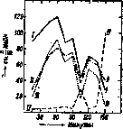
The correlation between electrical conductivity, acidity, and content is actually extremely close.
The curves of electrical conductivity, total acidity, and free hydrochloric acid (Fig. 1) repeat each other.
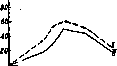
In normal conditions, the changes in total acidity and the amount of free HCl after a test breakfast run in parallel (Fig. 2); the total amount of chlorides, apparently, does not undergo fluctuations.
The presence of hydrochloric acid in gastric juice causes the swelling of protein, which undergoes further breakdown, and the bactericidal action of gastric juice.
Hydrochloric acid also causes the breakdown of disaccharides, which is observed, although to an insignificant degree, during gastric digestion.
In view of the fact that the inverting effect belongs equally to both boiled and unboiled gastric juice, this process cannot be considered enzymatic. (On the constancy of the concentration of free HCl in secreted juice, see Gastric juice.)
A fine regulation of the active reaction of gastric contents is observed within the limits most favorable for peptic digestion.
Carlson provides the following table of the composition of normal human gastric juice (obtained from a gastric fistula):
Acidity { free . . . . . . . . . . 0.40–0.50%
Acidity { total . . . . . . . . . . 0.45–0.60%
Solid components { organic . . . .
{ inorganic . . .
0.051-0.075%. Amino acid nitrogen: 3-10 mg%. Ammonia: 2-8 mg%. Chlorides: 0.50-0.68%. The material for the formation of HCl in gastric juice is provided by chlorides contained in the gastric mucosa, which apparently possesses a selective ability to retain chlorides brought by the blood. It is believed that during the secretion of gastric juice, the amount of chlorides in the blood decreases, the reserve alkalinity increases, and the concentration of hydrogen ions decreases. Indeed, a number of authors who investigated the blood composition of dogs with gastric and esophageal fistulas were constantly able to experimentally prove the changes described above after sham feeding. However, if one examines an animal under normal conditions, when the acidic gastric content is not discharged outward but undergoes neutralization and absorption in the lower segments of the digestive tract, then no noticeable changes occur in the active reaction of the blood, and data on the total amount of CO2 in the blood and the reserve alkalinity of the blood are contradictory. Gastric juice contains enzymes: pepsin, chymosin, and lipase (see Gastric juice); the most important of these is pepsin. In the stomach of newborn animals, there is an independent enzyme, chymase, which has a curdling effect and is a true protease capable of breaking down not only casein but also some other proteins (for example, legumin, syntonin) in a weakly acidic reaction. There is no chymase in the stomach of adult animals, and the curdling effect belongs to pepsin, which, like any protease, when breaking down casein, first converts it into paracasein. This action of pepsin is noticeable when there are no favorable conditions for peptic protein breakdown, i.e., at pH > 4. Gastric lipase, studied in detail by Volhard, is of little importance in the digestive processes of humans; it is limited to the breakdown of only emulsified fats. The optimum action of an unpurified preparation lies at pH = 5.5-6.3, the minimum at 8.6. However, after purification by electrodialysis and adsorption with kaolin, the optimum shifts to the alkaline side and lies at pH = 7.1-7.9. The food bolus, upon entering the stomach cavity, undergoes changes mainly in relation to its protein components. Pepsin brings the breakdown to the stage of complex polypeptides. The products of the first stage of hydrolytic protein breakdown by gastric juice are acid albuminates. The final products of gastric digestion are usually considered to be albumoses (see) and peptones (see). In different parts of the stomach, albumoses and peptones are formed unevenly: peptones predominate in the pyloric part, and albumoses in the fundic part. For the course of the egg protein hydrolysis reaction by the action of pepsin, a rule (Schütz-Borisov) has been established, according to which the amounts of peptone formed are directly proportional to the square roots of the amounts of enzyme. This rule was subsequently confirmed by numerous researchers, although it cannot be considered valid for all cases without exception. Nucleoproteins in the stomach are broken down into so-called nuclein and protein. When investigating the distribution of nitrogen by fractions in the gastric contents of healthy people, the following data were obtained: of the nitrogen in solution, 12% belonged to substances capable of coagulating, and 4.5-5.5% to acid albuminates. The nitrogen of non-coagulating substances was mostly (55-65%) in the form of albumoses. When eating meat, about 0.88% N of non-coagulating substances was found in the form of NH3 and about 1.34% in the form of amino nitrogen; when eating bread and meat, 1.2% and 3.4% respectively, i.e., extremely insignificant amounts. Ammonia, always detected in gastric contents, is likely a product of the metabolism of gastric gland cells. The study of the final products of gastric digestion is complicated by the constant reflux into the stomach of the contents of the upper segment of the intestine: bile, intestinal and pancreatic juices, which are rich in enzymes and the products of their activity. This reflux should be considered a normal constituent part of the act of digestion. If, during the reflux of duodenal contents and bile, hydrochloric acid is neutralized and the reaction becomes weakly acidic or neutral, then trypsin, erepsin, and pancreatic lipase come into action. In the composition of gastric contents extracted after a test breakfast, trypsin was successfully detected in 85% of cases, with significant amounts in 55% (Fig. 1). In animals, both carnivorous and especially herbivorous, enzymatic processes caused by microorganisms are of very great importance. The role of microorganisms and enzymes contained in food substances for human gastric digestion is negligible. The duration of food stay in the stomach is very different, however, in the same subject, it is longest for fats, less for proteins, and finally shortest for carbohydrate food. Using the method of fluoroscopy, Demuth showed that the duration of stay for carbohydrate food ranges from 2 hours 50 minutes to 5 hours 10 minutes, and for protein food from 3 hours 30 minutes to 6 hours 30 minutes. The stomach empties particularly quickly of raw vegetables (1-2 hours). The food mass of liquid or mushy consistency, upon the opening of the pyloric sphincter, enters the upper section of the intestine in portions, where it is subjected to the action of intestinal juice, pancreatic juice, and bile. Human intestinal juice, produced by the Lieberkühn and Brunner glands, was subjected to investigation after the introduction of the technique of obtaining intestinal contents via a thin probe (Einhorn) into clinical practice. After a test breakfast (a glass of milk diluted with water by half), a viscous liquid was obtained with a specific gravity of 1.006-1.010, with a neutral reaction to litmus, and with a constant admixture of bile. The dry residue constituted 2.15% of the initial material, ash 0.67%, chlorine 0.38%, P2O5 0.045%, and nitrogen titratable by formol titration 0.03%. According to Einhorn and Rosenbloom, intestinal juice contains coagulable proteins as well as peptones. The freezing point of intestinal juice is 0.620°. The enzymes in intestinal juice include lipase, which primarily breaks down emulsified fats; proteases, which break down polypeptides and do not act on native protein; sucrase, maltase, and lactase, found in children and also in adults on a milk diet. Intestinal juice also possesses a very weakly expressed amylolytic action and contains enzymes that break down nucleoproteins and acids, as well as anti-pepsin, anti-trypsin, enterokinase, and prosecretin. The secretion of two most important digestive glands—the pancreas and the liver—also flows into the upper segment of the small intestine, the duodenum. Human pancreatic juice is a colorless, odorless, salty-tasting liquid with a specific gravity from 1.006 to 1.008 and a weakly alkaline reaction (on average pH = 8.3). The osmotic concentration is slightly higher than in the blood (on average Δ = 0.62). Authors who described a significantly lower value of depression (Δ = 0.42-0.49; Glaessner and Wohlgemuth), in the opinion of Rosemann, were dealing with a malfunctioning gland. The surface tension of pancreatic juice and gastric juice is the same. The quantity and composition of the juice secreted by the pancreas are diverse and depend to a significant extent on the composition of the food. A fasting person secretes on average 10-15 cm3; after eating, from 30-50 cm3 to 84 cm3 per hour. The daily amount of secreted juice is from 420-850 cm3 to 1 liter. Table 1. Composition of pancreatic juice. Authors: Schumm, Glaessner, Ellinger u. Colin, Wohlgemuth. Solid constituents: 1.2389-1.5449, 0.8486-0.8667, 0.076-0.099, 0.0689-0.0939. Inorganic constituents: 1.2708-1.2494, 0.5662-0.6976, 0.1276-0.1744, 0.0842-0.0983. Protein coagulable upon boiling: 0.1374, 0.0765-0.0604. Pancreatic juice contains the enzymes: trypsin, lipase, amylase, maltase, peptidases (erepsin), and enzymes that break down nucleoproteins and nucleic acids. Table 2. Composition of the ash of human pancreatic juice (in %) (according to Wohlgemuth).
Na Cl SO3 P2O5 SO2 Ca Mg Fe 1.10 33.65 50.75 2.05 1.85 0.34 Traces. It is probable that the composition of pancreatic juice also includes a kinase (cotryptase), identical to intestinal enterokinase, but in an inactive form, in the form of prokinase (see below). Regarding the composition of bile and its fluctuations in humans, see Bile, bile acids, physiological significance of bile. Among the enzymes in the composition of bile, the following have been detected: amylase, oxidase, catalase, and a proteolytic enzyme. In view of the fact that all the most important enzymes participating in the processes of digestion are present simultaneously in the upper section of the intestine, the active reaction of the intestinal contents is of extremely great importance. However, there is no uniform opinion regarding the acidity of the intestinal contents. Determining the pH of the intestinal contents, McClendon and his colleagues sometimes found a weakly alkaline reaction, more often an acidic one (pH=4.1–6.5). A number of other researchers obtained strongly fluctuating results, starting from a distinctly acidic reaction to a weakly alkaline one. Okada and Arai found fluctuations within the range of pH=6.6–7.9; the suggestion was made that the acidity of the intestinal contents is not dependent on the acidity of the gastric juice. From the data presented, it follows that despite the alkaline reaction of the secretions pouring into the upper segment of the intestine, the contents in the sections adjacent to the stomach may have an acidic reaction. The question arising in connection with this regarding the possibility of the continuation of peptic digestion in the small intestines was initially resolved in the negative sense, because, firstly, the active reaction, lying within the range of pH=3.2–7.9, was far from corresponding to the optimal conditions for the action of pepsin, and secondly, the compounds of protein with bile acids formed in the small intestines are incapable of swelling, which is necessary for the successful action of pepsin. However, the works of Abderhalden, who studied the action of pepsin on elastin, established that in these cases such a stable compound of substrate with enzyme is formed that peptic cleavage continues in an alkaline medium until the entire substrate is impregnated with alkali. Abderhalden succeeded in finding active pepsin in the contents even of the lower-lying segments of the small intestines (in the ileum). The question of the independent secretion of pepsin in the small intestines by Brunner's glands, which very much resemble pyloric glands in their structure, remains unresolved. In addition to pepsin, which retains its activity (although to an insignificant degree), enzymes of the pancreatic and intestinal juices act on proteins in the small intestines; of these, primarily trypsin. This enzyme is secreted by the pancreas, more often apparently in an inactive state—in the form of trypsinogen or protrypsin—and is activated by enterokinase, discovered by Pavlov. Pavlov considered enterokinase to be a separate enzyme produced by the intestinal mucosa and catalytically influencing the transition of trypsinogen into active trypsin. Hamburger and Hekma viewed the process of trypsin activation as the result of the combination of the proenzyme with the kinase. Waldschmidt-Leitz brought significantly greater clarity to the question of the action of enterokinase; he succeeded, by methods of adsorption, in again separating an active trypsin preparation into zymogen and kinase. Furthermore, in the opinion of Waldschmidt-Leitz, trypsin is capable, even without any activation, of exerting a proteolytic effect on protamines and peptones; consequently, enterokinase is responsible only for the activation of trypsinogen in the sense of extending its action to specific structures—namely, high-molecular-weight proteins. The site of formation of enterokinase has not yet been precisely established; on one hand, it has long been noted that enterokinase is produced by the glands of the small intestines, and on the other hand, it has been possible to obtain active pancreatic juice after extraction of a preparation of the latter that has been left to stand for some time. These data established with certainty the possibility of the formation of enterokinase in the tissue of the gland itself. Waldschmidt-Leitz expresses the hypothesis that enterokinase is always formed in the cells of the pancreas, but in the form of a preliminary stage—prokinase. Prokinase is activated by erepsin in the intestine into active enterokinase; enterokinase, by forming an unstable compound with trypsinogen, significantly expands the range of its proteolytic action on various proteins. An idea of the action of various proteases can be obtained from Table 3. Despite the fact that in the upper segments of the intestine there may be a significantly more acidic reaction than corresponds to the optimal action of trypsin, tryptic cleavage of protein can nevertheless take place here. Heller attributes a major role to bile, which, while not being an activator of trypsin, sharply changes the degree of dispersion of proteins, significantly increases their surface area, and thereby facilitates the interaction of enzyme and substrate. This action of bile is of especially great importance for the cleavage of proteins in the intestines of newborns, where, in view of the only weakly acidic reaction of the gastric contents (pH=4.5–5.5), the role of peptic digestion is insignificant. The second protein enzyme of the intestinal contents—erepsin, produced both by the pancreas and by the intestinal glands—is identical in both cases. The previously recognized difference turned out to be non-existent. Erepsin is not capable of cleaving proteins, nor protamines, histones, or peptones formed as a result of peptic digestion. In those cases where such phenomena were described, the researchers were dealing with an insufficiently pure preparation containing trypsin. Erepsin catalyzes only the cleavage of ordinary natural or synthetic polypeptides. The optimum for the action of animal erepsin lies at pH=7.8, and the course of hydrolysis corresponds to the equation of a monomolecular reaction. The cleavage of protein in the intestine proceeds in stages: first, tyrosine and tryptophan are split off, then glutamic acid, aspartic acid, leucine, alanine, etc. The cleavage of proteins to the amino acid stage in the intestine proceeds very energetically: Abderhalden and Kramm indicate that already after 1 hour a significant amount of amino nitrogen is formed, corresponding after 6 hours to 2/3 of the amount that can be obtained upon hydrolysis with fuming hydrochloric acid. In the contents of the small intestines, it is possible to detect very little unchanged protein, few albumoses, somewhat more peptones, and an insignificant amount of products of deeper protein cleavage (amino acids). The latter is explained by the energetically proceeding absorption. Trypsin and erepsin do not act on certain proteins, for example, on serum and egg albumins, or raw connective tissue protein. However, after treatment of these substances with gastric juice, they easily undergo further cleavage. In general, preliminary peptic digestion significantly facilitates and accelerates tryptic digestion. In the intestine, the cleavage of nucleoproteins that began in the stomach continues. The enzymes of this group are still little studied; in particular, the enzymes that cleave pyrimidine nucleosides are completely unknown. Nucleosides are not subjected to the action of intestinal juice; nucleosidases are apparently only an intracellular enzyme of tissues. In the small intestines, deep cleavage occurs not only of protein substances but also of carbohydrates and fats. Polysaccharides are subjected here to the action of amylase of the pancreas, intestine, and bile, and disaccharides, depending on their structure, to the action of sucrase, maltase, and lactase (mainly in children) of the intestine. Sucrase (invertin) is found in different segments of the small intestines in unequal amounts: it is most abundant in the upper part of the jejunum, then in the lower part of the jejunum, then in the duodenum, and finally in the ileum. Sucrase of the human small intestines is inactivated by half within an hour at 51°, and is completely destroyed at 55°; the optimal pH lies between 6 and 8; sucrase, just like maltase (and in newborns, lactase), is found in significantly greater quantity in the walls of the intestine than in the secretion secreted by them. For a long time, the question of the continuation of the action of salivary amylase in the intestine remained controversial. It should apparently be acknowledged that only a part of ptyalin is destroyed in the stomach, and consequently, the amylolytic action of salivary diastase can continue in the intestine. The most strongly expressed action is possessed by the amylase of pancreatic juice, which is activated by bile, amino acids, and salts.
It is possible, however, that bile does not have an activating effect on amylase, and its significance is limited to changing the active reaction of the medium. Pancreatic juice amylase belongs to α-amylases. The products of carbohydrate breakdown are: 1) maltose, which is further broken down by maltase into 2 particles of glucose; 2) glucose and fructose, resulting from the breakdown of sucrose; and 3) glucose and galactose, formed during the breakdown of lactose. All listed monosaccharides undergo absorption. Cellulose and pentosans are not broken down by the enzymes of the intestine of higher animals. Fat breakdown occurs in the small intestine due to the presence of lipase in the pancreatic secretion. For a long time, it was believed that the lipases of gastric, intestinal, and pancreatic juices were independent, distinct enzymes. In defense of this view, data were cited regarding the different pH optimum of these esterases and their completely different action on different substrates. However, studies by Willstätter established that the diversity in the action of lipases depends on the substances accompanying them. After purification, the optimum action of lipases was found to lie at the same pH = about 8.0. Willstätter explains the influence of accompanying substances by the phenomenon of adsorption: if both the enzyme and the substrate are adsorbed simultaneously, this leads to an acceleration of hydrolysis; if only the substrate or only the enzyme is adsorbed, this slows down the breakdown. This explanation, however, is not generally accepted. The amount of lipase in the pancreas is significantly greater than in the stomach: from 1,000 g of gastric mucosa, one can obtain the same amount of lipase as from 10 g of the pancreas (Willstätter). Neutral fats undergo breakdown by lipase with the formation of glycerin and free fatty acids, which, due to the alkaline reaction of the intestinal contents, turn into soaps. Lipase catalyzes the breakdown not only of neutral fats but also of many other esters. Bile has a great influence on fat breakdown, which accelerates the hydrolytic breakdown of fat by pancreatic juice, emulsifies fats, makes high-molecular fatty acids soluble in water in the presence of soaps, etc. (see Bile—physiological significance of bile). The great importance of bile in the digestion of lecithin has also been noted. As for the significance of intestinal flora for the processes of digestion, for humans, it is insignificant in the upper sections of the small intestine, but greater in the lower sections, where microorganisms causing carbohydrate fermentation are mainly present. However, it should be noted that the intestinal flora varies greatly depending on the diet and the reaction of the medium. Regarding the digestive significance of intestinal flora, expressed in the breakdown of food components, products of its digestion, and components of digestive juices under the influence of fermentation and putrefaction processes, there are two opposing opinions: some authors see these processes as an inevitable evil for animals, others believe that the intestinal flora participates in the act of digestion with its enzymes, and some authors even believe that in the absence of microbes, there cannot be a proper course of digestion. However, studies by Nuttall and Thierfelder and Metchnikoff showed that proper nutrition of not only lower animals but also mammals is possible in the absence of intestinal flora. In various animals, the significance of fermentation processes is very different. For example, in herbivores, the fermentation of the main components of plant food—cellulose and pentosans—with the assimilation of the products formed during this has a very important significance, whereas in humans, only young, tender fiber of fruits and vegetables can be partially assimilated. The contents of the small intestine pass gradually into the large intestine [with carbohydrate food on average after 4 hours, with fatty food after 5, and with protein food after 6 (Cannon)]. The secretion of the mucous membrane of the large intestine is rich in mucus and contains, in humans, diastatic, lipolytic, and apparently proteolytic enzymes. The significance of these enzymes is insignificant. In the large intestine, the enzymes of the small intestine continue their action; as a rule, they are also included in the composition of excrement (for example, trypsin, erepsin, diastase, lipase). In the large intestine, there is a large number of bacteria causing carbohydrate fermentation and protein putrefaction. Protein putrefaction is caused by both anaerobic and aerobic bacteria. Anaerobes (among them primarily Bac. putrificus) can break down native proteins; they contain a mixed protease (pepsinase + tryptinase) with optimal activity at pH=6 (Bernby) and peptidase. During protein putrefaction under the influence of anaerobic bacteria, amino acids and products of deeper protein breakdown are formed as intermediate products: NH3, indole, organic acids—propionic, butyric, caproic, lactic, para-oxyphenylpropionic, acetic; from gases—H2S, CO2, and H2. The formation of phenol has not been established with precision. Aerobic bacteria (Bact. coli group) produce peptidases. Among the products of putrefaction formed as a result of the vital activity of this group of bacteria, indole, skatole, phenol, histamine, fatty acids (valeric, caproic, and succinic), NH3, and H2S have been found. As a result of the vital activity of Bac. proteus vulgaris in the absence of carbohydrates, protein breakdown occurs with the formation of NH3, primary amines, H2S, aromatic hydroxy acids, indole, and indoleacetic acid; phenol, mercaptan, alcohols, aldehydes, and ketones are not formed. During protein putrefaction in the intestine, so-called ptomaines—animal alkaloids—can form; some of them have a poisonous effect. Cadaverine and putrescine are not formed under normal conditions (Baumann). With the intensification of putrefaction processes, phenomena of intestinal autointoxication can develop. In the large intestine, reduction processes also take place: cholesterol turns into coprosterol, bilirubin into urobilin and urobilinogen. Regarding the fate of the formed phenol, cresol, indole, and skatole, it is known that, upon entering the circulatory system, they turn into ethereal sulfuric acids in the liver, which are then excreted with urine. Fermentation processes are caused by very different bacteria (Bact. coli, lactis aerogenes; pentoaceticus, butyricus, etc.). Among the products formed during various types of fermentation (lactic acid, acetic, formic, alcohols, CO2, H2, CH4), pyruvic acid and acetaldehyde have recently been found. In the large intestine, energetic absorption of water and the formation of excrement occur.
Related articles
Cite this page
“Digestion.” Soviet Medical Encyclopedia. English translation of Bolshaya Meditsinskaya Entsiklopediya, 1st ed. (Moscow, 1928–1936), ed. N. A. Semashko. https://sovietmedicalencyclopedia.pages.dev/article/digestion/