Mouth
Historical document, translated for reference. It reflects medical knowledge of the 1920s–30s and is not medical advice.
Summary
This article describes the embryonic development of the mouth in mammals, including the formation of oral structures, and provides detailed anatomical information about the oral cavity's boundaries, lining, and associated structures.
Encyclopedia article (1928–1936)
MOUTH, ORAL CAVITY. Development. The mouth in mammals develops from the intestinal canal, which is a derivative of the embryonic inner leaf (entoderm). In the earliest stages of development, the embryonic intestinal canal is a straight tube located on the ventral side of the embryo. At the front, this tube ends blindly, while at the back it passes to the dorsal part of the embryo into the neural tube via canalis neurentericus (see vol. X, p. 510, figs. 9 and 10). The latter is a temporary formation, as the intestinal canal soon transforms into a tube closed at both ends. The front part of the neural tube bends ventrally, so that a bulge forms at the front end of the embryo (fig. 1). The first rudiment of the mouth is an invagination of the ectoderm directly behind this bulge. This so-called oral pit (fig. 1, 3). It begins to grow toward the blind end of the intestinal canal, the contacting surfaces fuse, and then rupture, forming fragments - the so-called primitive palatal curtains, which exist only temporarily. The external shape of the oral opening is determined by five surrounding ridges (fig. 2). The anterior ridge, limiting the oral opening from the brain side, is called the frontal tubercle or process (fig. 2, 12), behind which lie paired maxillary processes (fig. 2, 11). The groove that separates them from the frontal tubercle is called the eye slit. The groove lying between the maxillary and mandibular (fig. 2, 10) processes corresponds to the future corner of the mouth. To the side and behind the frontal process is the paired nasal slit (fig. 2, 1). Behind the oral pit in the neck region of the embryo, several slit-like openings facing the ventral side of the embryo form in the intestinal wall. They form as follows: deep invaginations (internal pharyngeal grooves) appear in the intestine toward the ectoderm, and the ectoderm in turn forms slight invaginations (external pharyngeal grooves) (fig. 2,3, 4, 5 and 6) directed backward. These invaginations and protrusions come into contact, forming, as with the mouth, a thin membrane, which then ruptures. Thus, in higher vertebrates, four pharyngeal gill slits arise. They are bounded by pharyngeal gill arches. In fish, the mucous membrane of the gill arches forms folds, beneath which are numerous blood vessels. In higher vertebrates, the gill slits soon close again, leaving grooves bounded by gill arches. The first gill arch - the jaw, or mandibular - is called the arch lying in front of the first gill slit (fig. 2) [see also the separate table (vol. X, pp. 507-508), fig. 2], between it and the mouth. The second gill arch - the sublingual, hyoid - is called the arch lying between the first and second gill slits. Followed by the third and fourth gill arches. From the frontal process and the "mandibular arch" the face is formed. In the 3rd week, the frontal process forms on each side one process extending downward and forward - nasal processes (fig. 3, 8). The mandibular arch limits the nasal-oral cavity from below. Already in an embryo of 2-3 mm, on the upper surfaces of the first and second gill arches, a protrusion forms, rising upward and forward toward the nasal process. Thus,
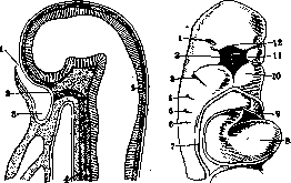
Figure 1.
Figure 2.
Figure 1. Median section through a rabbit embryo: 1-oral opening; 2-first rudiment of brain appendage; 3-oral invagination; 4-chorda dorsalis; 5-ectoderm. Figure 2. Head half of a human embryo 4.2 mm long, view from the front: 7-nasal slit; 2--nasal-oral pit; 3-b-correspondingly 1st, 2nd, 3rd, 4th external gill grooves; 7- place of reflection of the embryonic amnion (amnion); «-heart; 9-cardiac sac (pericardium); 10-lower jaw; 11-upper jaw; 12-frontal process. The mandibular arch divides, forming two branches or processes: the upper and lower jaw (fig. 3,6 and 7). The slit separating the maxillary processes from the lower ones is called the intermaxillary slit and corresponds to the primary oral opening. From each maxillary process a palatal plate and palatine process grow. Appearing in the 7th week, the palatal processes grow in a horizontal direction toward each other and meet along the median line at the end of the 3rd month. Thus the palatal septum is formed, posteriorly supplemented by the palatine processes, dividing the nasal-oral cavity into two cavities: the upper - nasal and the lower - oral. The maxillary processes with the internal nasal processes give rise to the upper lip and upper jaw, fusing along the median line. The mandibular processes, fusing along the median line at the beginning of the 2nd month, give rise to the lower lip and lower jaw with their surrounding soft parts. The lining covering all tubes and cavities of internal organs is called the mucous membrane (tunica mucosa). Teeth can be considered as calcified papillae of the mucous membrane. Salivary glands appear as protrusions of the oral mucous membrane; these protrusions, due to the gradual growth of individual outgrowths, become solid grape-like (acinous) glands. The tongue is formed from an unpaired anterior and paired rudiments. The anterior is a ridge rising from the floor of the oral cavity; it gives the entire anterior, papillae-equipped part of the tongue, each of the posterior rudiments consists of one tubercle located between the second and third gill arch. From these rudiments, the posterior, gland-equipped part of the tongue is formed. Anatomy. Under the oral cavity (cavum oris) is meant the initial part of the digestive tract, which extends from the oral fissure (rima oris) to the isthmus of the fauces (isthmus faucium); the latter is the boundary between the oral cavity and the pharynx (pharynx). With the mouth closed, i.e., at rest, the oral cavity can hardly be called such: in this state, the edges of the tongue adjoin the teeth of the upper jaw, the front part of its back is located directly on the hard palate, and only between the soft palate and the posterior part of the tongue's back is there a narrow, air-filled space. From this space, however, a true cavity is formed as soon as the lips and jaws move away from each other when the mouth opens. The oral cavity is bounded: in front - by the lips, on the sides - by the cheeks, the ascending branches of the lower jaw, and the medial pterygoid muscles, above - by the hard and soft palate, below - by the floor of the mouth, behind - by the anterior palatine arches and the uvula hanging between them. The alveolar processes divide the oral cavity into the part located outside the dental rows - the vestibule of the mouth (vestibulum oris, s. cavum buccale), and the part located between the dental rows and the isthmus - the proper oral cavity (cavum oris proprium). The oral cavity is lined with a dense mucous membrane, which on the lips passes into the outer skin, and at the isthmus - into the mucous membrane of the pharynx. On the alveolar process, the mucous membrane is tightly connected to the periosteum and forms immovable gums (gingivae) surrounding the teeth with a slightly raised edge. This edge, tightly encircling the neck of the tooth, is called the circular ligament of the tooth (lig. circulare dentis, s. lig. annulare). The connection with the periosteum is also very tight on the hard palate. In addition to the excretory ducts located in the submucosal layer of sebaceous and mucous glands, the paired salivary glands' ducts also open into the oral cavity: sublingual, submandibular, and parotid. In the so-called corners of the mouth (anguli oris), both lips pass into each other via the labial commissures. In the middle of the edge of the upper lip there is a small thickening (tuberculum labii superioris). To this thickening in the middle of the lower lip corresponds a slight depression. On each lip, the outer skin part (pars cutanea), transitional part (pars intermedia), and mucous part (p. mucosa) are distinguished. The red border of the lip (mar- 2-frenulum linguae; z-pli-gO labll) СОСТОИТ ИЗ пе- sa sublingualis; 4-carun-реХОДНОЙчастИИСМеж- cula sublingualis; 5-frenu-НЫХ СЛИЗИСТЫХ Обла- SUbmaXIllaris; 'V-glandula стеи. B переходной ча- sublingualis; 8-tn. genio-СТИ B большом КОЛИ- glossus; 9-m. longitudina-честве имеются саль- lis inU lluf^1* Un" ные железы: если слегка потянуть кожу, то их можно увидеть просвечивающими в виде желтоватых точек. B слизистой части рассеяны многочисленные слизистые железы (gl. labiales); место впадения их выводных протоков находится в слизистой оболочке. Слизистая часть губ переходит на челюсть, причем место перехода так и называется переходной складкой слизистой обо
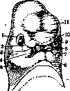
Figure 3. Head of a human embryo 13.7 mm long: 1-eye; 2-eye-nasal slit-tear duct; 3-nasal slit-rudiment of nasal passage; 4 - oral opening; 5-first ex-
-„
-ternal gill groove; b-lower jaw; 7- upper jaw; S-internal nasal process; 9- external nasal process; 10-frontal process; 11- anterior cerebral vesicle (cerebral hemispheres).
Figure 4. Lower surface of the tongue: 1-plica fimbriata; 2-frenulum linguae; 3-glandula sublingualis; 4-caruncula sublingualis; 5-frenulum submaxillaris; 6-glandula sublingualis; 8-m. genioglossus; 9-m. longitudinalis inU Un* linguae; 10-m. transversus inU linguae; 11-glandula submandibularis; 12-ductus submandibularis; 13-ductus sublingualis; 14-ductus parotideus; 15-plica submandibularis; 16-plica sublingualis; 17-plica palatoglossa; 18-plica palatopharyngea; 19-tonsilla palatina; 20-uvula. The red border of the lip consists of the transitional part and the adjacent mucous areas. In the transitional part, there are numerous sebaceous glands: if the skin is slightly pulled, they can be seen as yellowish dots. In the mucous part, numerous mucous glands (gl. labiales) are scattered; the place of their excretory ducts opening is located in the mucous membrane. The mucous part of the lips passes onto the jaw, and the place of transition is called the mucous fold.
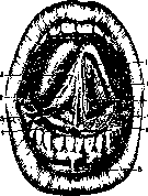
the lips. Over the middle incisors of the upper jaw and under the middle incisors of the lower jaw at the transition of the mucous membrane, a frenulum (frenulum labii superioris, resp. inferioris) is formed (fig. 4), which in the upper jaw sometimes extends to the interdental papilla (gingival thickening between the teeth). The mucous membrane of the cheek (buccaa) contains sebaceous and mucous glands: they are located mainly in the area of the molars and are collectively called gl. molares, s. buccales. Opposite the crown of the upper second molar, a small, wart-like elevation can always be seen - the opening of the excretory duct of the parotid salivary gland (papilla salivalis superior). The
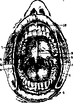
Figure 5. Walls of the oral cavity: 1-raphe palati; 2- arcus glosso-palatinus; 3- tonsilla palatina; 4-arcus pharyngo-palatinus; 5-frenulum labii inf.; 6-isthmus faucium; 7-plica pterygo-mandibularis; 8-uvula; 9-foveola palatina; 10-frenulum labii sup.
posterior border of the vestibule of the M. is formed by a fold of the mucous membrane (plica pterygo-mandibularis, fig. 5), extending to the soft palate and well palpable when the M. is open. The palate (see. forms the roof of the oral cavity (fig. 6).
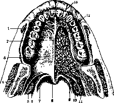
Figure 6. Hard and soft palate: 1-plicae palatinae transversae; 2-raphe palati; 3-foveola palatina; 4-fossa supratonsillaris; 5-arcus glosso-palatinus; 6 and 10-tonsilla palatina; 7-arcus pharyngo-palatinus; 8-uvula (palatina); 9-m. pharyngo-palatinus; 11-m. glosso-palatinus; 12-glandulae palatinae; 13-papilla incisiva.
The salivary glands of the oral cavity are divided into two groups - large and small (see. Salivary glands, Parotid gland, Submandibular gland, Sublingual gland). Histology. The mucous membrane of the oral cavity, particularly of the lips, differs little in structure from the skin. The multilayered flat epithelium of the skin directly passes into the same epithelium of the mucous membrane. Similarly, the basal membrane of the skin, without interruption, directly continues into the basal membrane of the mucous membrane. The same can be said of the fibrous connective
tissue covering the surface of the mucous membrane, are embedded in the sub-
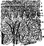
mucosa, which is due to a thin layer of bundles of the skin's own layer (corium, s. derma), continuing directly into the propria layer of the mucous membrane (tunica propria) (fig. 7 and 8). Hair and sweat glands are absent in the mucous membrane of the lips and oral cavity; sebaceous glands are present only in the mucous membrane of the lips, occurring in other parts of the oral mucosa only occasionally. The place of sweat glands in the oral mucosa is taken by numerous mucous and serous glands (fig. 7 and 8). The epithelium of the mucous membrane of the lips is multilayered flat, its layers are arranged the same as in the skin, but it does not have a horny layer, because its plate-like cells are washed away by saliva and gradually exfoliate. Numerous salivary glands of the lips, the excretory _ _,.
ducts of which open Fig. 7. Perpendicular ^ttt,otltlggg „„ „p_ to the surface cross-section of the mucous membrane of a child's lip: 1-layer of cornified cells, gradually flattening toward the surface chiotom glor gogtoya. and exfoliating; 2-granular layer; a-layer of many-sided LOKNISTOY SOvDINI-cells with processes (stratum „,„*„„» „,pt, /„„„ spinosum); 4-maternal layer of connective tissue (fig. epithelium of the mucous 8). The mucous membrane; 5-papillae of the gums' own membrane; 6-connective tissue base of the mucous membrane; 7-mucous gland; 8-bundles of striated muscle „„„„g,,^,,,, „„„„„ fibers.
its base, covering without an underlying layer over the periosteum of the jaws; it contains no glands at all. The mucous membrane of the cheeks is homogeneous in structure with that of the lips, but has no submucosal layer, therefore the bodies of the numerous complex tubular glands (gl. buccales et molares) contained in it are embedded in a layer of loose fibrous connective tissue between the bundles of fibers of the underlying muscle (m. buccinator). In the area where the excretory duct of the parotid salivary gland passes, there are larger salivary glands of the mucous
membrane. The mucous membrane of the palate, with its base covering without an underlying layer over the periosteum of the jaws; it contains no glands at all. The mucous membrane of the cheeks is homogeneous in structure with that of the lips, but has no submucosal layer, therefore the bodies of the numerous complex tubular glands (gl. buccales et molares) contained in it are embedded in a layer of loose fibrous connective tissue between the bundles of fibers of the underlying muscle (m. buccinator). In the area where the excretory duct of the parotid salivary gland passes, there are larger salivary glands of the mucous membrane (glandulae labiales). The mucous glands of the palate, by their location and origin, are due to the epithelium that covers the edges of the two fusing (right and left) palatal processes of the upper jaw; in structure this epithelium does not differ from that of neighboring parts. Only at the edge of the palatal veil and palatal arches does the epithelium of the mucous membrane transform from multilayered flat to single-layered ciliated with cilia, characteristic of the respiratory area of the nasal cavity. In the thickness of the mucous membrane of the palatal veil lie numerous complex tubular glands - mucous and mixed. Blood vessels are located in the mucous membrane of the oral cavity both in its main layer and in the papillary layer. Lymph vessels begin in the papillary basis of the mucous membrane, under which they form dense networks,
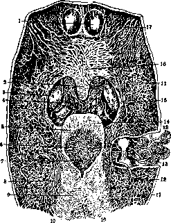
Figure 9. Lymphatic vessels of the walls of the pharynx and larynx: 1-posterior nasal openings (choanae); 2- uvula; 3-tonsils (tonsillae palatinae); 4- root of the tongue; 5-epiglottis; 6-lymphatic network of the larynx and its trunks; 7-entrance to the larynx; 8-greater horns of the thyroid cartilage, connected to the hyoid bone lig. thyreo-hyoid.; 9- larynx from behind; 10-lymphatic network of the upper part of the esophageal wall; 11-lymphatic network of the upper-lateral walls of the pharynx; 12-trunk-collector of the lower-lateral wall of the pharynx; 13-lymphatic nodes to which all lymphatic vessels of the pharynx and larynx go; 14-lymphatic network of the epiglottis; 15-descending trunk; 16-lymphatic network of the posterior surface of the palatal veil; 17-lymphatic vessels of this network. (According to Sappey.)
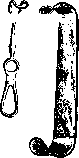
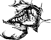
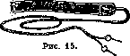
from which branches pass through lymph nodes (fig. 9). For the histology of the glands, see Glands and Salivary Glands. Physiology. The mouth participates in the following processes: food intake, respiration, formation of voice and speech. In addition to the sense of taste (see), the mucous membrane of the mouth undoubtedly possesses pain, tactile, and thermal sensitivity. Healthy teeth do not possess pain sensitivity: the latter appears only in pathological conditions of the teeth (see Teeth). However, teeth possess clear temperature sensitivity. A light touch is felt by teeth quite distinctly, and its location is determined with sufficient accuracy. Under the influence of mechanical and chemical irritations of the mucous membrane of the mouth, saliva is secreted into the latter. From the oral cavity originate a number of reflexes: chewing (see Chewing), vomiting (see Vomiting), and coughing. For respiration, the oral cavity has great significance only in lower animals (amphibians, fish), in humans, however, breathing through the mouth appears only in pathological conditions, mainly in diseases of the nose and nasopharynx, while under normal conditions, oral breathing appears only as vicarious: during intense conversation, fast walking, running, and generally during all stronger bodily movements when the need for air is very great. For the participation of the mouth in the formation of voice and speech, see Voice and Speech. Bacteriology and immunity. The mouth is free from microbes only in the first days of life. Through food substances, breathing, and contact with fingers, microbes are constantly introduced into the mouth (see Human Microflora). With an abundance of microbes and wide possibilities for their penetration into the body, rapid healing of wounds in the mouth is observed, apparently as a result of the presence in the mouth of still little-studied reactions of local immunity, the bactericidal properties of potassium thiocyanate in saliva, the bactericidal properties of leukocytes, the regenerative ability of the oral epithelium, mechanical cleansing of the mouth by saliva, etc. But under pathological conditions, with the presence of large defects of the mucous membrane and the influx of new portions of pathogenic microbes, especially highly virulent ones, these protective adaptations prove insufficient, and the mouth becomes infected. The same occurs under the influence of certain poisons (mercury), in the presence of general infections (e.g., typhoid) or constitutional diseases (e.g., diabetes). The floor of the mouth is especially sensitive to the effects of all kinds of harmful influences due to the delicacy of its mucous covering, the looseness of the submucous layer, and the richness of its lymphatic vessels. A dentist during a thorough examination of the mouth can often detect diseases, the first manifestations of which are precisely in the mouth. On the other hand, the dentist must always remember about the rich microflora of the mouth, so that his hands and instruments do not become a means of transferring all kinds of infections. Methods of medical examination. Examination of the mouth itself must be preceded by an examination of the external parts of the face and neck. Attention should be paid to their relationship to each other and to the condition of the lymph nodes, tumors, etc. The examination of the external parts and tumors clarifies our understanding of the condition of the organs of the mouth and its surroundings. It is especially important to examine the condition of the jaw lymph nodes, which often react first to the pathological condition of the mouth organs. Very valuable data can be obtained by bimanual (two-handed) palpation, especially for detecting concretions located in the thickness of soft tissues (in glands, in excretory ducts). Examination of the mouth itself is best done in daylight, when the slightest changes in the color of the mucous membrane are distinguishable. But in our latitudes and under the conditions of evening work in our medical institutions, it is also necessary to use artificial lighting. Most often, Telsehow's reflector is used (fig. 10); one can also use an ordinary frontal reflector, reflecting light into the mouth from an ordinary (matte) electric light bulb. Even more convenient is the frontal electric lamp shown in fig. 11. To move aside the soft parts—lips, cheeks, tongue—all kinds of spatulas, tongue depressors, and hooks (fig. 12 and 13) are used, as well as dental mouth mirrors [see separate table, vol. XI (pp. 83-84), fig. 2], which at the same time reflect light onto the part of the mouth being examined and also examine parts of the mouth inaccessible to direct visual inspection. For examining the translucency of teeth and accessory cavities, the apparatus shown in fig. 14 and 15 is used. X-ray photographs (extraoral, as well as intraoral, obtained on small films wrapped in black paper and inserted into the appropriate place in the oral cavity) are a very valuable aid for examining the condition of the bony parts of the jaws and their processes. For examining the patency of the excretory duct of the parotid gland, thin button-shaped probes are used. It is very important to determine the condition of the transitional fold of the mucous membrane, which is smoothed out in the presence of inflammatory processes and bulges out in the form of a tumor in the presence of submucosal abscesses. For examining teeth, in addition to the dental mouth mirror, a sharp-pointed dental probe is used, with which all surfaces of the teeth are examined, the presence of caries, dental calculus, the condition of the floor and walls of the carious cavity, and its sensitivity in different places are detected. For examining the vitality of the dental pulp, induced current is readily used, with one electrode placed on the skin of the cheek or hand and the other on the enamel of the tooth being examined; teeth with a living pulp react to a current of a certain strength, teeth with an inflamed pulp react more strongly to the same current, while teeth with a dead pulp do not react at all even to a strong current. For examining gingival pockets, Neumann advises using a dental syringe, the air stream of which very gently retracts the edge of the gum from the tooth surface.
Fig. 11.
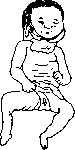
Pathology. Malformations of the mouth. Among symmetrical malformations of the mouth, it is necessary to mention so-called epignathus, where the parasite sits on the palate of the autosite; in this case, several varieties of this malformation are observed (according to Schwalbe): 1) in the mouth of the autosite (on the palate) there is the umbilical cord of the parasite, 2) parts of the parasite's body hang down from the mouth of the autosite, 3) from the mouth of the autosite the parasite protrudes in the form of an amorphous mass, sometimes in the form of a more organized tissue mass. A series of single malformations in the oral cavity arise due to various deviations in the development of individual parts of the embryo. This includes hare lip (see) and cleft palate (see). If finally no connection occurs between the maxillary process and the first gill arch, then the picture of a transverse facial cleft (fissura buccalis transversalis, s. macrostomia) results. Very often in the upper jaw (rarely in the lower) along the median line, a larger or smaller gap is observed between the central incisors—diastema. The cause of its appearance is the presence behind it of supernumerary teeth, the development of dense bone tissue at the place of connection of both maxillary bones, or the low attachment of the frenulum. Among malformations of the lower jaw, it is necessary to mention its duplication, when next to the normal jaw there is a second—smaller one (fig. 16), one of the forms of epignathus. Complete absence of the lower jaw is rarely observed (fig. 17)—agnathia (see), and in such cases both ears often fuse on the side of the absent lower jaw—synotia (see). This may also be associated with the absence of the mouth—atresia (see). Among malformations of the tongue, it is necessary to mention cleft tongue—lingua bifida—and the rare absence of the tongue. In a normally developed tongue, there may be a short frenulum, which prevents its movements and interferes with speech. A too long frenulum can lead to suffocation attacks in children. For all kinds of malformations and anomalies in the dental system, see Teeth. Among atrophic processes, in addition to physiological senile atrophy of the alveolar process and gums, it is necessary to mention the atrophy that occurs in so-called alveolar pyorrhea (see). Among progressive processes, it is necessary to stop at gum hypertrophy (see Gingivitis). In rare cases, overgrowth of the gums takes on colossal dimensions and extends to the palate: the picture of so-called elephantiasis of the gums (elephantiasis gingivae) results. Similar forms of hypertrophy are observed in late stages of syphilis. A special form of gum hypertrophy occurs during pregnancy, and the process usually begins in the middle of pregnancy and persists after childbirth during lactation.
Fig. 17. Agnathia with synotia.



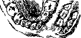
opinions. Therapy is powerless in such cases, but the disease passes on its own after feeding ceases. - Traumatic injuries. More or less significant penetrating wounds of the oral cavity disrupt the possibility of hermetic closure of the mouth, which disrupts the functions of nutrition, speech, spitting out saliva, and wound drainage. Wounding of the parotid gland and its excretory duct leads to the formation of salivary fistulas. Wounds of the lips and cheeks, after healing, result in disfiguring scar strictures of the mouth. Wounds of the tongue occur in the form of transverse and oblique incised wounds or even tears of the tongue, sometimes with complete detachment of a larger or smaller anterior part. In gunshot wounds of the oral cavity, bullets or fragments can become lodged in the accessory cavities, causing profuse bleeding. Penetrating wounds of the oral cavity are accompanied by fractures of the jaw bones, fractures and comminution of teeth, and teeth and bone fragments, embedding themselves in the tongue, cause sharp inflammatory phenomena in it. A frequent complication in gunshot injuries to the oral cavity is the formation of phlegmon of the submandibular area and the floor of the mouth. In some cases, acute osteomyelitis arises. Inflammatory processes are observed in all organs of the oral cavity: in its mucous membrane, mucous membrane of the gums, in the dental pulp (pulp), periodontium, salivary glands, tongue (see Stomatitis, Gingivitis, Pulpitis, Periodontitis, Glossitis). Among acute inflammatory processes, attention should be paid to erysipelas of the mucous membrane of the oral cavity - stomatitis erysipelatosa. Here it is a partial manifestation of erysipelas of the face and head, but sometimes it occurs isolated (primarily) in the oral cavity and pharynx. In the oral cavity, where erysipelas also frequently spreads from the accessory cavities of the nose, bright redness of the mucous membrane and its edema spread to the tongue, pharynx, and soft palate. The process can take a threatening course if it spreads deeper to the larynx, due to dangerous edema of the glottis. Any local treatment of erysipelas of the oral cavity is futile. Among the methods of general treatment, mention should be made of injections of scarlatinal antitoxic or anti-erysipelatous serum, autogenous blood therapy, and treatment with a quartz lamp. In the latter case, ultraviolet rays should be directed directly into the oral cavity. With poor care of the oral cavity in persons with severe nutritional disorders (scrofula, scurvy), a severe disease of the oral mucous membrane, known as oral decay or stomatace, s. stomatitis necro-membranacea (see Stomatitis), arises as a result of infection (often present in the mouth and of healthy individuals) by bacilli and spirochetes of Vincent. For water cancer, see Noma. Difficult eruption of wisdom teeth often leads to severe inflammatory phenomena in the oral cavity. This tooth, due to phylogenetic reduction, erupts from the lingual side of the ascending ramus, and the gum in this place cannot spread apart and surround the crown of the tooth. The development of the tooth in the ascending ramus and in an inclined (up to horizontal) position further hinders its eruption. In such cases, a pocket forms above the crown, which becomes the source of the inflammatory process due to the accumulation of food residues and secretions in it, which undergo decay and cause an ulcerative process in it. The process is initially local, but later the inflammation spreads to neighboring tissues, masticatory muscles, the submandibular cellular tissue, the mucous membrane of the pharynx, and even the pharynx: severe perimandibular phlegmonas arise with marked trismus and complete inability not only to take food but also to swallow liquids, with severe general condition and high temperature. In the initial stages of the process, it is possible to achieve subsidence of inflammatory phenomena by systematic displacement of the overhanging gum with iodoform gargles. In later stages, the removal of the wisdom tooth is indicated. For phlegmonas of the floor of the oral cavity, see Angina Ludovici. Diseases of the lips - see Lips. Among chronic inflammatory processes, attention should be paid to syphilis of the oral cavity, which can manifest here as a primary lesion (affection), secondary manifestations, and in the form of gummatous syphilis. The primary lesion appears as a result of infection from an infected person through kissing, infected dental and other instruments, a pencil, unnatural sexual intercourse, but occurs only if the carrier of the infection has a fresh virus. The site of primary infection is predominantly the lips, tip of the tongue, tonsils, less frequently the gums, hard palate, cheeks. The clinical picture is similar to that in syphilis of the genital organs. Initially, there is a superficial defect of the epithelium with hard, roll-like edges. The hardening spreads deeper, and a nodule arises, which can reach the size of a cherry. The defect is covered with a firmly adherent crust on the underlying tissue. A dense and painless swelling of the regional lymph glands is always observed, less frequently the glands are painful. Subjective phenomena are surprisingly insignificant. - Secondary symptoms of syphilis on the mucous membrane of the oral cavity manifest as erythematous spots on the palate and tonsils, with a picture resembling ordinary angina; the afebrile course and simultaneous roseolar rash on the skin make it possible to make a differential diagnosis. To the papulous syphilis of the skin correspond papules of the mucous membrane (plaques muqueuses): rounded, sharply delimited flat elevations with a gray-white surface (as if cauterized with silver nitrate) on the lips, tongue, palate, mucous membrane of the cheeks and tonsils. Subjective phenomena caused by papules are mostly insignificant and express themselves as more or less slight pain. Papules are very contagious, therefore carriers of them represent a constant danger to those around them. - Tertiary symptoms appear only many years after the primary lesion in the form of focal lesions with a tendency to disintegration - gummas - or in the form of various infiltrates. Gummas are found mainly on the hard and soft palate and on the tongue, less frequently on the lips and very rarely on the mucous membrane of the cheeks. On the hard palate, gummas are usually on the median line and after some time show a tendency to ulcerative disintegration. The ulcers are characterized by a yellowish, fatty base. The process soon spreads to the bone, and a sequestrum forms, which separates, establishing communication between the oral and nasal cavities. Sequestration of the alveolar process in its frontal part with loss of the corresponding teeth may also occur. In the most severe cases, a large part of the nasal skeleton dies off together with the soft parts and parts of the upper lip. On the lips, besides gummas of the ordinary form, diffuse hypertrophy of the lips (cheilitis luetica) is encountered: hardening of the lips, dark red coloring of the mucous membrane, ragades (cracks) in the angles of the mouth; later - nodular thickenings with retractions between them. The so-called Hutchinson's teeth (see Hutchinson's triad) should not be considered as tertiary manifestations of syphilis: they arise as a result of the influence of the secondary stage on the parathyroid glands in the embryonic period. Tertiary syphilis can easily be confused with cancer and tuberculosis, especially on the tongue and lips. For tuberculosis, the presence of small grayish tubercles around the ulcer (see below) and the involvement of lymph glands are characteristic. In doubtful cases, excision is resorted to. For cancer of the oral cavity - see below. Treatment of syphilis of the oral cavity is specific. In defects of the palate, obturating dentures and surgical closure of defects are also used (see Obturators). For thrush (soor), gonorrhea of the oral cavity, leukoplakia, foot-and-mouth disease, glanders (malleus), xerostomia, scleroma, actinomycosis, leprosy, rhinoscleroma - see the respective words. - Tuberculous diseases of the oral cavity can be primary and secondary. The oral cavity apparently possesses some immunity to tubercle bacilli, since despite the fact that in patients with pulmonary tuberculosis many bacilli enter the mouth with sputum, tuberculous lesions of the oral mucosa in pulmonary patients are still observed very rarely. Even rarer are primary tuberculous lesions of the oral cavity (except for lupus). Primary tuberculosis in the oral cavity occurs in the form: a) lupus, b) tuberculous fissures, ulcers, and infiltrates, and c) in the form of tuberculosis of the jaw bones. - For lupus - see Tuberculous diseases of the skin. Infiltrates, ulcers, and fissures are most often observed on the lips, palate, and tongue. Ulcers - with a characteristic uneven, dirty-colored base, undermined edges, and numerous warty tubercles on the periphery. Treatment - cauterization with thermo-cautery or 50% lactic acid; radiotherapy. Primary tuberculosis of the jaws, in its symptoms, differs in no way from the secondary one described below. Carious teeth with disintegrated pulp allow Koch's bacillus to penetrate through the canal to the apical foramen. As a result, tuberculous periodontitis arises, which in course and symptoms is no different from ordinary periodontitis, but quickly leads to severe loosening of the teeth. At the same time, a sharp swelling of the submandibular and upper group of deep cervical lymph glands is observed. Treatment: extraction of the tooth, curettage of the socket, and radiotherapy of the glands. When it spreads to the jaw bone, a picture of chronic tuberculous osteomyelitis of the jaws and their processes arises with the formation of sequestra, fistulas, and bone-adherent scars.
The teeth become loose and fall out. Treatment of osteomyelitis according to generally accepted principles, in addition to radiotherapy, tuberculinization, calciotherapy, etc. Balz's disease is characterized by painless swelling of the lip glands and infiltration of the periglandular tissue. The disease runs a chronic course; suppuration and finally scarring occur in the glands. There is no swelling of the submandibular lymph glands. Treatment: internal and external application of iodine. Among dermatoses of the soft tissues of the mouth, the following are encountered. Eczema of the lips occurs in the form of erosions, arrosions, and brown crusts; the lips are greatly swollen. In lichen planus, nodules also appear on the lips, buccal mucosa, palate, and tongue, along with the skin rash. Involvement of the oral mucosa is sometimes also observed in pemphigus and various forms of erythema. Drug exanthems are sometimes observed even from small doses of antipyrine, amidopyrine, phenacetin, and even aspirin; the mucous membrane swells like a blister and a sensation of tension and burning occurs; swallowing is difficult. Herpes zoster occurs on the buccal mucosa, tongue, and gums, with blisters appearing on the reddened mucosa, which soon rupture. Among animal parasites, mention should be made of Trichina spiralis, cysticercus, and echinococcus. The first is usually localized in the muscles of the tongue, with the disease being accompanied by severe swelling of the tongue. Cysticerci and echinococci are observed on the tongue, lips, and salivary glands; clinically they appear as cysts. During or immediately after menstruation, aphthous ulcers sometimes appear on the oral mucosa. There are reports of vicarious bleeding from the gums during this period; after tooth extraction, severe consecutive bleeding may occur. In pregnant women, the appearance of gingivitis is often observed, sometimes with severe hypertrophy of the papillae and a great tendency to bleeding from the gums. Tumors of the mouth. Papillomas—proliferations of normal papillae—are most often observed on the mucosa of the uvula, palatal arches, and base of the tongue in the form of warts, often pedunculated, sometimes in the form of growths resembling cauliflower. On the outer side of the lips, papillomas with a pronounced tendency to keratinization occur—cutis simplex. Adenomas originate from various glands of the mouth, more often being mixed tumors. They usually grow slowly, more rarely take on a malignant character. With retention of secretion, cysts—cystadenomas—may form. Fibromas of the mouth are most often found on the alveolar processes in the form of epulis. Myxomas are encountered in the mouth almost exclusively as mixed tumors—myxosarcomas or myxochondrosarcomas. Lipomas are most often found on the tongue (see Lipoma), more rarely are observed on the floor of the mouth, where they are usually located symmetrically on both sides of the midline. More rarely, lipomas are found on the cheeks, where they are often injured; very rarely—on the lips, gums, palate, and tonsils. Cavernous angiomas are most often found on the tongue (see), more rarely—on the lips (see), palate, and uvula. Small angiomas often bleed, large ones lead to difficulties in speaking and eating. Lymphangiomas are found in the mouth, mainly on the tongue (see), in three forms: nodular, diffuse, and cystic; more rarely they are found on the cheeks, lips, and floor of the mouth. Myomas are very rarely encountered in the mouth as leiomyomas, even more rarely as rhabdomyomas. Chondromas are observed in young age on the lips, gums, and salivary glands. Osteomas, single and multiple, are encountered in the mouth in the form of exostoses on the alveolar processes and adjacent parts of the jaws. Regarding carcinomas of the tongue and lips—see Tongue, Lips. Carcinomas of the floor of the mouth are very rare; they originate from the epithelium of glands (submandibular), initially appearing in the form of ulcers, but then transforming into extensive infiltrates that greatly hinder tongue movements. Carcinomas of the cheeks often develop under the influence of irritation of the mucosa by sharp edges of teeth, as well as on the basis of leukoplakia. They should not be confused with simple bedsores that heal quickly after eliminating the cause. Primary carcinomas of the palate are very rare; when they develop, they can lead to perforation into the nasal cavity or to ingrowth into the maxillary cavity. Secondary ones occur when a neoplasm spreads from adjacent parts. Regarding carcinomas of the jaws—see Jaws. Regarding sarcomas—see Tongue, Jaws. Cysts (see) are observed both in the area of the soft tissues of the mouth and in the jaw bones. Small glandular retention cysts are found in the mouth everywhere there are glands, especially on the inner side of the lips and on the mucosa of the cheeks and tongue (see). More important are the cysts located on the floor of the mouth, which sometimes reach considerable size and are known by the name ranula (see). Along with retention cysts on the floor of the mouth, dermoids (see) are encountered in the form of sublingual and mental cysts, always located along the midline (unlike ranula). They sometimes reach considerable size and cause disorders of swallowing and speech. Regarding follicular cysts, adamantinomas, cystomas, and odontomas—see the respective words. Regarding salivary stones—see Concretions, Parotid gland, Submandibular gland, and Sublingual gland.—Treatment of tumors and cysts—surgical. For malignant tumors of the oral cavity, radiotherapy and radiotherapy, as well as diathermy (electrocoagulation) are successfully used. Among occupational diseases of the mouth, mercurial stomatitis should be noted (see Mercury). Microflora of the mouth—see Human microflora.
Related articles
Mentioned in
Cite this page
“Mouth.” Soviet Medical Encyclopedia. English translation of Bolshaya Meditsinskaya Entsiklopediya, 1st ed. (Moscow, 1928–1936), ed. N. A. Semashko. https://sovietmedicalencyclopedia.pages.dev/article/mouth/