Cleft Palate
Historical document, translated for reference. It reflects medical knowledge of the 1920s–30s and is not medical advice.
Summary
This article from the 1928-1936 Soviet Medical Encyclopedia describes the embryological development of the facial processes and the anatomical origins of congenital cleft lip and palate. It details the fusion timeline of the frontal, nasal, and maxillary processes during embryonic development and discusses proposed causes of the arrest in fusion.
Encyclopedia article (1928–1936)
CLEFT PALATE (faux lupina, cheilo-gnatho-palatoschisis), the name for a congenital cleft involving the upper lip, the alveolar process of the upper jaw, the hard and soft palate, which occurs as a result of delayed fusion of the embryonic processes forming this part of the face. In a normally developing embryo, the frontal tubercle, which becomes paired after the 15th day of development, grows downward from the head end. By the end of the third week, an olfactory pit (future nasal cavity) forms on each frontal tubercle, dividing the tubercle into median and lateral parts—the median and lateral nasal processes. The median nasal processes are separated from each other by a small notch. These tubercles, growing and proliferating, wedge themselves between the maxillary processes of the 1st branchial arch (see Figure 1), which begin to grow in the 2nd week of development. During subsequent growth, the maxillary processes approach each other at their anterior ends, connect with the lateral nasal processes at the wings of the nose and with the median nasal processes, which have fused into the interma-

Figure 1. 1 - fusing first branchial arches, lower jaw; 2 - maxillary processes; 3 - lateral nasal processes.
xillary (incisive) bone, connected with the nasal septum (end of the 6th week). By the 8th week, a projection—the palatine process—begins to grow toward the midline on the median surface of the maxillary process (see Figure 2); fusing at the beginning of the 3rd month along the midline, the palatine processes form the hard palate, toward which the vomer, growing out of the frontal process, descends from above, from the side of the nasal cavity, and connects with the hard palate along the suture line. Later, the halves of the soft palate (also from the palatine processes of the upper jaw) fuse along the midline. The incisive bone (intermaxillary bone), filling the gap between the anterior ends of the maxillary processes, forms part of the alveolar arch with 4 upper incisors, corresponding to the segment of the upper lip and a small part of the hard palate up to the incisive foramen. The fusion of the incisive (intermaxillary) bone with the alveolar process of the upper jaw occurs at the boundary between the lateral incisors and canines (Goethe, Kölliker, Merckel). The causes producing the delay in the fusion of the aforementioned processes are still not precisely known, which leads to one or another type of disfiguring cleft. There are indications of the influence of heredity, insufficiency of plastic forces, amni- Figure 2. 1 - palatine processes
otic bands, fusion of the maxil-
lary bone; 2 - alveolar arch
of the upper jaw; 3 - intermaxillary
bone fused with the upper jaw.
of the embryonal processes and by its traction preventing its normal fusion with the neighboring one, excessive development of the tongue wedging between the maxillary processes, preventing them from coming together and deflecting upward the median edges of the growing palatine plates; it is pointed out, as with other congenital malformations, that hereditary syphilis has an influence. Depending on the severity of the defects in embryonic development, various degrees of malformation are produced. In the mildest cases, one can observe so-called simple harelip (soft tissue cleft - cheiloschisis) or one connected with a cleft of the alveolar process (unilateral non-fusion of the incisive bone - cheilognathoschisis); in more pronounced cases - bilateral harelip, most often associated with a double cleft of the alveolar process (see Harelip) and hard palate. Finally, the most sharply pronounced malformation is a complete bilateral cleft, strictly a cleft palate, when the palatine plates and vomer have not fused throughout their entire extent, the premaxillary bone protrudes forward, there is a bilateral harelip and a cleft of the soft palate along the midline. To this category must be referred cases of underdevelopment of the incisive bone (underdevelopment of the median nasal processes) and vomer; in this case, a defect is produced involving the middle part of the upper lip, alveolar process, hard and soft palate (prosoposchisis, i.e., facial cleft). Complete cleft of the lip, alveolar process, and palate can also be unilateral, when one side of the premaxillary bone is fused with the maxillary process, and the vomer with the palatine process of the same side. The cleft of the soft palate always runs along the midline. Incomplete congenital clefts of only the hard palate are very rare. The hard palate can sometimes be closed in the presence of even a double cleft of the alveolar process and a simultaneous cleft of the soft palate. Unilateral clefts, complete and partial, are more frequently on the left. The width of the clefts varies and is related, chiefly, to the degree of underdevelopment of the palatine processes of the upper jaws. Sometimes the defect of the muscular part of the soft palate is covered by a developed mucous membrane; in other cases, so-called intrauterine healing of the hard palate cleft occurs, and there is a bone defect beneath the mucous membrane. The soft palate in this case is usually very short, and speech has a nasal tone. According to Edberg, labiopalatine defects are more often observed in males (57%); combined lesions account for 50%, clefts of the hard and soft palate - 25%, and of the lip - 25%. The premaxillary bone in bilateral complete cleft, owing to the enhanced, unrestrained growth of the vomer, protrudes more or less sharply forward. It is covered with mucous membrane, sometimes with scars; anteriorly there is a small area of skin on it (underdeveloped middle part of the upper lip), the nose is flattened, the nasal skin septum is almost unexpressed, the wings of the nose are stretched over the clefts. Instead of the hard palate, nasal passages are visible with more or less developed conchae and nasal septum; on the sides - at the alveolar processes - are underdeveloped palatine plates directed often obliquely upward, further posteriorly - the halves of the underdeveloped soft palate hanging down on the sides. The pharynx is wider than usual. In unilateral complete cleft, the upper jaw on the side of the cleft lags in growth, its alveolar process stands more posteriorly than on the other side. The premaxillary bone, grown forward due to the growth of the vomer and the absence of connection with the upper jaw, further emphasizes the retarded growth of the upper jaw on the affected side. In time, 2 or 4 incisors appear on the protruding premaxillary bone, while lateral incisors or canines are present on the median edges of the alveolar processes of the upper jaw. Sometimes 4 incisors can be observed on the premaxillary bone and one more incisor on the alveolar processes of the upper jaw. According to Albrecht, in these cases the extreme incisors on the premaxillary bone are supernumerary (atavism). Teeth on the incisive bone and on the alveolar processes of the upper jaw usually stand incorrectly, are crooked, rotated along the long axis, etc. The tongue in patients with these malformations is larger than usual, sometimes not very agile. Nutrition of newborns in the presence of a cleft is impaired the more, the more sharply expressed the malformation is. The disturbance is especially severe in cleft palate with the premaxillary bone protruding far forward. Due to the cleft of the palate, there is no proper suction, the child cannot suck the breast or nipple. Sometimes it helps to insert the nipple under the tongue, which with its back fills the palatine defect, thanks to which suction can be accomplished; in other cases, the nipple is pushed deeper toward the root of the tongue and by the pressure of the tongue on the nipple, milk is injected into the pharynx; sometimes from the very beginning it is necessary to feed with a spoon. In all cases, normal nutrition suffers, children often choke, gag, coughing and vomiting begin. Food easily enters the nasal cavity, stagnates in pockets and depressions, decomposing and causing irritation and inflammation of the mucous membrane; upon swallowing the decomposed food, intestinal disorders develop; the entry of food deeper into the respiratory tracts causes inflammation of the bronchi and lungs. These complications and the lack of proper nutrition cause significant mortality. According to Fröbelus (Tikhov), in the St. Petersburg Foundling Home out of 67 children with facial and palatine clefts, 47 died. According to Kaerger, even among those treated, 30% die during the first 2 years. Mortality is also high due to insufficient care and the negligent, inattentive, and sometimes indifferent attitude of parents and surroundings toward deformed children. At a later age, nutrition also suffers from sometimes incorrect bite, due to the improper development of the upper jaw and its teeth. If children survive, speech deformity is subsequently added: the inability to demarcate the oral cavity from the nasal cavity makes correct pronunciation of all sounds impossible except "m" and "n"; an unintelligible, barely understandable, nasal speech is produced, moreover dull, since there is no resonance of the closed nasal cavity. All speech defects are the sharper, the more developed and expressed the cleft is. The children themselves poorly hear their speech defects, it seems to them that they pronounce correctly, but the incomprehension by others, the mockery of peers make children withdrawn, gloomy, silent. Attempts to use prostheses to improve nutrition and speech are far from always feasible in view of the frequent necessity of changing the prosthesis as the child grows. Prosthetics at a later age also encounters difficulties, since it is difficult to make a prosthesis convenient simultaneously for both nutrition and speech. Care for the prosthesis, the need for replacement, repair, etc. constantly bind the patient and constantly remind him of the defect. Operative treatment gives more satisfaction, as it eliminates,
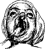
Figure 3.
deformity and largely restores proper functions. Its tasks are reduced first of all to improving feeding conditions in newborns, which is achieved to a certain extent by closing the defect of the upper lip by one of the existing methods (see Harelip). In unilateral cleft with slight protrusion of the intermaxillary bone, this is achieved more easily than in bilateral cleft with a far protruding incisor bone (see figure 3). In the latter case, one has to transect the vomer subperiosteally (Wangensteen's method) and press the incisor bone back into the gap between the alveolar processes of the upper jaw, after carefully freshening the adjacent surfaces. In case of discrepancy between the sizes of the incisor bone and the alveolar process defect, one has to trim the incisor bone from the sides to the required size. The repositioned incisor bone is held in the new position by the restored upper lip. Suturing the incisor bone to the alveolar process of the upper jaw is usually not used, since a bone fusion is obtained very rarely, and the stitches can damage the tooth germs. Some recommend removing the intermaxillary bone altogether, replacing the defect later with a prosthesis. In unilateral cleft with a strongly protruding incisor bone, one can fracture the alveolar process on the healthy side between the canine and the incisor, break it posteriorly and level it with the alveolar process of the other side. Most surgeons close the cleft of the upper lip in the first days after birth, since the operation presents no danger and can be done even without anesthesia. If it is necessary to put the incisor bone in place, one must reckon with greater trauma and heavier bleeding. This operation in the early period is done only in stronger children, while in weak ones it is preferred to wait until the child gets stronger. Early closure of the lip defect contributes to the narrowing of the hard palate cleft (sometimes to complete disappearance—Drachter, Payr) and thereby facilitates the future operation on it. This is also facilitated sometimes by the suture of the soft palate, which Berry recommends sewing before the hard palate in wide clefts of the hard palate. For sewing the palate (uranostaphyloplastica), the main method is Langenbeck's operation (see figures 4 and 5): the edges of the cleft are freshened with a sharp knife along the entire extent of the intended suture; then lateral incisions are made, starting from the hamulus of the pterygoid process, anteriorly, along the alveolar process, keeping to the bases of the alveoli.
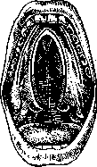
Figure 4.
ends, depending on the circumstances, either at the incisors or between the canine and the premolar, but always with such a calculation that the anterior leg of the future flap is not too narrow and weak. These lateral incisions within the boundaries of the hard palate penetrate to the bone. With special palatal elevators, through the lateral incisions, the outlined mucoperiosteal flaps are then separated from the hard palate along their entire length, carefully preserving the integrity of the anterior and posterior legs. With an elevator and scissors, the attachments of the soft palate to the edge of the bony part of the hard palate are separated. If the soft palate is insufficiently mobilized, the hamuli of the pterygoid processes of the sphenoid bone are fractured (Billroth), which is more expedient than the transection of the palatal muscles proposed by Langenbeck, the integrity of which it is important to preserve for the best function of the soft palate. The hemorrhage arising during the separation of the flaps is stopped by temporary tamponade and pressure. Separating the flaps of the hard palate, they try not to damage the palatal artery, which is not always successful. After mobilization of the flaps until they touch without tension and after stopping the bleeding, the median edges of the flaps are sutured with interrupted sutures (silk, horsehair, thin silkworm gut, wire). Special needles (Langenbeck, Sklifosovsky) are proposed for more convenient placement of sutures. In young children, to reduce the danger of sometimes quite heavy bleeding, Langenbeck's operation can be performed in two stages (Monnier, Wolff)—first, the flaps are separated and tamponaded, and after 4-8 days the edges are freshened and sutured. To prevent blood from entering the bronchi and lungs, operations are usually performed with the head tilted back and hanging over the edge of the table. Older patients can be operated on in a sitting position under local anesthesia. In children, general anesthesia is usually used (with a Braun, Junker, Roth-Dräger apparatus) through a rubber catheter passed into the pharynx through the nose. It is less convenient to use the usual Esmarch mask. For free access to the palate, a wide opening of the mouth and depression of the tongue are necessary, which is achieved by one of the mouth gags proposed for this purpose (Trélat, Whitehead, etc.). In cleft palate, after suturing the hard palate, a small triangular defect may remain in the anterior part, which then requires additional
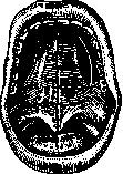
Figure 5.
al operation. If for some reason this cannot be done, then in extreme cases a prosthesis-obturator can be fitted, which usually performs its purpose well. Good contact of the freshened edges of the flaps, their good nutrition, and the absence of tension are necessary, otherwise the sutures easily diverge (most often at the border of the hard and soft palate). It is especially difficult to achieve success with poorly developed palatal processes and a wide cleft. To facilitate the suturing of the hard palate (uranoplastica), various operations have been proposed aimed at bringing the maxillary bones closer together either by bloody mobilization of the upper jaw (Codivilla), a part of it (Helbing, Schoemaker) or by forcible compression of the upper jaws, sometimes with incision of the still soft bone (Brophy, Karger) and with subsequent approximating wire suture through the entire jaw. Other bloodless methods of bringing the bones together boil down to the use of bandages, clamps (Neumann, Kredel, and others). To close a wide defect of the hard palate, it was also proposed to use the nasal conchae (Kraske), the vomer (Sabatier) or a flap from it in a unilateral cleft (Lannelongue, Eiselsberg), the little finger (Eiselsberg), as well as various soft tissues: a flap from the lip (Rosé), cheek (Thiersch, Delorme), tongue (Gersuny), skin flaps (Eiselsberg, Nussbaum, Rotter, etc.). Kirschner sews not only the hard and soft palate, but also the previously detached mucous membrane of the palatal plates from the nose side, detaches the pharyngeal muscles from the median plates of the pterygoid process, and separates the tensor veli palatini muscle from the pyramidal process of the palatal bone. Sewing the soft palate (staphylorrhaphia) is sometimes easier than the hard palate, but anatomical relationships sufficient for proper function are rarely obtained. In most cases, the soft palate turns out to be short, inactive, does not fit snugly against the posterior wall of the pharynx, does not provide sufficient separation of the oral and nasal cavities, and thereby the speech defect is not eliminated. To improve the function of the soft palate, it was proposed to suture its edge to the posterior wall of the pharynx, to sew a pedicled flap from the posterior wall of the pharynx into the soft palate defect, to inject paraffin under the mucosa of the posterior wall of the pharynx (to increase the so-called tubercle formed by the contraction of the muscles of the posterior wall of the pharynx and contributing to its complete closure by its contact with the soft palate and lateral walls). Recently, methods have been proposed for lengthening the soft palate by shifting it backward (retrotranspositio) together with the separated mucoperiosteal flaps of the hard palate (Ganzer, Ernst, Lvov, Dorrance), as well as detachment of the lateral walls of the middle part of the pharynx (mesopharyngoconstrictio), along the border between the pharyngeal muscles and the internal pterygoid muscle (Ernst), to narrow a pharynx that is wider than usual. In Ernst's operation (see figure 6), an incision of the mucosa is made from the posterior end of Langenbeck's incision along the pterygomandibular raphe to the lower posterior molars. Then the loose cellular tissue between the pharyngeal muscles and the internal pterygoid muscle is bluntly divided to the prevertebral fascia and the resulting lateral pockets are tamponaded. The palatal artery is freed from the can-
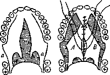
Figure 6.
Figure 7.
Ernst's incision (a), Flaps (A, B) shifted according to Limberg.
inwards and backwards. or by resection of a part of its wall (Lvov) or by osteotomy (Limberg), or it is opened at the first moment of the operation, to be resumed upon the restoration of blood circulation through the soft palate. Sometimes Ernst adds a backward displacement of the palate (see figure 7). Otherwise, it is the Langenbeck operation. To hold in place the tampons used until the pockets fill with granulations, Ernst uses a celluloid plate-prosthesis, which also protects the suture line. The latter operations, supplementing Langenbeck's operation, yield a good functional result (Limberg, Rauer) and apparently have a future. There are many advocates of closing the cleft of the hard and soft palate in the first months of life, before the child begins to speak. The purpose of early surgery is to teach speech with the restored anatomy of the palate and pharynx and to improve feeding conditions. Others prefer to operate no earlier than 2-3 years of age and older (after 5-10 years), when both the anatomical proportions are larger and children are more hardy. Later surgery does not affect subsequent function, but it protects against the late deformations of the upper jaw observed as a result of early operations. Every operation must, of course, be preceded by careful preparation of the oral and nasal cavities—eliminating, as far as possible, inflammatory processes of the mucous membranes and tonsils, carious teeth, etc. The postoperative period must be conducted with great care, eliminating all harmful factors (e.g., talking, solid food), with washing of the nasal and oral cavities, gargling, etc. The stitches are removed on the 5th-7th day. If the sutures part, it is useless to restore them immediately. It is better to wait until the inflammatory phenomena caused by the operation subside; small defects heal on their own. In case of complete divergence of the sutures, reoperation is recommended no earlier than a few months (5-10). Upon healing of the wound, it is necessary to take care of improving the patient's speech, for which persistent systematic training in the correct reproduction of sounds (logopedics) using the method of teaching the deaf-mute is required. In this way, it has sometimes been possible to achieve significant speech improvement even with incompletely closed palatal defects. Training is hindered by the fact that patients have already acquired a number of incorrect movements (of the lips, tongue, soft palate, pharynx) for speech reproduction, and also by the fact that patients do not hear the flaws in their speech during conversation. Only the reproduction of a gramophone recording gives them an exact idea of speech defects and helps in correct training. Massage and gymnastics of the soft palate also help to develop the usually atrophied muscles placed in new conditions. The soft palate can be massaged with a wooden, rubber, or metal instrument given a rounded T-shape. For the final elimination of speech defects, it is necessary to correct dental malpositions that disrupt articulation (Babitskaya).
Related articles
Mentioned in
Cite this page
“Cleft Palate.” Soviet Medical Encyclopedia. English translation of Bolshaya Meditsinskaya Entsiklopediya, 1st ed. (Moscow, 1928–1936), ed. N. A. Semashko. https://sovietmedicalencyclopedia.pages.dev/article/cleft-palate/