Harelip
Historical document, translated for reference. It reflects medical knowledge of the 1920s–30s and is not medical advice.
Summary
This article from the 1928–1936 Soviet medical encyclopedia describes the etiology, clinical presentation, and surgical treatment of harelip (cleft lip). It details the embryological origins of the deformity and discusses the functional complications, such as feeding and speech difficulties, as well as the surgical techniques used at the time.
Encyclopedia article (1928–1936)
HARELIP (labium leporinum, cheilo-schisis), a genotypic deformity consisting of a cleft of the upper lip of varying degrees. Among the deformities known as congenital facial clefts, harelip occurs more frequently than all others as an independent condition and, in comparison with a complete cleft of the alveolar process of the maxilla combined with a cleft of the hard and soft palate, constitutes a milder degree of deformity. Harelip is generally distinguished by the constancy of its form and location, being the result of the failure of the cleft between the medial and lateral nasal processes to close by the sixth week of embryonic life on one or both sides. The absence of the regular fusion of the processes forming the face may depend on insufficient growth energy of embryonic tissues under the influence of a certain genotypic defect, the inheritance pattern of which cannot yet be considered definitively established (sufficiently extensive pedigrees are lacking). The mechanism of the formation of upper lip clefts is understandable from the diagram of a human embryo of the first weeks (Fig. 1). In particular, the nose and the philtrum of the upper lip (philtrum) are formed from the medial nasal process, on the lower edge of which there are rudimentary elements of these organs in the form of a central notch and lateral tubercles. The clefts between the medial nasal and lateral processes gradually decrease due to the growth of the lateral nasal processes, and from them rema-
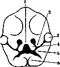
Figure 1. Face of a 31-day-old embryo; 1-medial nasal process; 2-lateral nasal process; 3-eye; 4-processus globularis; 5-maxillary process; 6-lower jaw. (After B. Seifert.) only the openings of the nasal passages, the outer edge of which is formed by the lateral nasal processes. The formation of the upper lip ends with fusion with the maxillary process. From the failure of the cleft between the medial nasal and maxillary process to close on one or both sides, different types and degrees of upper lip clefts occur, as well as deeper defects of the alveolar arch, hard and soft palate (see Cleft Palate). Thus, the cleft of the upper lip can be unilateral or bilateral. A cleft on one side is located to the side of the midline in a somewhat oblique direction toward the eye (more often on the left side); it can be complete, when it passes through the entire lip and connects with the nasal opening, or incomplete, reaching half or two-thirds of the lip, with a thinned skin bridge remaining between the lip and the nasal opening, containing no muscle tissue. Usually, the position of the cleft corresponds to the line between the canine and the 1st incisor. In cases of bilateral clefts, the middle part of the lip, corresponding to the intermaxillary (incisive) bone, usually lags in development. Less common are cases where the growing vomer pushes the intermaxillary bone forward, as a result of which the middle part appears in the form of a protruding trunk (Fig. 2). In unilateral clefts, the intermaxillary bone can also be pushed forward and stand obliquely. Sometimes a lip cleft can be limited to a slight depression along the edge of the lip. In the first two stages of clefts, disfigurement of the nose is observed in the form of a displaced and flattened nasal wing on the corresponding side. As with both complete and incomplete lip clefts, anomalies in the position and number of teeth are also observed. Harelip, in addition to the striking disfigurement of the face, causes a whole series of functional disorders related to nutrition and respiration. First of all, in infants, sucking is impaired during breastfeeding or bottle-feeding due to the inability to hermetically close the oral cavity and create negative pressure. Liquids introduced into the mouth enter the nasal cavity, causing irritation and choking. Aspiration pneumonia arises from the suction of food particles into the bronchi. Air entering the lungs unheated also contributes to the occurrence of pneumo-
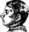
Figure 2.


nia and bronchitis. In children who have survived the first months of life, these difficulties and dangers are smoothed out due to adaptation. In growing children, harelip causes incorrect speech articulation. Operation for a lip cleft in healthy children is indicated in the first months of life, as with proper technique it gives good results; furthermore, the restoration of the lip contributes to the formation of the upper jaw arch and the narrowing or closure of the cleft
of the alveolar process; however, the operation for clefts of the hard and soft palate is currently postponed by most surgeons until the age of 2–3 years—until the time of eruption of the milk teeth. The operation aims to restore the function and normal shape of the lip and to correct the usually simultaneously existing disfigurement of the stretched nasal wing. It is necessary to suture not only the skin and muscles but also to pay attention to the careful suturing of the mucous membrane to form the vestibule of the mouth. This is achieved by various methods, of which the most widespread are the old method of Mirault for unilateral cleft and that of König for unilateral and bilateral. (From Figs. 3 and 4, the lines of incisions and sutures in both methods are visible.) Asymmetry of the nasal wing is eliminated by mobilizing it, detaching it from the edge of Figure 3. Operation according to König.
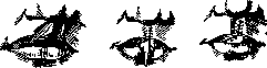
Figure 4. Three sequential stages of the operation according to Mirault. the apertura pyriformis, and shifting it toward the midline using the plate wire suture of Brown: the needle is inserted at the edge of the nasal wing, the wire is passed along the edge of the bony floor of the nose under the mucous membrane, and brought out at the edge of the healthy wing; the ends of the wire are secured with lead seals on aluminum or zinc plates, under which pieces of adhesive plaster are placed. This suture places the wing in a normal position and, in addition, prevents the sutured edges of the wound from separating. The wound is left open. To protect the wound from separation and the cutting through of sutures, Limberg recommends the Hagemann adhesive bandage. The plaster is rolled in the middle part into a tube, which is placed under the lip, and the ends are cut and spread out on the cheeks. In cases of a far-protruding intermaxillary bone, the operation presents great difficulty due to the impossibility of suturing the lip over the protruding process. Here, a submucosal vertical incision of the cartilaginous part of the vomer and setting it back (according to Bardeleben) or submucosal excision of a plate of corresponding size is recommended. In any case, one should not remove the protruding intermaxillary bone, which can be adjusted and fixed into the cleft of the alveolar arch. Otherwise, the formation of the upper jaw will be delayed.
Related articles
Mentioned in
Cite this page
“Harelip.” Soviet Medical Encyclopedia. English translation of Bolshaya Meditsinskaya Entsiklopediya, 1st ed. (Moscow, 1928–1936), ed. N. A. Semashko. https://sovietmedicalencyclopedia.pages.dev/article/hare-lip/