Lips
Historical document, translated for reference. It reflects medical knowledge of the 1920s–30s and is not medical advice.
Summary
This article describes the anatomy, embryology, and histology of the lips, including their muscular structure, vascularization, and innervation. It also discusses developmental defects such as harelip, cleft palate, and microstomia.
Encyclopedia article (1928–1936)
LIPS (Latin labium—lip), a term used to denote certain anatomical formations, for example: labium anterius et posterius orificii uteri externi (anterior and posterior lip of the external orifice of the cervix); labia pudendi majora et minora (greater and lesser pudendal lips, see Genitals); labium ostii pharyngei tubae auditivae s. Eustachianae anterius et posterius [anterior and posterior lip of the pharyngeal orifice of the auditory (Eustachian) tube]; labium valvulae coli superius et inferius [superior and inferior lip of the Bauhin valve (see)]; labium vocale (vocal lip, formed by the muscle of the same name and the fold of the laryngeal mucous membrane covering it); labium intermedium, laterale et mediale cristae iliacae (lips of the iliac crest). Labia oris (lips of the mouth), closing the mouth opening (rima oris) with two skin folds, are the beginning of the digestive tract (Rauber). They are formed differently: while the lower lip originates on the left and right sides from the mandibular process of the first visceral arch, with both processes joining at the midline, the upper lip is formed from three processes; both lateral ones originate from the same first visceral arch as the maxillary processes, while the median process originates from the frontal prominence, which forms the frontonasal process. In the embryo, there is a cleft between the median process and the lateral ones on each side, the so-called nasal groove, extending from the lateral part of the edge of the future upper lip to the nasal pit. These data on the developmental processes in the lips are very important for understanding some of their developmental defects (see Harelip, Cleft palate). Developmental defects of the lips include cases of synecheilia, when the fusion of the lips goes too far toward the midline; this defect causes a very small mouth, the so-called microstomia. Absence of the lips (acheilia) is a rare deformity. In their final form, the lips are separated from other parts of the face by shallow but definitely expressed grooves (nasolabial, labiomandibular, labiomental). A distinction is made between the upper and lower lip; they have the same structure. The skin of the lips is peculiar in its structure; it is supplied with hair, forming a mustache and beard in men, and a delicate downy layer in women; the thickness of the skin is rich in sebaceous glands. On the inner oral side, the lips are covered with a mucous membrane, which is a continuation of the mucous membrane of the cheeks; the epithelial layer of this mucous membrane belongs to the stratified squamous type. In the loose submucosal tissue, numerous, easily palpable mucous (and occasionally sebaceous) glands are noted. The mucous membrane, passing at the edge of the lips to the outer surface, forms here a ribbon-like red border of the mouth, which ends sharply at the place of fusion with the skin. In the thickness of the lips, muscles are located. A distinction is made between the strong fibers of the orbicularis oris muscle (m. orbicularis oris), serving as a sphincter, then muscle fibers originating from the muscles elevating the upper lip (mm. quadratus labii superioris, zygomaticus, caninus), from the muscles depressing the lower lip (mm. quadratus labii inferioris, triangularis), and from the muscles retracting the corner of the mouth (m. risorius). The lips are abundantly supplied with blood through a fairly large arterial branch of the external maxillary artery (art. maxil. externa), the circular artery (a. orbicularis), and through small branches of the surrounding arteries from the nasal, buccal, transverse facial, infraorbital, mental, and submental arteries. The venous network is also very rich; blood drains into the veins of the face and chin. Here it is necessary to point out the connection of the upper lip with the venous system of the orbit and venous sinuses at the base of the skull through the buccal veins (vv. buccales), which has great clinical significance in the spread of septic processes (see Figure 1). Lymphatic vessels drain into the lymph nodes located in front of the ear, and into the submandibular lymph nodes, as well as into the lymph node behind the angle of the lower jaw, on the lateral wall of the pharynx. In addition, the submental lymph nodes receive lymph from the lower lip (see Figure 2). The nerves of the lips originate: a) motor—from branches of the facial nerve, b) sensory—from the ramifications of the 2nd and 3rd branches of the trigeminal nerve, c) sympathetic—from the branches of the superior cervical ganglion of the cervical part of the sympathetic nerve. The mucous membrane at the base of the lips folds and passes to the mucous membrane of the gums. Along the midline it
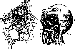
Figure 1.
Figure 2. Figure 1. Anastomoses between the veins of the face and the ophthalmic veins: 1-superior ophthalmic vein; 2-cavernous sinus; 3-inferior ophthalmic vein; 4-pterygoid plexus; 5-superficial temporal vein; 6-posterior facial vein; 7-internal jugular vein; 8-common facial vein; 9-submental vein; 10-anterior facial vein; 11-anastomosis between the inferior ophthalmic vein and the pterygoid plexus; 12-angular vein; 13-nasofrontal vein. (According to Corning.) Figure 2. Lymphatic vessels and regional lymph nodes of the lips: 1-submandibular lymph nodes; 2-hyoid bone; 3-internal jugular vein; 4-internal carotid artery; 5-deep cervical lymph nodes; 6-masseter muscle. (According to Corning.) [The lip] forms a fold of varying length and size in different individuals—the frenulum of the upper and lower lip, containing a small arterial branch. The function of the lips consists not only in closing the oral cavity during eating and sucking; they also play an important role in proper breathing and speech. Pathology. The shape and size of the lips vary even within the limits of the norm, and deviations are especially great in various kinds of pathological processes. In a pathological state, the lips can sharply change their original appearance. On one hand, there are very unsightly double lips, when (usually on the upper lip) the mucous membrane and its glandular layer hypertrophy; the altered part of the mucosa protrudes in the form of a large roll below the edge of the lip, especially when laughing. A rational treatment for this disfigurement is the excision of the excess part of the mucosa. On the other hand, hypertrophy of the lips occurs due to an increase in the number of lymphatic fissures contained in them, and due to lymph stasis in these fissures, signs of chronic inflammation and the formation of cracks on the free edge of the lip are simultaneously detected. Such a condition is observed mainly in scrofulosis, predominantly on the upper lip. Acquired elephantiasis (see Elephantiasis), which is the result of frequently recurring inflammations (especially erysipelas), approaches this form of lip enlargement. Rarely, an increase in the size of the lips of congenital origin is also encountered due to the dilation of lymphatic fissures; such a change is called macrocheilia; cavernous spongy tissue permeates all layers, the entire thickness of the lip, and spreads to the cheeks; a disfiguring elephantiasis results, mainly of the lower lip, which is based on a soft simple or cavernous lymphangioma. Finally, edematous doughy enlargements of the lips are encountered in acromegaly, as well as in cases of myxedema and cretinism. Sometimes the shape and structure of the lips depend on various types of trauma. Injuries are encountered on the lips very often in the form of incised and contused wounds, as well as in the form of crushing and bites. These wounds, although they are always infected, but in view of the fact that the tissues of the lips are very well vascularized, they rarely undergo wound infection; therefore, it is always desirable after cleaning the wound and stopping the bleeding to apply sutures to restore the correct shape of the lip. Special attention must be paid to the precise alignment of the edges of the vermilion border of the lip with sutures. Severe disfigurements of the lips are left behind by burns, especially 3rd-degree burns. In individual cases, after burns, the mouth opening is tightened by scars, the lips are everted, the corner of the mouth is pulled to the side, etc. For the treatment of these disfiguring scars, one has to resort to a number of plastic surgeries. Among the inflammatory processes of the lips, one must mention completely superficial diseases (eczema, sycosis, herpes labialis, etc.). Deeper and more serious acute inflammatory processes on the lips are the furuncle and carbuncle. Here it should only be pointed out the severity of these two diseases on the upper lip. Sometimes from the very beginning, the clinical picture of the disease takes on a severe character with high temperature; an infectious purulent thrombophlebitis of the facial veins joins the local purulent process; thrombophlebitis through the angular vein and the superior ophthalmic vein can spread further and higher, to the cavernous sinus; the latter complication is fatal. Treatment at first should be conservative; when an abscess has formed, one resorts to opening it either by an incision or by a Paquelin cautery. But with a carbuncle and in a malignant course, an early incision is necessary, splitting the entire inflammatory infiltrate, usually permeated with small abscesses; one should warn against squeezing out the cores. Primary infection with anthrax is observed on the lips and can create difficulties in terms of differential diagnosis with a carbuncle. A pustule that has turned into a black scab, surrounded by transparent vesicles, serous discharge from under the scab, and colossal edema around it (see Figure 3), and the finding of anthrax bacteria in a smear clarify the matter. Among chronic diseases, only three types have special significance: tuberculosis, syphilis, and scleroma. The transition of the process to the lips in rhinoscleroma is in some cases considered a secondary phenomenon. Tuberculosis in the vast majority of cases is secondary: the infection is usually introduced by the sputum of a patient suffering from pulmonary tuberculosis and having a wound or crack on the lip. In this case, a stubbornly non-healing ulcer develops, expanding, having undermined edges with a soft bleeding floor; it is painful, a wide rim of chronic edema is visible around it; individual tubercles are observed on the surface (see Figure 4). Syphilis is observed on the lips in the form of a primary lesion due to extra-genital infection, usually after a kiss with a sick person or after sharing dishes with them.

Figure 4. Tuberculosis of the lower lip. Figure 5. Hard chancre of the upper lip.
(According to Kappis.) According to the data of Chatenever at the State Venereological Institute, out of 50 extra-genital scleroses registered over 5 years, 25 were localized on the upper and lower lip. Relatively rapid development, painlessness, density, lardaceous floor of the ulcer (see Figure 5), and early swelling of the regional lymph nodes distinguish the hard chancre of the lip from a tubercular ulcer. Lesions of the secondary period are noted mainly at the corners of the mouth in the form of mucous patches (plaques muqueuses) and weeping papules. Tertiary lesions are manifested in the form of gummatous ulcers with a chronic course on an infiltrated base; these ulcers secrete dirty, grayish pus. But besides this, there is a sclerotic form, when the entire lip is permeated with dense fibrous tissue, the surface of the lip becomes dry, and cracks, so-called rhagades, appear on it. Syphilis of the lip is treated according to general rules (see Syphilis). Scleroma of the lip is a complication of scleroma of the pharynx and nose. Among neoplasms on the lips, a cyst of the mucous membrane of the lip, haemangioma, and cancer are most often encountered. The appearance of the cyst is very peculiar: on the inner surface, usually of the lower lip, above the even and pink level of the mucosa, a spherical, painless bulge rises; palpation determines an elastic formation the size of a pea or a cherry in the submucosal space; the apex of the protrusion shines through with a bluish tint; inflammatory phenomena are completely absent. The cyst is a retention cyst of a mucous gland and develops very slowly. To destroy the easily recognizable cyst, it is not enough to make an incision, because a recurrence soon occurs. Complete excision of the cyst under local anesthesia, with subsequent suturing, is necessary. The above-mentioned lymphangioma and haemangioma are related diseases. Both of them form in some cases more diffuse, in others more limited tumors. Haemangiomas of the lip are encountered in the form of telangiectasias (see Figure 6) and in the form of cavernous haemangiomas; often the first form passes into the second, i.e., is the beginning of the latter. The indicated vascular tumors

Figure 6. Haemangioma
of the upper lip and the left half of the face. (According to Stich.) Figure 7. Skin cancer of the upper lip. (According to Karpis.) are of congenital origin. Treatment initially, while the tumor is limited, consists of its excision; with its extensive spread, radical treatment is already difficult to apply; one has to resort to cauterization through the thickness of the tumor with a Paquelin apparatus or an electrocautery, sometimes with preliminary ligation of a large artery of the given region (carotid, external maxillary, etc.). Finally, sometimes fluids that coagulate blood and damage the endothelium of the cavity walls (alcohol, Clauden) are injected into the hemangioma cavity. The result is not always successful. Cancer of the Lips occurs in two forms: a) non-keratinizing and b) keratinizing. The first corresponds to the so-called Krompecher's basalioma and the concept of ulcus rodens; the second to the spinocellular cancer of the French. The first affects mainly the upper lip (see Figure 7), the second the lower (see Figure 8). The first develops very slowly, the second gives early metastases. Cancer of the lower Lip is encountered incomparably more often (more often in men than in women). In the USSR, this form is common and occupies one of the first places in frequency; it often develops on whitish spots (leukoplakia) of the mucous membrane of the Lips. A patient with cancer of the lip usually turns to a doctor when he has an ulcer of various sizes, covered with crusts, with infiltrated hard edges, with an uneven bottom, pitted with plugs and cores; the ulcer is painless and inevitably destroys the underlying tissues of the Lips. 3-6 months after the onset of cancer of the Lips, enlarged, hard lymphatic glands appear in the submental and submandibular regions. Under

Figure 8. Initial stages of cancer
a microscope, these enlarged lymphatic glands do not always turn out to be affected by cancer, but in practical terms, it is more expedient to consider them as a metastasis of the lip cancer ulcer and remove them. The occurrence of cancer of the lower Lip is sometimes linked to chronic, frequently repeated irritation of the epithelium on the marginal part of the Lip by various factors: the impact of cold and dry air, often leading to cracks in the mucous membrane of the Lips, the harmful effect of very hot food, tobacco combustion products, the pressure of a smoking pipe on the Lips, etc. An operation performed in a timely manner, before the formation of metastases in the lymph glands, yields up to 70-95% of complete recoveries according to the statistics of foreign clinics (Mayo, Balfour, Bloodgood clinics); in the presence of metastases, even insignificant ones, in the mobile regional lymph glands, this percentage drops sharply to 40-50. The operation itself consists of a wedge-shaped or trapezoidal excision of the diseased section of the Lip with subsequent direct suturing connecting the edges of the wound, or with the performance of plastic surgery of the Lip from the tissues of the cheek according to Dieffenbach. Depending on the presence of swollen regional lymph glands, it is recommended, if possible, to completely remove the regional lymph glands together with the cellular tissue and sometimes together with the salivary glands (Herzen). Another method of treating cancer of the Lips is the application of radiant energy according to the method of the Regaud Institute (Paris) in the form of larding the tumor tissues or the area around the tumor with metal needles containing radium or, which is undoubtedly worse, its emanation. Against metastases on the neck, tubes with radium enclosed in a special collar are used. At the present time, mixed radiological and surgical treatment is often used. The very encouraging results obtained with the use of one or the other method prove that cancer of the Lips is a completely curable disease if attention is paid to it in a timely manner.
Related articles
Cite this page
“Lips.” Soviet Medical Encyclopedia. English translation of Bolshaya Meditsinskaya Entsiklopediya, 1st ed. (Moscow, 1928–1936), ed. N. A. Semashko. https://sovietmedicalencyclopedia.pages.dev/article/lips/