Trigeminal Nerve
Historical document, translated for reference. It reflects medical knowledge of the 1920s–30s and is not medical advice.
Summary
The trigeminal nerve is the fifth cranial nerve, a mixed sensory-motor nerve that originates from the ventral surface of the middle third of the pons. It consists of a larger sensory root and a smaller motor root, with sensory fibers providing innervation to the face, mouth, and other structures, while motor fibers control the masticatory muscles.
Encyclopedia article (1928–1936)
TRIGEMINAL NERVE, trigeminal nerve, V pair of cranial nerves, mixed sensory-motor nerve. The trigeminal nerve originates on the ventral surface of the middle third of the pons and is represented there by two roots: an outer, thicker, sensory root (portio major of the trigeminal nerve, or sensory root), and an inner, thin, motor root (portio minor of the trigeminal nerve, or motor root). In the sensory root pass fibers conducting sensation from the skin of the face, anterior part of the hairy scalp, eye membranes, nose and mouth, cerebral dura mater, teeth and periosteum of facial bones. From the motor root are formed fibers for the masticatory muscles (masseter, temporal, and pterygoid muscles) and for m. mylohyoideus, anterior belly of m. digastricus, m. tensor veli palatini and tensor tympani. | The sensory root of the trigeminal nerve begins from the Gasserian ganglion (see). The cells of the ganglion are unipolar, their processes divide into peripheral and central branches. From the latter the sensory root is formed. Entering the pons, they divide into short ascending and long descending fibers. All ascending fibers end in the sensory nucleus (nucleus sensibilis principalis of the trigeminal nerve, or pontine sensory nucleus of the trigeminal nerve, fig. 1). The nucleus has an extension of 4-5 mm, consists of cells of various sizes, sometimes scattered, sometimes collected in groups. The long fibers descend through the medulla oblongata to the cervical region and form the descending, or spinal root of the trigeminal nerve (radix descendens, or spinalis of the trigeminal nerve). Throughout its
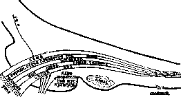
Figure 1. Diagram of the location of the motor roots and nuclei of n. trigemini in the pons and medulla oblongata.
Along its course, it is accompanied by cells forming the sensory nucleus of the spinal root (nucleus sensibilis radicis spinalis T. p.). On the periphery of the descending root, the marginal cells of Waldeyer (Marginalzolleii Waldeyer) are grouped. In the spinal root, fibers conducting sensitivity from different levels of the face and mucous membranes do not mix, but are topographically separated. Fibers of the spinal root give numerous collaterals to the nuclei of the facial and vagus nerves and to the motor nucleus of the T. p., forming the anatomical basis for numerous reflexes (see below). Part of the sensory fibers participates in the formation of the mesencephalic root of the T. p. (tractus mesencephalicus, s. cerebralis T. p.), a small bundle extending along the lateral wall of the Sylvian aqueduct to the level of the anterior colliculus. The root is accompanied by ovoid cells with homogeneous substance (nucl. radicis mesencephalici T. p.). The nuclei of the mesencephalic and spinal roots are considered as intervertebral ganglia that have penetrated into the brain. Part of the fibers of the mesencephalic root descends, forming the bundle of Probst (fasc. Probst), which ends in the nuclei of Deiters, glossopharyngeal, and vagus nerves. Individual fibers descend to the motor nucleus of the T. p. It is located at the level of the entry of the sensory root into the pons Varolii (nucl. motorius, s. masticatorius T. p.). The nucleus has an ovoid shape, 2.5-3 mm long, and is divided into a dorsal cellular group for innervation of the masticatory muscles (mm. masseter et temporalis) and a ventral group for mm. mylohyoideus, pterygoidei, and digastricus. The central pathway for sensory fibers of the T. p. is constructed in two ways. One part of the fibers begins from the large cells of the inner part of the sensory nucleus and goes backward. Crossing the midline ventrally from the nucleus of the hypoglossal nerve, then located dorsally above it, these fibers go in the oral direction. At the level of the pons, they mix with spino- and bulbo-thalamic fibers and end in the ventral nucleus of the thalamus. The second part of the central pathway begins from the frontal part of the sensory nucleus. The fibers ascend as part of the medial loop and also end in the ventral nucleus of the thalamus. They pass their entire path separately from the first. It is not established whether the spinal root participates in the formation of central pathways. The central motor neuron for the T. p. begins in the lowest part of the anterior central gyrus. The axons of the cells located here pass through the internal capsule and cerebral peduncle. At the level of the posterior colliculus, they exit from the pyramidal bundle and descend partially to the motor nucleus of the opposite side, partially to the homonymous one. The motor root, after exiting the pons, pierces the dura mater together with the sensory root at the point of attachment of the tentorium cerebelli to the upper surface of the pyramid, lies between both layers of the dura mater, and approaches the Gasserian ganglion from its lower surface. From the Gasserian ganglion, three primary branches of the T. p. emerge. The first two contain exclusively sensory fibers, while the third contains both sensory and motor fibers from the motor root (Fig. 2). The first branch closest to the midline is called the first, the one farthest from it is called the third. The first branch is the orbital nerve (n. orbitalis, s. ophthalmicus), a sensory nerve for the dura mater, skin of the forehead, bridge of the nose, upper eyelid, eye membranes, and part of the nasal mucosa. Still within the cranial cavity, a recurrent nerve (nervus recurrens) branches off from it, turning backward and ending in the tentorium cerebelli. The n. ophthalmicus itself penetrates through the superior orbital fissure into the orbit, where it divides into three large branches: 1) the nasociliary nerve (n. nasociliaris), participating in the formation of the ciliary ganglion, 2) the ethmoidal nerve (n. ethmoidalis), innervating the mucous membrane of the anterior ends of the first and second conchae and the skin of the bridge of the nose, 3) the supraorbital nerve (n. supraorbitalis), which, after passing through the orbit, bends at its edge and innervates the skin of the forehead and anterior part of the hairy part of the head. The second branch is the maxillary nerve (n. maxillaris), thicker than the first, and exits the skull through the foramen rotundum. It also gives a recurrent branch spreading along the dura mater together with the a. meningea media. After exiting the foramen rotundum, the second branch goes to the pterygopalatine fossa, where it divides into the following main branches: 1) the superior dental nerve (n. alveolaris sup.), branching in the teeth of the upper jaw, 2) the infraorbital nerve, entering the orbit through the inferior orbital fissure (n. infraorbitalis). Its branches in the bony canal of the maxillary sinus connect with the n. alveolaris sup. and form the superior dental plexus. After exiting the orbit, the infraorbital nerve branches into the skin of the lower eyelid, cheek, and upper lip. The second branch participates in the formation of the sphenopalatine ganglion (ganglion sphenopalatinum), giving sensory fibers to the nasal nerves (nn. nasales), branching in the mucous membranes of the choanae, posterior ends of the two superior conchae, and to the palatine nerves (nn. palatini). The third branch is the mandibular nerve (n. mandibularis, s. inframaxillaris), exiting the skull through the foramen ovale. Like the first two, it also gives a recurrent branch to the dura mater of the brain. After its exit from the oval foramen, the auriculotemporal nerve (n. auriculotemporalis) branches off, which curves around the articular process of the mandible, passes through the parotid gland, and ascends to the temple. The motor fibers of the third branch separate directly at the oval foramen (n. masticatorius) and go to the muscles of mastication, bearing the name of the innervated muscles: temporal (nn. temporales), masseteric (nn. masseterici), and pterygoid (nn. pterygoidei). A special branch, n. mylohyoideus, innervates the muscle of the same name and the anterior belly of m. digastricus. After giving off the motor branches, the third branch enters the mandibular canal, passes through it, exits through the mental foramen, and branches into the skin of the chin and lower lip. Along the way in the canal, fibers branch off to the gums and teeth of the lower jaw. Individual branches of the T. p. anastomose with each other and with other nerves of the head. The T. p. gives sensory fibers to the ciliary, sphenopalatine, and lingual ganglia and motor fibers to the ganglion oticum. Through the T. p. system, a large number of reflexes are realized. For most of them, only receptors pass through the T. p., while the effectors go through the facial nerve. These are the corneal, conjunctival, palatoglossal, from the bridge of the nose, from the auditory canal, sucking, and other reflexes. Pathology. The large area occupied by the nuclei and roots of the T. p. in the pons and medulla oblongata determines its involvement in a number of pathological processes occurring in these areas. Paralysis of the masticatory muscles occurs in bulbar paralyses, poliomyelitis, and hemorrhages into the pons. The spasmodic closure of the jaws (trismus), caused by irritation of the cells of the motor nucleus of the T. p., is a cardinal sign of tetanus. When one half of the pons is affected, a crossed sensory syndrome sometimes occurs, when anesthesia is localized on the side of the face, homonymous with the focus, and on the opposite limbs (hemianaesthesia cruciata). In syringobulbia and less often in tabes dorsalis, the spinal root of the T. p. sometimes suffers, which manifests as a dissociated disorder of facial sensitivity (see Syringomyelia) with boundaries forming a semicircle encompassing the nasal and oral openings. In tumors of the pontocerebellar angle, weakening or disappearance of the corneal reflex is an early and valuable symptom. Meningitis of the base of the brain, especially syphilitic, often causes lesions of the T. p., mainly its sensory fibers. Motor paralyses of the trigeminal nerve of this etiology are rare; they are of a flaccid nature, i.e., accompanied by atrophy, areflexia, and reaction of degeneration of the masticatory muscles. Bullet wounds to the base of the skull during the imperialist war caused lesions of the T. p. in approximately 10% of cases, more often its sensory fibers. The most common are inflammatory diseases of the Gasserian ganglion and the branches of the T. p., which form the basis of a huge number of neuralgias of the T. p. (see Neuralgia). In neuralgia of the T. p., severe pains of boring or shooting nature are sometimes concentrated in one place, sometimes spread along the course of individual branches. At their height, the pains sometimes irradiate beyond the face. Attacks of neuralgia occur almost exclusively during the day and last for several minutes. Prolonged neuralgic pains are caused by the involvement of the sympathetic nerve. On the side of the neuralgia during an attack, sometimes a painful tic (see Tics) and autonomic disorders in the form of vasodilation, increased secretion of tears, nasal mucus, and saliva occur.
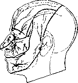
Between attacks, Ball's tender points are often found, which correspond to the sites where cutaneous branches emerge from bony canals (Fig. 3). In the area of neuralgia, hyperalgesia is not uncommon. Attacks of neuralgia occur either spontaneously or are triggered by cooling of the face, chewing large pieces, etc. Acute neuralgias caused by malaria, influenza, are more often localized in the first branch, while chronic ones more frequently affect the second and third branches of the trigeminal nerve. In diagnosing neuralgia, differential diagnosis should be conducted from eye diseases (for example, glaucoma, iritis) and diseases of the paranasal sinuses (frontitis, sinusitis). The latter sometimes spread to the trigeminal nerve and cause neuralgia. The prognosis of trigeminal neuralgia is determined by the etiology and duration of the disease. Neuralgias that occur after acute infections often, especially in young age, have a favorable course and end in several days or weeks. Neuralgias in the elderly, especially the toothless, usually take a chronic course. Neuralgia begins with one branch, and subsequently either remains limited to it or spreads to other branches, but does not cross to the opposite side of the face. The left half of the face is affected more often than the right.
Treatment of neuralgia usually comes down to therapy of the underlying disease (malaria, syphilis, diabetes, etc.). In symptomatic therapy of neuralgia, thermal procedures and the application of galvanic current play a major role. During an attack, analgesics are used, mainly of the salicylate series. Drugs of the morphine series are permitted only as an exception in acute cases; their use in chronic neuralgias usually leads to morphinism.
Surgical treatment of trigeminal neuralgia. All modern methods of surgical treatment of trigeminal neuralgia can be divided into two large groups: 1) palliative operations and 2) radical operations. The first group can in turn be divided into two subgroups: bloodless treatment-injections and bloody-excision of peripheral branches at the site of emergence from bony canals (canalis supraorbitalis, canalis infraorbitalis, canalis mentalis). The second group includes operations of sectioning branches in front of the Gasserian ganglion-extracranial and intracranial operations on the Gasserian gang itself. The latter in turn include partial resection of the ganglion, its complete extirpation, section of the central root behind the Gasserian ganglion from the middle cranial fossa, and finally section of the root in the posterior cranial fossa.
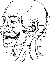
From the operations of the first group, alcohol injections deserve attention, developed mainly by surgeons. For the first time, treatment of trigeminal neuralgia by alcohol injections was described by Schlosser in 1903; subsequently this method was thoroughly developed and described by Hartel, Kulenkampff, Sicard, Kirchner, and others in our Union by Gutnikov, Irger, and others. At present, the method of alcohol injections has taken the following form: injections on the periphery at the site of emergence of the trigeminal nerve, along its trunk, so-called intermediate injections, basal injections, and intracranial injections (Fig. 4). Here we are already talking about injection into the Gasserian ganglion. For proper production of the injection, a clear understanding of the topography of the course and relationships of the branches of the trigeminal nerve is required. In addition, this method required the study of the action of chemical substances proposed for injection, in particular alcohol, and the physiological and pathological phenomena observed in this connection. Only after taking into account all these data did the method receive its modern form: a) alcohol of 70-80% concentration is used - a concentration that guarantees, on the one hand, bactericidal properties, and on the other, ensures functional interruption of the nerve. On the basis of experiment and clinical observations, it has been established that alcohol at this concentration destroys nerve fibers, causing secondary degeneration. Most authors indicate that the degenerative process does not spread to the central segment of the nerve. The injection technique is not complicated. Injection is performed with ordinary needles, but necessarily thin ones. For intermediate injections into the base of the skull and for injections into the Gasserian ganglion, long needles with a certain graduation of their length are used. Some authors (Braun) propose having a sliding sleeve on the needle, however this seems superfluous. The diameter of the needle is recommended not to exceed 0.7 mm, and the needle should be elastic and to a certain extent
Fig. 4. Points for injection of alcohol into the branches of the trigeminal nerve: 1-for the first branch through the supraorbital foramen; 2 and 3-for the second branch through the orbit; 4 and 5-for the second branch through the zygomatic bone; 6-for the second branch from 1/3 to A of the maxillary bone; 7-for the Gasserian ganglion or ordinary node (according to Hartel); 8-paths to the peripheral branches of the second branch (according to Hoffa); 9-paths to the third branch (according to Braun); 10-paths to the third branch (according to Miehausen); 11-paths to the mandibular nerve; 12-paths to the Gasserian ganglion; 13-paths to the mandibular nerve.
A special or modified eccentrically positioned needle adapter (syringe with a needle) is used. The patient is given morphine or veronal orally (0.5 g the day before). The operation is performed under local anesthesia. For peripheral injections, the anesthesia is superficial; for intermediate and ganglion injections, the path of the needle is anesthetized, but the trunks themselves are not anesthetized in any of these three cases, which allows one to judge by the pain sensation the proper location for alcohol injection. The peripheral trunks are easily anesthetized; they are found at the site of exit through the bone openings: nn. supraorbitalis, infraorbitalis, mentalis, zygomatico-orbitalis, nasalis externus. From 0.5 to 1.0 of alcohol is injected. Intermediate injections are made on the nerves passing through the orbit, nasal and oral cavities: n. ethmoidalis, ram. naso-lacrimalis, spheno-palatinus, n. alveolaris post., n. palatinus, n. alveolaris and lingualis. The amount of alcohol injected ranges from a few drops to 0.5-0.8. Basal injections of the branches of the trigeminal nerve are mainly limited to the II and III branches; regarding the first branch, most authors hold a negative opinion. Injection of the II branch is made in the area of the foramen rotundum; for access to which three routes have been proposed (Fig. 4): 1) orbital (Hertel's)-orbital, 2) through the fossa pterygo-palatina-pterygo-palatine fossa, and 3) the route from the oral cavity (Ostwald's), which was for a long time very common. The site of puncture in this case is the area behind the wisdom tooth, from where the needle is directed toward the vault, and when the needle reaches the planum intratemporalis, its tip is advanced slightly forward until it no longer glides along the bone; now the needle enters the area of the fossa spheno-palatina, from where when it is advanced 6-8 mm upward, the needle can enter the foramen rotundum. The amount of alcohol in these injections is from 1.0 to 1.5 cm3. The III branch is reached: 1) by Hertel's method, which is a modification of Schlesser's method through the cheek; 2) by the so-called transverse route in the area of the zygomatic bone (Braun, Offerhaus, etc.), and 3) by Ostwald's method. The second method is most often used, which is as follows: the needle is punctured at the middle of the zygomatic bone, advanced 4-5 cm in depth to the lamina pterygoidea externa, where the distance between the tip of the needle and the foramen ovale is approximately 1 cm. Further, the needle is withdrawn slightly and with gliding movements is directed somewhat backward, remaining at the same depth of immersion and causing characteristic paresthesias. Hertel's method is as follows: the needle is punctured on the cheek in the area of the molar teeth without piercing the mucous membrane (under the control of the left hand finger, introduced into the oral cavity). The direction of the needle is determined by the sagittal plane passing through the pupil and the line passing through the articular tubercles of the lower jaw. Immersing 5-6 cm, the needle reaches the depth where the oval opening is located. Hertel's method was subjected by various authors to minor modifications that did not change its essence. All methods of basal intracranial injections, regardless of their effectiveness, are not without danger of damage to neighboring organs and various complications, which requires the development of precise and serious indications for their performance. Only in cases of persistent recurrent attacks and after unsuccessful therapeutic measures-sanitation of the oral cavity, nasopharynx, accessory sinuses, etc.-is the use of alcohol injections necessary. Into the Gasserian ganglion, from 1.0-2.0 to 4.0 of alcohol is injected. The rate of injection into the Gasserian ganglion and in basal injections should be slow-from 2 to 15-20 minutes (Külenkampf). For basal and intracranial injections, hospitalization of the patient is recommended due to the possibility of severe phenomena. The therapeutic effect of peripheral, intermediate, and basal injections varies among different authors. Complete cure is observed by some authors in 26% (Patrick), in 10% (Fleisch), while others report somewhat higher rates (Ostwald). The effect of alcoholization of the Gasserian ganglion is relatively persistent. According to Hertel's data, a favorable effect was noted by him in 50% with observation of patients for several years; in 24% there were recurrences. Külenkampf and Gutnikov (77%) had approximately the same data. According to Gutnikov's latest statistics from 1920, the percentage of cures is 68.5, recurrences 31.5. Among the complications of this method, sclerotic and trophic changes, disturbances of sympathetic innervation of the eye up to neuroparalytic keratitis, and finally fatal outcomes are described (Gutnikov).-Bloody, non-radical operations include excisions. The unreliability of the results obtained in these operations is explained to a large extent not so much by the imperfection of the surgical intervention itself as by the fact that until recently it was not possible to determine with sufficient accuracy the site of the origin of pain (locus morbi). The definite effect obtained by various authors can probably be explained by the fact that in some cases the locus morbi was located peripherally, and in others, when the locus morbi could not be removed, yet a therapeutic effect, even if temporary, was still obtained, the latter can be attributed to trauma in the complex understanding of this word. Here, among other moments of trauma, such as hemorrhage and its absorption, the phenomenon to which Monakov gave the name diaschisis probably also plays a role. According to Dege's material, only in 14% are the results favorable; in other cases, recurrences were observed after two to three years; in 27% the recurrences were with not particularly severe pains. More than 50% underwent reoperation. Radical operations include neurotomies at the base of the skull and intracranial neurotomies. The former are performed mainly on the III branch. Intracranial neurotomies are performed on all three branches, but most often on the III branch and very rarely on the I. Access to the III branch does not essentially differ from access to all three branches in their intracranial section. Among the operative methods proposed for this purpose (Krause, Hartley, Quenu, Poirier, Lexer, Cushing, etc.), Lexer's method is the most widespread, which in essence is as follows. The skin incision begins in front of the tragus not lower than the beginning of the earlobe, is carried upward, goes in an arc along the line connecting the upper edge of the auricle with the eyebrow on the same side, and descends past the outer edge of the palpebral fissure. Thus, both parallel vertical incisions do not descend below the place where the corresponding branches of the facial nerve pass. Further, the zygomatic bone is exposed, from which a piece of it corresponding to the width of the skin incision is removed with a Gigli saw. Now the skin incision is deepened through the muscles to the periosteum, which is cut and carefully separated with a periosteal elevator together with the m. pterygoideus ext., attached to the crista infratemporalis. Then the skin-muscle flap, consisting of the m. masseter with part of the excised zygomatic bone, the lower part of the temporal muscle, the lateral pterygoid muscle together with the periosteum separated from the skull, is turned downward. Thus, the infratemporal fossa and the III branch of the trigeminal nerve passing through it are exposed. Behind the branching of the III branch (n. buccinatorius, lingualis and alveolaris inf.), the a. meningea media is visible in depth. The found branches are taken on a small blunt hook and cut with a knife; some authors suggest performing a physiological section by means of endoneural alcohol injection. Intervention on the trunks of the branches did not always give lasting results; cases of recurrence of pain were often observed both in the operated branch and in neighboring branches. The cause of this is either regeneration of the trunks, or central neuromas, or unrecognized disease of the Gasserian ganglion, which in the first period of the disease manifests itself as involvement of only one branch, or insufficient study of the patient in terms of general diseases, such as neurosis, diabetes, etc. (Adson). Often after this operation, it is necessary to intervene again and perform resection of the 7S0 Gasserian ganglion or section of the root between the Gasserian ganglion and the medulla oblongata. Removal of the Gasserian ganglion is a serious operative intervention not only due to the difficulty of access to it but also due to its complex relationships with the venous plexuses of the base of the skull, as well as due to the proximity of the a. meningea media, sinus cavernosus, a. carotis interna. The methods of access to the Gasserian ganglion most frequently used were developed by Lexer and Krause. Each of them has its own advantages and disadvantages. By the method described above, one reaches the III branch of the n. trigemini and exposes the area of the cristae infraorbitalis, in which a hole is drilled, which is expanded with rongeurs upward. At this point, the dura mater and the a. meningea med. passing here are exposed. The dura mater is carefully separated from the base of the skull and the hole in the bone is expanded to the foramen ovale. The a. meningea med. is isolated and after ligatures are applied, it is cut at this place. The II and III branches are well visible at this point, although the foramen rotundum is still separated from the trepanation hole by a narrow strip of bone.
The temporal lobe of the brain, covered by the dura mater, is lifted upward with a spatula, and after an incision in the dura mater, Gasser's ganglion is exposed, which is separated from the cavum Meckeli. The latter is recommended to be done bluntly, while at the same time pulling the third branch, under which a thread has been passed for this purpose. The separation of Gasser's ganglion is complicated by adhesions with the dura mater, which must be separated with a knife. Having cut the second branch and the trunk of the trigeminal nerve, Lexer turns the ganglion forward and also cuts the first branch, which is stretched in the process, and only at the very end is the third branch cut. The bleeding that occurs after removal of Gasser's ganglion can be stopped by tamponade. To prevent subsequent bleeding, a strip of gauze is left for several days. Krause's operation differs not only in the incision of soft tissues and bone (the incision, unlike Lexer's method, is made higher above the base of the zygomatic bone with the downward displacement of the skin-muscle-bone flap of the temporal region), but also in intracranial techniques. The temporal lobe must be lifted upward with a spatula significantly higher than in Lexer's method. Next, Krause grasps Gasser's ganglion near the trunk with a Tyrshel hook and, carefully cutting the second and third branches at their passage through the foramen ovale and foramen rotundum, also carefully and slowly turns the ganglion, which is still connected to its trunk and first branch. With gradual turns, it is possible to separate this connection and then remove Gasser's ganglion. In Krause's method, a major inconvenience is the need to lift the lower-lateral surface of the hemisphere, which often leads to compression of the brain. In addition to very severe bleeding, which requires a lot of time to stop, a serious complication is paralysis of the muscles innervated by the third branch (mm. masseter, temporalis, pterygoideus internus), and finally severe eye conditions (keratitis neuroparalytica) due to loss of corneal sensitivity and disruption of sympathetic innervation, since a significant part of the sympathetic fibers going to the eye pass through Gasser's ganglion (Burdenko-Rapoport). The severity of the operation is characterized by a high mortality rate - from 13 to 20 (Krause). The final results are very good in terms of relief from pain. However, with this operation too, a recurrence of pain is observed, which French authors partly attribute to centrally originated pain, partly to pain caused by an advanced sclerotic process. To avoid harmful complications from the eye - partial extraction of Gasser's ganglion was proposed with the intention of preserving the first branch, if it is not involved in the pathological process (Frazier, Stookey, Hutchlnson). The cutting of the root behind Gasser's ganglion is performed from the middle cranial fossa and from the posterior. The root was initially cut completely (Horsley, 1891), but in 1919 Frazier published his proposal to preserve the motor root, which can be separated from the mass of the root. Cushing, Kanavel, Davis (Gushing, Kanavel and Davis) developed this proposal, and since 1922 this operation has become widespread, mainly in America, where many surgeons have performed hundreds of such operations. Cushing performed 375 such operations, Frazier-511, Edson-371. The results of the operations are stable, but the technique is difficult, since it is generally difficult even when accessing Gasser's ganglion, but also requires special lighting equipment, without which it cannot be performed. With this operation, in a small number of cases, severe general cerebral phenomena and the above-mentioned eye complications were observed. In 2%, cases of death from shock and cerebral hemorrhages are described. Attempts to divide the root in such a way as to leave the fibers of the first branch untouched have not yet become widespread. The operation of transplanting the bulbar root, proposed as early as 1903, was rarely practiced. Only recently (1932) Dandy drew attention to it; the operation is still little known, and its technique is being developed in the direction of clarifying the topography of the motor, tactile, and painful parts of the nerve in this section. It is recommended to cut only the painful part of the root, which is successfully achieved (Dandy).
N. Burdenko. TRIGONITIS, trigonitis, inflammation of the mucous membrane of the trigone of the bladder (trigo-num Lieutaudi). Clinically, inflammation of the neck of the bladder (cystitis colli) is usually observed simultaneously. The rest of the mucous membrane of the bladder remains unchanged in this case. Acute T. occurs as a result of the spread of the inflammatory infectious process from the posterior part of the urethra to the neck of the bladder and the trigone, for example, in acute posterior gonorrheal urethritis. Inflammation of the prostate gland is also usually accompanied by acute T. Acute T. is accompanied by severe dysuria, and sometimes terminal hematuria. The urine contains a large number of leukocytes. During cystoscopy, a picture of acute inflammation of the trigone mucosa can be detected, but cystoscopy should be considered contraindicated in acute T., since the introduction of instruments in acute T. threatens complications and worsens the course of the process. Chronic T. is observed mainly in women and usually has the character of a non-infectious, but stagnant process. It is based on a circulatory disorder in the area of exit from the bladder, depending either on the incorrect position of the uterus (retroversio or retroflexio), in which the cervix presses on the area of the trigone of the bladder, or on the prolapse of the anterior vaginal wall, usually insignificant, or on a chronic inflammatory process in the parametrium. In chronic T., subjective disorders are insignificant, often completely absent, and manifest as a certain urgency and increased frequency of urination, sometimes as unpleasant sensations during urination itself. All these phenomena are not constant, but occur periodically, appearing or intensifying with the approach of menstruation as a result of the resulting hyperemia of the pelvic organs. The urine usually shows no deviation from normal. During cystoscopy, the trigone mucosa is found to be loosened, dull, and edematous. Sometimes individual white spots are found, rising above the rest of the mucosa and representing areas of thickened, metaplastic, desquamating epithelium (T. areata alba). In the area of the sphincter, growths in the form of villi are often found. Treatment of acute T. consists in treating the main infectious process in the urethra or prostate gland; in chronic T. in women, symptomatic treatment consists of instillations of silver nitrate solutions (x/a-1%) into the neck of the bladder, while radical therapy consists in treating the main gynecological disorder.
i. Epstein. TRISMUS, tonic spasm of the masticatory muscles (mm. masseteres, temporales, pterygoidei). In severe cases, the teeth are so tightly clenched in T. that they cannot be separated by either active or passive attempts, the masticatory muscles are sharply tense and hard to the touch. Usually, the muscles of both sides are involved in T., in rarer cases, for example, when the pterygoideus muscle of only one side is in a state of spasm, there is a simultaneous deviation of the lower jaw to the opposite side. T. depends on irritation of the motor portion of the trigeminal nerve, the centers of the masticatory muscles in the cerebral cortex, and the pathways going from them, arising both directly and reflexively. T. is one of the earliest and most characteristic symptoms in tetanus, then in meningitis, but it also occurs in a number of other diseases of the nervous system, both more general and more localized, for example, in tetany, in the tonic stage of an epileptic seizure, in diseases in the area of the Varolius bridge (tumors, acute bulbar paralysis). Irritation of the corresponding cortical centers by any pathological process can also give a picture of T. In all these cases, T. is usually accompanied by other symptoms characteristic of the given disease. Cases of isolated T. are rare and occur mainly reflexively, due to a local focus of irritation in the area of the lower jaw, for example, in inflammatory processes in the area of the temporomandibular joint or the mucous membrane of the latter, in periostitis of the lower jaw, in carious processes in the wisdom tooth, in inflammatory processes in the masticatory muscles. Finally, T. can occur not only in organic, but also in functional diseases of the nervous system; it has been repeatedly described, for example, in hysteria.
Related articles
Mentioned in
- Anastomoses
- Blinking
- Brown-Séquard Syndrome
- Brown-Séquard Tract
- Conjunctival Reflex
- Convulsions
- Corneal Reflex
- Destructive
- Enophthalmus
- Facial Nerve (facial nerve, the seventh pair of cranial)
- Flechsig's Tract
- Gasserian Ganglion
- Glaucoma
- Glycogeusia
- Gradenigo Syndrome
- Graduated Cylinder
- Hemispasmus
- Herpes
- Jaws
- LACRIMATION
Cite this page
“Trigeminal Nerve.” Soviet Medical Encyclopedia. English translation of Bolshaya Meditsinskaya Entsiklopediya, 1st ed. (Moscow, 1928–1936), ed. N. A. Semashko. https://sovietmedicalencyclopedia.pages.dev/article/trigeminal-nerve/