Electric Organs
Historical document, translated for reference. It reflects medical knowledge of the 1920s–30s and is not medical advice.
Summary
Electric organs are specialized structures found in certain fish that generate electrical discharges. These organs, modified from muscle tissue or glandular elements, vary in structure and function among different electric fish species.
Encyclopedia article (1928–1936)
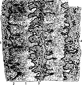
Electric organs are characteristic of certain fish, which are therefore called electric fish (fig. 1); these include the electric ray (Torpedo marmorata), the electric catfish (Malopterurus), and the electric eel (Gymnotus electricus). Weaker electric organs are found in Raja and Mormyrus (so-called pseudo-electric fish). Electric organs are modified muscles. In the electric eel, they exist as two pairs of elongated structures, one pair located along the dorsal side of the tail, and the other along the ventral side of the caudal fin; they extend for 4/5 of the length of the body. The degree of their development can be judged by the fact that the weight of the electric organs relates to the weight of the electric eel's body as 1:2.66, with these fish reaching 20 kg in weight and 2 m in length. Electric organs consist of approximately 6,000 plates lying one after another (fig. 2). The plates of the electric organ (fig. 3) have irregularly wavy outlines on their surfaces and on the side facing forward form a papillary layer; the surface facing the tail of the fish is covered with villi. The plate is covered on the outside with a thin membrane - the electrolemma. To the papillary layer of each plate is adjacent a richly developed plexus of nerve fibers with nerve endings. It is located in
Fig. 2. Cross-section of the electric eel: E-electric organs.
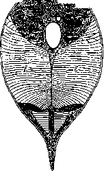
the intermediate layers between the plates of the electric organ. As a whole, they roughly resemble a voltaic column. The electric organs of the electric eel are innervated by approximately 350 nerves, which arise from ganglion cells located in various parts of the spinal cord behind the central canal. The nerve fibers of the electric organs exit through the anterior roots of the spinal cord. During an electric discharge, the papillary (anterior) surface of the plate is negative, while the villous (posterior) surface is positive; consequently, during an electric shock, the current in the electric eel flows from the tail to the head of the fish. The electric organs of the marble ray (fig. 4) are located in the anterior half of the body on the sides of the skull, filling the entire thickness of the flattened body of the ray. Each electric organ, after removal of the skin from above,
Fig. 3. Cross-section of the plates (1) of the electric organ of the electric eel; 2-papillary surface; 3-villous surface; 4-caudal end; 5-anterior end of the fish's body.
has the appearance of a kidney-shaped formation consisting of 507-610 vertically standing and adjacent columns. Each column in turn is formed by 375 electric plates. The nerve trunks innervating them emerge from paired electric lobes located on the dorsal surface of the medulla oblongata on either side of the midline. The direction of the electric current when the electric organ of the marble ray acts is from the ventral surface to the back of the fish. Electric organs of the electric catfish have a different origin; according to Fritsch's data, they are not connected to muscles and are apparently derivatives of glandular elements of the skin cover. Electric organs in the thickness of the skin cover the entire body of the electric catfish. They are innervated on each side of the body by one powerful lateral nerve, which comes from giant ganglion cells located in the initial part of the spinal cord. The direction of the current during a shock is from the head to the tail of the fish. Electric fish inhabit hot and warm countries: the electric eel lives in the warm fresh waters of Guiana and Brazil, the electric catfish in the rivers of North Africa, Torpedo (various species) in the Atlantic Ocean, in the Mediterranean Sea. An electric shock is obtained if one touches, for example, the tail of an electric eel. At this time, a strong shock occurs, stunning a person. The first shocks from large electric eels are particularly sensitive. Subsequent shocks weaken due to fatigue of the fish. Even strong shocks cannot kill a person or larger animals. Nevertheless, for a short time, a shock can knock a person off their feet and as it were paralyze the muscles. This explains the local name of the electric eel 'arimna,' which in the language of the Tamanaque Indians means 'depriving of motion.' The electric eel paralyzes small animals, fish, crayfish, etc., and devours them. Production of electricity
The usual alternating current used in technology changes direction 100 times per second (50 periods); high-frequency currents used in radio engineering, as well as in medicine (for example, d'Arsonval currents, diatherapy), up to 10^6 times per second.

Fig. 4. Cross-section of the body of Torpedo ocellata. O-electric organs.
is not a specific feature of electric organs, since bioelectric phenomena are characteristic of working muscles, the heart, smooth muscles, nerves, etc., but in electric organs, due to their special structure, the production of electricity increases greatly.
E. Panlovsky.
Related articles
Mentioned in
Cite this page
“Electric Organs.” Soviet Medical Encyclopedia. English translation of Bolshaya Meditsinskaya Entsiklopediya, 1st ed. (Moscow, 1928–1936), ed. N. A. Semashko. https://sovietmedicalencyclopedia.pages.dev/article/electric-organs/