Medulla Oblongata
Historical document, translated for reference. It reflects medical knowledge of the 1920s–30s and is not medical advice.
Summary
The medulla oblongata is the lowest part of the brain, serving as a pathway between the brain and spinal cord while housing vital reflex centers for functions like swallowing and breathing.
Encyclopedia article (1928–1936)
Medulla Oblongata (syn. medulla oblongata, s. bulbus medullae spinalis), the lowest part of the brain (myelencephalon), very complex in structure and having important functional significance: 1) it serves as a conductor for fibers connecting various parts of the brain with the spinal cord, and through it with the periphery; 2) it has significance as a central apparatus—in it are located reflex centers, moreover it is the site of origin of complex combined acts, such as swallowing, breathing. Together with the intermediate, middle, and hindbrain, it forms the brainstem. It develops from the primary hindbrain vesicle (see Brain). The medulla oblongata lies between the pons Varolii above, from which it is sharply demarcated, and the spinal cord below, into which it passes without sharp boundaries; the boundary between them is considered to be the decussation of the pyramids and the exit of the first pair of cervical roots. At the place where the medulla oblongata transitions into the spinal cord there is a bend (occipital). On the skeleton, the lower boundary of the medulla oblongata corresponds to the upper
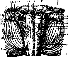
Figure 1. Anterior surface of the medulla oblongata: 1- spinal cord; 2- decussatio pyramidum, boundary between medulla oblongata and spinal cord; 3- cerebellum; 4- medulla oblongata; 5- fasciculus lateralis ant.; 6- fasciculus mediana ant.; 7- olive of the medulla oblongata; 8- pyramid of the medulla oblongata; 9- flocculus of the cerebellum; 10- pedunculus cerebelli ad pontem; 11- pons Varoli; 12- foramen caecum; 13- n. abducens; 14- n. facialis; 15- n. Wrisbergi; 16- n. acusticus; 17- n. glosso-pharyngeus; 18- n. vagus; 19- n. hypoglossus; 20- n. accessorius-bulbar and spinal (20') its root; 21- first cervical root.
which in the medulla oblongata receive the name fasciculus gracilis (gracile fascicle) and fasciculus cuneatus (cuneate fascicle); they are separated from each other by septum paramedianum. Fasciculus gracilis ends with an enlargement-clava (clava), fasciculus cuneatus-with a tubercle (tuberculum cuneatum). In the upper part, the posterior surface of the medulla oblongata forms the floor of the fourth ventricle (ventriculus quartus) or rhomboid fossa, its lower triangle; the apex of this triangle passes into the spinal canal; the narrowest part of the triangle is called calamus scriptorius (writing pen), above it passes the transverse lamina-obex (bolt). The base of the triangle corresponds to the widest part of the fourth ventricle, which, expanding outward, forms a recess or recessus lateralis. Along the floor of the fourth ventricle, on the border of the lower and upper triangles (corresponding to the posterior surface of the pons Varoli), pass striae medullares, s. acusticae. On the sides, the rhomboid fossa is bounded by the restiform bodies (corpora restiformia). Along the median line of the rhomboid fossa runs a groove (sulcus medianus, fossae rhomboideae); on each side of it is a triangular elevation (trigonum hypoglossi), corresponding to the nucleus of the XII nerve; its apex is turned downward, base upward; outward from it lies another triangle, turned, conversely, with apex upward, of smaller size, grayish in color-ala cinerea or fovea vagi; in this place is located the nucleus of the X nerve; this place is also called the vital node, since its destruction (nucleus of the X nerve) leads to death. In the outer angle of the floor of the fourth ventricle,
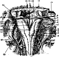
Figure 2. Posterior Surface of the medulla oblongata; floor of the fourth ventricle: 1- spinal cord; 2- medulla oblongata; 3- n. accessorius and 3'-its spinal root; 4- nucleus fasciculi gracilis; 5- nucleus fasciculi cuneati; 6- n. vagus; 7 and 8- n. glosso-pharyngeus; 9- recessus lateralis of the fourth ventricle; 10- n. acusticus; 11- n. abducens; 12- pons Varoli; 13- a. basilaris; 14- eminentia teres; 15- pedunculus cerebelli ad pontem; 16- tuberculum acusticum; 17- fissura mediana post.; 18- trigonum n. hypoglossi; 19- ala cinerea; 20- calamus scriptorius; 21- obex; 22- a. vertebralis; 23- dura mater (cut).
in the so-called area acustica, there is an elevation (tuberculum acusticum, or auditory tubercle), from which white stripes (striae acusticae) go to the median line. From the cerebellum to the outer walls of the lower part of fossae rhomboideae goes an epithelial plate (velum medullare posticum-posterior medullary velum); above its edge Ct. The length of the medulla oblongata is from 2.5 to 3 cm, the anteroposterior diameter is 12-15 mm, the transverse diameter is 10-12 mm below and 22-25 mm above. Weight is 6-7 g (1/216 of the weight of the entire brain).- The medulla oblongata has the shape of a truncated cone, slightly flattened in the anteroposterior direction: in it are distinguished anterior, posterior, lateral surfaces, base and apex. On the anterior surface (fig. 1) runs a groove (fissura mediana ant.), which is a continuation of the same groove of the spinal cord and ending at the pons Varolii with a blind opening (foramen caecum inf.); below at the border with the spinal cord it is interrupted for 6-8 mm by the decussation of the pyramidal fibers occurring here; along its sides lie thick bundles-pyramids, bounded outward by a groove (fissura lateralis ant., or antero-lateralis, s. fissura praeolivaris, s. fissura n. hypoglossi). On the lateral surface of the medulla oblongata is located the olive (oliva inf., olive of the medulla oblongata), of oval shape, white color, 12-15 mm in length, 4-5 mm in width; behind it passes a groove (fissura lateralis post., s. fissura retroolivaris). Along the entire length of this groove emerge the roots of three cranial nerves IX, X and XI (n. glosso-pharyngeus, vagus, accessorius); the root of n. XII emerges in the fissura lateralis ant. Between the medulla oblongata and the pons Varolii, at the level of the pyramids, emerges n. abducens (VI), and in the angle near the cerebellum-pp. facialis and acusticus (VII, VIII). The posterior surface of the medulla oblongata in its lower and upper parts shows a significant difference (fig. 2). In the lower part along the median line runs fissura mediana post., s. fissura longitudinalis post.; on each side of it are located continuations of the posterior columns of the spinal cord-fasciculi of Goll and Burdach,
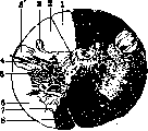
Figure 3. Cross section of the medulla oblongata at the level of its lower border: 1- fasciculus of Goll; 2- fasciculus of Burdach; 3- posterior horn; 3'- substantia gelatinosa Rolandi; 4- posterior roots; 5- crossed pyramidal tract; 6- lateral column; 7- anterior horn; 8- direct pyramidal tract.
the rhomboid fossa is covered by tela chorioidea, and then by the cerebellum. The microscopic structure of the medulla oblongata changes depending on the level: in the lower parts it somewhat resembles the spinal cord in structure (fig. 3); characteristic of this part is the decussation of the pyramidal fibers (decussatio pyramidum), occurring on the anterior surface of the medulla oblongata, as a result of which the fissura med. ant. decreases in depth, being filled with fibers passing from one side to the other. The anterior horns are still intact but have lost their connection with the posterior ones; in them originates n. XI; substantia gelatinosa Rolandi increases in size, around its periphery is located the descending, or spinal, root of n. V; around the periphery of the lateral column lie the bundles of Flechsig, Gowers and fasc. vestibulo-spinalis (fig. 4), more inwardly-fasc. rubro-spinalis. The posterior columns are sharply delimited into 2 bundles-funiculus gracilis and funic. cuneatus; in these bundles appear nuclei-nucl. funic. gracilis and nucl. funic. cuneati; in them ends the first sensory neuron; substantia reticularis has increased in size; the central canal is located posterior to the decussation, near it is the dorsal or bulbar nucleus of n. accessorii. Somewhat higher the pyramids form powerful bundles (pyramis medullae oblongatae), located on either side of the fissura mediana anterioris; on the surface of the pyramid lies the arcuate nucleus' (nucl. arcuatus, s. arciformis, s. praepyrimalis). Somewhat posterior and lateral to the pyramids are the main olives (olivae inf., s. nucl. olivaris inf.), medial to them is the accessory olive (oliva accessoria medialis, s. interna, s. nucl. olivaris accessorius ant.). The nuclei of the posterior columns have significantly increased in size, while their white matter has decreased, since it gradually ends in these nuclei; in them begins the second sensory neuron-the internal arcuate fibers (fibrae arcuatae internae); they curve around the central canal and anterior to it decussate with fibers of the opposite side, forming the sensory or superior decussation (decussatio lemniscorum); after the decussation the fibers are located in the interolivary layer, forming lemniscus medialis, or the medial loop. Anterior to the central canal is fasc. longitudin. post., or medialis and the nucleus of n. XII, lateral to it-n. X, and posterior and lateral to it-n. XI. The bundle of Flechsig begins to deviate backward, forming fibrae arcuatae ext. ant., which play a large part in the formation of corp. restiformia; anterior lies the bundle of Gowers, inwardly-substantia gelatinosa Rolandi and the descending root of n. V, fasc. rubro-spinalis, vestibulo-spinalis. The remaining part is occupied by substantia reticularis, in which nuclei and fibers are located; a large accumulation of nuclei in the lateral parts is выделено under the name nucl. reticularis lateralis, s. nucl. funic. lateralis. The median line is occupied by the raphe; nuclei lying near the raphe are designated as nucl. raphes. In the higher parts of the medulla oblongata at the level of the opening of the fourth ventricle, on its bottom, covered with ependyma, in the gray matter (stratum cinereum fossae rhomboideae) are located the nucleus of n. XII, the dorsal nucleus of n. X, fasc. longitudinalis post.; somewhat anterior to the nucleus of n. X lies fasc. or tractus solitarius (the solitary bundle), surrounded by the nucleus of the solitary bundle (nucl. funic. or tracti solitarii, s. nucl. terminalis, s. subst. gelatinosa vagi et glossopharyngei); in this nucleus gradually end the fibers of funic. solitarii. Nucl. funic. gracilis and cuneati decrease in size, as do the bundles themselves; lateral to the cuneate nucleus appears a new nucleus, the so-called external nucleus of funic. cuneati (syn. nucleus of Monakow, nucl. corp. restiformis); it is as it were a center around which corp. restiforme is formed; the bundle of Flechsig also plays a large part in the formation of corp. restiforme, the fibers of which in the form of fibr. arcuatae externae ant. deviate backward and lie around the external nucleus of funic. cuneati; in addition, a small number of fibers from the posterior columns (fibrae arcuatae ext. post.) enter corp. restiforme. From the nuclei of the posterior columns still exit fibrae arcuatae int., forming the decussation and joining those previously decussated in lemnis. medialis, which is located along the median line in the interolivary layer. From nucl. arcuatus depart fibrae arcuatae ext. ant., which pass along the anterior surface of the pyramids, heading toward corp. restiforme. Near the inferior olive lie already two accessory olives (olivae accessoriae, s. nucl. olivaris accessorius medialis et dorsalis); fibers from the olives
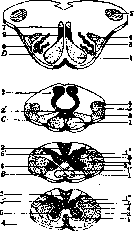
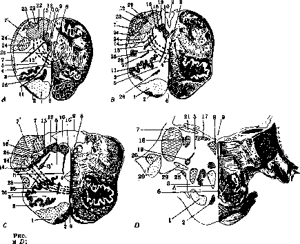
Figure 4. Cross-sections through the cervical part of the spinal cord (A) and through the medulla oblongata (B, C and D) at different levels. Comparative arrangement of various bundles in the spinal and medulla oblongata: i-pyramidal bundle; II-decussation; III-decussated pyramidal bundle; 4-posterior columns; 4'-decussation of fibers from the nuclei of the posterior columns (decussatio lemniscorum); 5-bundle of Gowers; 6-its sensory part (fasc. spino-thalamicus); s-corpus restiforme; 6-anterior horns of the spinal cord; 6'-posterior horns; 7-fasc. longitud. post.; 8-oliva inf. participating in the formation of corp. restif., mainly of the opposite side. In subst. reticularis, besides other nuclei, appears nucl. ambiguus-motor nucleus of n. vagi. Gradually upward the bottom of the fourth ventricle expands (fig. 5); corpus restif. increases. On the bottom of the ventricle, starting from the median line, lie the following formations: fasc longitudinalis medialis, s. post., fasc. tecto-spinalis, nucleus funiculi teretis, s. nucl. eminentiae med., nucl. n. XII, nucl. n. X; between these last nuclei is wedged a nucleus of very small cells-nucl. intercalatus Staderini; in the outer angle of the ventricle is nucl. vestibularis med. (Schwalbe), and near corp. restiforme-radix descendens n. vestibularis. The other formations are the same as in the lower parts, only in substantia reticularis, between n. ambiguus and the olive, lies the sympathetic nucleus-nucl. sympathicus medullae oblongatae, s. nucl. salivatorius inferior Kohnstamm; its fibers enter into the composition of n. IX. At the very highest levels of the medulla oblongata the fourth ventricle significantly expands, forming lateral recesses (recessus lat.); the transverse diameter of the med. oblongata becomes wider due to the strong development of the corpora restiformia; on their outer surface is located the acoustic tubercle (tuberculum acusticum), the place of termination of part
5. Cross-sections of the medulla oblongata at different levels A, B, C 1-pyramids; 2-nucl. arcuatus; 3-oliva inf. and 3'-its hilus; 4-decussatio lemniscorum and 4'-lemniscus medialis; 5-substantia reticularis; 6-raphe; 7-bundle of Flechsig and 7'-corpus restiforme (its inner part); 8-fasciculus longitudinalis post.; 9-central canal and 9'-IV ventricle; 10-nucl. n. hypoglossi and 10'-nucl. funiculi teretis; 11-n. hypoglossus; 12-nucl. n. accessorii and 12'-n. accessorius; 13-nucleus dorsalis n. vagi and 13'-nucl. ambiguus; 14-n. vagus; 15-fasciculus solitarius; 16-nucl. n. glossopharyngei and 16'-n. glossopharyngeus; 17-nucl. vestibularis dorsalis; 18-nucl. Deiters'a; 19-nucl. n. facialis; 20-nucl. ventralis n. cochlea; 21-striae acusticae; 22-posterior columns and 22'-their nuclei; 23-nucl. Burdachi ext.; 24-substantia gelatinosa Rolandi and 24'-descending root of n. trigemini; 26-fibrae arcuatae ext.; 26-oliva accessorii medialis; 27-oliva accessorii dorsalis; 28-funiculus lat., medullae oblongatae and its nucleus; 29-corpus trapezoidus.
fibers of the auditory nerve; another part of them ends in the nucl. ventralis n. cochleae, located more ventrally. Along the floor of the fourth ventricle are located nucl. funic. teretis, nucl. praepositus; in front of them passes the fasc. longitud. post.; the rest of the floor of the fourth ventricle is occupied by the nucleus of the vestibular nerve [nucl. medialis (dorsalis) n. vestibularis]. In the inner part of the corp. restiform. is located nucl. p. vestibul. lat. (nucleus of Deiters) and tract, nucleo-cerebellaris, connecting these nuclei with the cerebellum. Along the very floor from the tubercul. acust. go the striae acusticae, which, approaching the midline, descend into the depth of the Medulla Oblongata. In the other parts of the Medulla Oblongata at this level, the changes are not significant: the lemniscus med. forms a more compact bundle, between it and the fasc. longit. post. lies the central nucleus of Roller (nucl. centralis inf.); the olives decrease in size; in the subst. reticul. instead of the p. ambiguus lies the nucleus of the p. facialis. In the lateral column is the bundle of Gowers and the fasc. rubro-spinalis; in front of the pyramidal fibers appear the own fibers of the bridge of Varolius. B l o o d s u p p l y o f t h e M e d u l l a O b l o n g a t a. The arteries of the Medulla Oblongata depart from the initial part of the a. basilaris, from the a. vertebralis and from its branches - the a. spinalis and a. cerebellaris inferior. These arteries divide into 1) aa. radiculares, entering together with the roots of the cranial nerves into the Medulla Oblongata, where they divide into two branches - one goes along the root to the periphery, the other - into the tissue of the Medulla Oblongata to the nuclei of the corresponding nerves; 2) aa. centrales, mediales, s. a. sulci bulbaris, passing through the entire thickness of the Medulla Oblongata to the fourth ventricle and nourishing the formations located near the midline - pyramids, lemniscus medialis, parts of the inferior olives. some nuclei of cranial nerves. Depending on the height of the aa. centrales, they depart from different arteries; the upper ones from the a. basilaris, the lower ones from the a. spinalis ant.; 3) aa. periphericae, departing from the arteries of the soft meninges and nourishing the white matter of the brain and nuclei in the substantia reticularis. The Arteriae chorioideae take origin from the a. cerebellaris, which nourishes the lateral parts of the medulla oblongata. Physiology of the Medulla Oblongata. Despite its comparatively small size, the Medulla Oblongata is an extremely important organ functionally, as indicated by its extremely complex structure. Due to the presence in the Medulla Oblongata of accumulations of nerve cells, it has the significance of a central apparatus; in it are laid the nuclei of the following cranial nerves: n. hypoglossi, n. accessorii, n. vagi, n. glossopharyngei, nervi acustici (vestibular and cochlear), then the nucleus of the n. facialis and the sensory nucleus of the n. trigemini; the nuclei of the last three nerves also extend into the pons (function of the nerves - see the corresponding nerves and Autonomic Nervous System). The motor nuclei of these nerves are the motor centers of individual muscle groups and muscles and associated with the function of simple reflexes, due to the termination near them of sensory fibers. But the activity of the medulla oblongata is not exhausted by only such reflexes, it is also the place of origin of complex coordinated movements, automatic acts, in the formation of which several motor and sensory systems participate; such are for example the acts of swallowing, breathing, chewing, sneezing, vomiting (see the corresponding words). These latter data led to assume the existence in the Medulla Oblongata of special coordinating mechanisms and for each complex act of special coordination centers; which would connect various simple centers for a certain physiological purpose, not being themselves directly connected with the muscles. In microscopic study of the Medulla Oblongata, they tried to find such groups of cells in which it would be possible to localize various centers, as for example the center of swallowing, the center of breathing, etc.; especially many works were devoted to the center of breathing; Flourens localized it in the angle of the rhomboid fossa, this place was called the vital node (noeud vital), then it was localized in the nucleus funic. solitarii, in the subst. reticul. etc., but even to this day no final conclusions have been reached about the localization in general of coordinating mechanisms for the above-mentioned acts; on the contrary, many scientists began to deny the possibility of the existence of such centers, and assumed that the connection, which undoubtedly exists between individual simple centers, occurs through association neurons, reaching a great development in the Medulla Oblongata, namely in the substantia reticularis, which is called the associative field of the Medulla Oblongata. The cells of the subst. reticul. are mostly with short axons, but there are also with long ones, connecting with all the motor centers of the Medulla Oblongata, the pons, and the spinal cord; the sensory fibers entering the Medulla Oblongata end with their collaterals in the substantia reticularis, near its cells, which for example explains why respiratory reflexes can be caused by irritation of the mucous membranes and skin. The occurrence of the respiratory reflex is conditioned not only by the transmission of irritation along the sensory nerves, but also by its own mechanisms, for example by the difference in the ratio of O and CO2 in the blood. The influence of the Medulla Oblongata on cardiac activity see Vagus nervus, Autonomic Nervous System. In the Medulla Oblongata there are groups of cells having a toning influence on the spinal centers of the vasomotor apparatus, but they in turn are under the influence of the main vasomotor centers in the diencephalon. An influence of the Medulla Oblongata on sweating, on the pupil is assumed due to a possible interruption of the central sweating conductors and the ocular sympathetic pathway in a group of cells located in the substantia reticularis of the Medulla Oblongata and playing the role of relay stations. It has been suggested that in the Medulla Oblongata there are centers for metabolism, because in diseases of the Medulla Oblongata the presence of sugar in the urine is often noted; a prick of the floor of the fourth ventricle (prick of C. Bernard) at the level of ala cinerea causes glycosuria; a prick somewhat higher, on the contrary, leads to a decrease in sugar in the blood. From these data it follows that in the posterior nucleus of the n. X (in ala cinerea) there are 2 groups of cells acting antagonistically, one parasympathetic (inhibiting the formation of sugar and standing in close connection with the pancreas), the other - sympathetic (causing the formation of sugar and having a connection with the liver).- Finally the Medulla Oblongata also has relation to salivation due to the presence in it of the nucl. salivatorius inf. The Medulla Oblongata also plays the role of a conductor, because in it pass fibers in various directions and different in function: In the Medulla Oblongata occurs the crossing of motor fibers; in it ends the first sensory neuron and begins the second, which also undergoes crossing. Through the Medulla Oblongata occurs communication between the periphery and the cerebellum and the brain1. Pathology of the Medulla Oblongata. Due to the complex anatomical structure, the pathology of the Medulla Oblongata is very complex. Disease of the Medulla Oblongata or the so-called bulbar syndrome is characterized by a disorder of the function of cranial nerves, the nuclei of which are located here, of movement, sensitivity, of the vasomotor apparatus, of the cerebellum, of cardiac activity, of metabolism, etc. Of the motor disorders, hemiplegia can be noted, and if the focus is close to the midline, then quadriplegia; crossed or alternating paralysis (paralysis of the arm on one side and of the leg on the other). The disorder of sensitivity can manifest in three different forms: 1) complete loss of sensitivity; 2) preservation of deep sensitivity; 3) dissociation of sensitivity of the syringomyelia type. Alternating disorders of sensitivity are also observed. Very often a change in sensitivity is accompanied by vasomotor disorders in the form of asymmetry in body temperature (up to 1° and more), in the filling of the vessels, in blood pressure (checked by means of a plethysmograph); all these changes are observed on the side opposite to the focus. Cerebellar phenomena are quite frequent in diseases of the Medulla Oblongata, they occur on the side of the focus, depend on the lesion of the corpus restiforme, of the cerebellar bundles or of the olivae inf. (giving fibers to the corp. restiforme). Of the cranial nerves, the nn. XII, XI, X, IX, VIII and partly the n. VII and VI can be affected (see Bulbar paralysis). Unilateral paralysis of the pharynx, soft palate is distinguished under the name of Avellis' syndrome, and if in addition there is paralysis of the n. XI, then Schmidt's syndrome results. In lesions of the Medulla Oblongata, on the side of the focus, Horner's syndrome or the sympathetic ophthalmic syndrome is observed (see Horner's symptom complex). Lesion of cranial nerves can be combined with paralysis and disorders of sensitivity on the opposite side, giving alternating syndromes. In the clinic, the most diverse combinations of the above-mentioned disorders are observed. Dissociated hemianesthesia and hemiplegia on one side, hemiasynergy, lateropulsion and miosis on the other - are distinguished as the alternating syndrome of Babinski and Nageotte. The syndrome of Cestan and Chenais is characterized by hemiplegia, hemianesthesia on one side, hemiasynergy, Horner's syndrome and Avellis' syndrome on the opposite.
Quite often the focus spreads to the pons or to the spinal cord, and in the first case there is a complex syndrome of the medulla oblongata and the pons (syndrome bulbo-protuberantiel), which, in addition to bulbar phenomena, is characterized by simultaneous involvement of the V, VI and VII pairs of cranial nerves or of the spinal cord. The retroolivary syndrome is characterized by disturbance of superficial sensitivity on one side, cerebellar phenomena, and paralysis of the pharynx, larynx, and soft palate on the other; paralysis of the extremities, as well as disturbance of deep and tactile sensitivity, are absent due to the integrity of the pyramids and the interolivary layer. When the nucleus of Deiters is involved in the process, a syndrome is observed, expressed by dizziness, oculomotor disturbances, weakness in the legs, nausea, disturbances of hearing, and pain in the temporal region. All these symptoms are explained by the connections that the nucleus of Deiters has with the surrounding formations. - Since the medulla oblongata contains the anatomical substrate for some important vital functions - for respiration, cardiac activity, metabolism, swallowing, etc., - damage to the medulla oblongata is accompanied by disturbance of these functions; various types of respiratory disturbance are observed - Cheyne-Stokes respiration, stertorous respiration, dyspnea of varying intensity with or without cyanosis; respiratory disturbance can lead to death. Of the cardiac disturbances, the Adams-Stokes syndrome, slow pulse (pouls lent permanent), tachycardia, embryocardia can be noted. - In many diseases of the medulla oblongata, glycosuria (syn. mellituria), simple polyuria (diabetes insipidus) or with albuminuria are observed. Finally, there is the so-called anxietas paroxysmalis (s. paroxystique)--anguish, appearing in the form of attacks, mostly nocturnal, with sudden onset, accompanied by trembling, cold sweats, marked pallor of the face, and a suffering expression; it depends on the disturbance of nutrition of the medulla oblongata. Special pathology. In the brain stem, the same processes are observed as in the brain: vascular diseases (softenings and hemorrhages), tumors, traumatic injuries, inflammations. Of the vascular diseases of the medulla oblongata, thrombosis, embolism leading to softening, and hemorrhage should be noted. Thrombosis of the arteries of the medulla oblongata is observed more often than embolism, and is a consequence of arterial disease (arteriosclerosis, atheromatosis, syphilitic arteritis, etc.). The focus of softening is most often unilateral, it can be both single and multiple; sometimes it simultaneously involves the pons or the spinal cord. Around the focus of softening, diffuse edema, dilatation of capillaries, phenomena of diapedesis are observed; as a result of the change in circulation, the nutrition of the tissue surrounding the focus also changes, sometimes over a considerable extent. The onset can be sudden or the disease may be preceded by prodromal symptoms - headaches, dizziness, nausea, brief losses of consciousness, syncope. The clinical picture depends on the localization of the process in the area of one or another artery - the above-mentioned symptoms are observed in various combinations. Two syndromes have been distinguished, observed: 1) in thrombosis of a. cerebellaris; it is characterized by hemianaesthesia alternans, paralysis of the X nerve and the cervical part of the sympathetic nerve (Horner's syndrome) on the side of the focus, disturbance of statics, more often of the cerebellar type, dizziness, and 2) in thrombosis of a. sulci bulbaris; it is expressed by tetraplegia, bilateral paralysis of the XII nerve, paralysis of the X nerve, disturbance of deep sensitivity leading to disturbance of statics. The development of the disease can be acute or subacute. More acute is observed in embolisms; embolism of a. vertebralis is accompanied by very severe bulbar phenomena and in most cases ends in death, either suddenly or after some time due to disturbance of swallowing and subsequent bronchopneumonia.
Hemorrhages into the medulla oblongata depend on the same causes as hemorrhages in other parts of the central nervous system - chronic arteritis, syphilis, intoxications, acute infectious diseases, injuries to the skull, falls from a height, rapid change in atmospheric pressure. Hemorrhage can be unilateral, bilateral, single and multiple. (Patho-anatomical changes - see Apoplexy.) The onset of the disease is sudden, apoplectic, with loss of consciousness. The clinical picture, as in softening, depends on the localization of the process. In extensive hemorrhages, instantaneous death occurs, but even small hemorrhages most often end in death after a short time. Hemorrhage rarely involves only the medulla oblongata, but spreads upward into the pons or downward into the spinal cord. In the presence of bulbar phenomena, the diagnosis presents no difficulty; it is necessary to differentiate from pseudobulbar paralysis (two successive strokes, disturbances of mentality, forced laughter and crying decide the question in favor of pseudobulbar paralysis) and from amyotrophic lateral sclerosis (see). The etiological diagnosis - softening or hemorrhage - is very difficult; knowledge of the development of the disease can help - a more sudden onset speaks for hemorrhage. The localization of the process can be determined by the combination of symptoms. Prevention and treatment - as in vascular diseases of other parts of the brain (see Brain - pathology, Apoplexy); with bulbar phenomena, special care is required (see Bulbar paralysis, Amyotrophic lateral sclerosis). Tumors of the medulla oblongata develop in the medulla oblongata itself or grow into it from neighboring formations. Tumors of the medulla oblongata itself are not particularly frequent; they can be single or multiple; tumors are of the same structure as in other parts of the central nervous system: gliomas, mostly of ependymal origin (more often in children), papillomas, epitheliomas (very rare). Even rarer than neuroepithelial tumors are mesenchymal tumors - sarcomas, endotheliomas, angiomas, fibromas, psammomas, tumors originating from plexus chorioideus; in addition, simple or parasitic cysts are observed - cysticercus, echinococcus. Simple cysts, mostly of traumatic origin, have a transparent thin membrane and contain a transparent fluid. Parasitic cysts (see Echinococcus) can be the size of a small nut. Cysticerci can cause extensive inflammatory and necrotic changes involving the membrane; sometimes they simulate gummatous processes. Of the infectious neoplasms, gumma, tuberculosis are very rare; abscesses have been described as metastases in suppurative foci in the lungs and in the liver. Tumors are more often localized on the posterior surface, masking the cerebellum; the tumor can bulge into the cavity of the fourth ventricle. - The clinical picture - as in tumors of other parts of the central nervous system, general cerebral symptoms and focal symptoms are observed. Of the general cerebral symptoms, headaches appear quite early, are localized in the occiput with irradiation
BLOWING
into the neck, shoulders, upper extremities, accompanied by rigidity when moving the head; vomiting can be considered both as a general cerebral symptom and as a focal one; in addition, drowsiness, lethargy, apathy, asthenia, decreased intellect, delusional ideas (delusions of grandeur, persecution), choked discs with rapidly occurring blindness are noted. Symptoms of increased intracranial pressure may also be absent. Focal symptoms depend on the location of the tumor, its size, and partly on its nature; bulbar phenomena, movement disorders, sensory disorders - unilateral, bilateral, alternating; deafness, neuralgia, dizziness, cerebellar disorders, choreiform convulsions, profuse salivation, taste disorders and finally metabolic disorders - sugar, water (glycosuria, albuminuria); cardiac and respiratory disorders.--The onset is slow and unnoticed; development is rather rapid, within several months, rarely a year. Death from bulbar phenomena. A tumor developing from the medulla oblongata into the cavity of the fourth ventricle is characterized by fluctuations during the disease - there are periods of improvement and worsening; headaches, dizziness, vomiting, hiccups, respiratory and pulse disorders alternate with periods of improvement, the patient feels well, sometimes only a slight nystagmus, cerebellar phenomena remain; during such a good state, sudden death is possible. Abscesses are accompanied by severe headaches; large fluctuations in temperature. - Diagnosis of a medulla oblongata tumor is very difficult, sometimes it is only established at autopsy if there are no general cerebral symptoms.-Treatment is possible only for gummata, for other tumors there is no treatment, since surgical intervention in this area is impossible. Traumatic injuries. Due to its location, the medulla oblongata is less protected from trauma than other parts of the central nervous system. Compression of the medulla oblongata is observed with dislocations of C1 or C2, then gunshot wounds or wounds with a sharp instrument. In all these cases, instantaneous death occurs. Slow compression of the medulla oblongata is observed with osteitis of the occipital bone,1 the first cervical vertebrae, with malum Potti suboccipitalis, with bone tumors, with tumors of the meninges or originating from surrounding formations, with aneurysms of the a. basilaris, a. vertebralis, with hemorrhages in the brain or ;in the cerebellum. The first symptom is neuralgic pain from compression of the roots, unilateral or bilateral, along the course of the occipital nerve or the first cervical roots; then sensory disorders in the areas innervated by these roots, rigidity of the neck, phenomena from the cranial nerves, more often the XII nerve (hemiatrophy of the tongue), the XI nerve (paralysis of the m. cucullaris and sterno-cleido-mastoidei), and other symptoms described above. In an aneurysm, it is sometimes possible to hear a noise on the posterior surface of the skull.-Diagnosis of compression in some cases does not present major difficulties, in others it is difficult to recognize the cause of compression; in these cases, the development of the disease, various: studies (lumbar puncture, encephalography, X-ray, RW in blood and cerebrospinal fluid and others) can help with the diagnosis.--The prognosis is serious, sudden death is not uncommon.-Treatment depends on the cause of compression. Inflammatory diseases of the medulla oblongata.-see Poliomyelitis. See also Bulbar paralysis.
Related articles
Mentioned in
- Abducens Nerve
- Alternating Syndromes
- Ambiguous Nucleus
- Analgesia
- Anarthria
- Anesthesia
- Anesthetic Agents
- Apomorphine
- Arbor Vitae
- Area
- Atropine
- Auditory Pathways
- Avellis Syndrome
- Barium
- Bernard's Sugar Injection
- Blinking
- Brain (551 General diagnostics of brain diseases)
- Brain (cerebrum, a comprehensive term for the entire)
- Brushtein
- Bulbar Palsy
Cite this page
“Medulla Oblongata.” Soviet Medical Encyclopedia. English translation of Bolshaya Meditsinskaya Entsiklopediya, 1st ed. (Moscow, 1928–1936), ed. N. A. Semashko. https://sovietmedicalencyclopedia.pages.dev/article/medulla-oblongata/