Area
Historical document, translated for reference. It reflects medical knowledge of the 1920s–30s and is not medical advice.
Summary
This article defines the term "area" in the context of neurology, referring to specific regions of the central nervous system, and in embryology, referring to the vascular field in developing embryos. It details anatomical structures such as the area acustica and area striata, as well as the biological process of blood vessel formation in the area vasculosa.
Encyclopedia article (1928–1936)
AREA (Lat.), field, area, place—in neurology, this term designates certain areas of the central nervous system (see below). Area acustica, an area located along the floor of the IV ventricle, in its outer corner, at the border of the pons Varolii and the medulla oblongata; due to the protrusion of the IV ventricle existing in this place, which captures the upper and outer surface of the restiform body, the area acustica extends onto the restiform body, forming the acoustic tubercle (see Tuberculum acusticum). Area olfactoria (parolfactoria Broca), a small gyrus on the inner surface of the frontal lobe of the brain under the rostrum corporis callosi, between the sulcus olfactorius anterior and posterior; by means of this gyrus, the lobus olfactorius anterior connects with the central area of the olfactory brain (gyrus cinguli). Area parabigemina, an area located in the tegmentum of the lower sections of the cerebral peduncle at the periphery, between the lateral lemniscus, the posterior colliculus, and the decussation of the superior cerebellar peduncles; a portion of the fibers of the lateral lemniscus terminates here. Area striata, an area of the cerebral cortex characterized by the presence in its 4th layer of a stripe of myelin fibers—the stripe of Vicq d'Azyr or Gennari; the cortex with such a structure corresponds to the visual centers, located on the inner surface of the occipital lobe around the fissura.
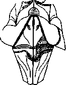
VIII (blackened area)—area acustica. calcarina and at its junction with the fissura parieto-occipitalis; sometimes it also extends to
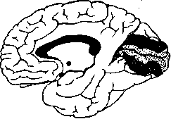
S—area striata (lightly hatched area).
the outer surface; it corresponds to Brodmann area 17; it is found in all classes of mammals.
E. Kononova. AREA VASCULOSA (from Lat. area—field, area and vasculosus—vascular), vascular field, an area of the extraembryonic blastoderm in meroblastic eggs (reptiles, birds) and in mammals, in which the development of blood vessels and blood elements primarily begins. At the very beginning of development (in a chicken on the second day of incubation), in the area of the area opaca adjacent to the area pellucida, one can notice the formation of cell clusters, in which the outer
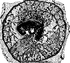
Rabbit embryo at 11 days; yolk vessels are visible—arteries and veins connected to the marginal sinus.
cells then turn into elements of the vascular wall (endothelium), and the inner ones into blood corpuscles (white and red, initially without Hb), containing nuclei. These cell clusters—blood islands—merging, form a network (primary vascular network), in which two arteries (vitelline arteries) and two veins (vitelline veins) are formed, which enter into connection with the primordium of the heart. Along the edge of the A. vasculosa, a marginal venous sinus is formed, sharply delimiting it from the section of the area opaca of the yolk field lying outward. While inside the embryo the formation of blood vessels and blood elements proceeds entirely at the expense of the mesenchyme, in the A. v. some authors (Hertwig) allow for the partial participation of the entoblast as well. In mammals, the formation of the A. v. and the development of primary vessels and blood elements also occur initially in the extraembryonic blastoderm and according to the same type (see figure).
Related articles
Mentioned in
Cite this page
“Area.” Soviet Medical Encyclopedia. English translation of Bolshaya Meditsinskaya Entsiklopediya, 1st ed. (Moscow, 1928–1936), ed. N. A. Semashko. https://sovietmedicalencyclopedia.pages.dev/article/area/