Ventriculi Cerebri
Historical document, translated for reference. It reflects medical knowledge of the 1920s–30s and is not medical advice.
Summary
The article describes the anatomy of the cerebral ventricles, including their structure, connections, and pathological conditions such as hemorrhages, tumors, and inflammatory processes. It details the development of the ventricles from primitive cerebral vesicles and discusses clinical manifestations associated with pathological changes in these structures.
Encyclopedia article (1928–1936)
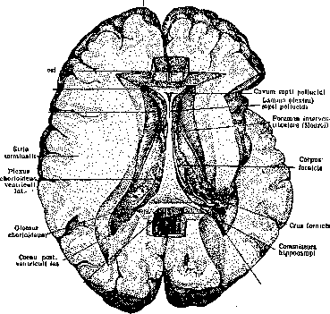
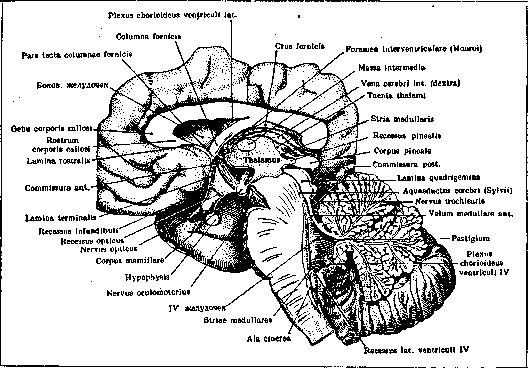
VENTRICULI CEREBRI (ventricles of the brain), cavities in the central nervous system. In the brain, the following ventricles are known: two lateral, III, IV, and V; in the spinal cord - ventriculus terminalis, or Krause's ventricle.-The lateral ventricles 23 7U are located in the cerebral hemispheres, partially reproducing their shape. They consist of a central part - cella media and processes extending from this part - horns (cornua): anterior, posterior, inferior (see figure 1). Each part of the ventricle corresponds to one of the lobes of the hemisphere: the anterior horn to the frontal lobe, the posterior to the occipital, the inferior to the temporal, and the central part to the parietal lobe. The anterior horn, triangular in cross-section, is bounded internally by septum pellucidum, externally and inferiorly by caput nuclei caudati, Polus frontalis Genu corporis callosi Cornu ant ventriculi lat Caput nuclei caudati, Splenium corporis callosi Polus occipitalis Figure 1. Ventriculi laterales cerebri. Commissura hippocampi, forming the roof of the third ventricle. anteriorly and superiorly by the corpus callosum. Between the two septa pellucida is the V ventricle, or cavum septi pellucidi. Cella media of the lateral ventricle, in cross-section, appears as a shallow fissure covered superiorly by corpus callosum; its floor is formed by the body of nuclei caudati, striae terminalis, and the outer part of the superior surface of thalami optici; internally it is closed by lamina epithelialis plexus chorioidei. From the central part inferiorly extends the inferior horn, and posteriorly the posterior horn; the transition point is called trigonum ventriculi lateralis. The posterior horn, triangular in shape, gradually narrows posteriorly, has a convex outer and concave inner surface, is covered by corpus callosum, while its other walls are formed by the white matter of the occipital lobe; on its inner side lie two longitudinal elevations: the lower one - calcar avis (bird's spur), corresponding to fissura calcarina, and the upper one - bulbus cornu posterioris (bulb of the posterior horn), formed by fibers of the corpus callosum surrounding the fissura parieto-occipitalis. The inferior horn extends from cella media in an arc downward and forward into the temporal lobe and ends 12 mm short of the temporal pole; its floor is a continuation of the posterior horn, the superior wall is formed by cauda nucl. caudati and stria terminalis; on the inferior and inner surfaces are elevations: 1) cornu Ammonis (Ammon's horn, pes hippocampi major), resulting from the indentation of the hippocampal fissure and ending anteriorly with a thickening having several notches - digitationes hippocampi, - separated by grooves; 2) a triangular elevation - eminentia collateralis Meckel, - corresponding to the collateral fissure. In the inferior horn, on the gyrus hippocampi and fascia dentata lies the fimbria - a continuation of the fornix. The lateral ventricles are enclosed on all sides except at one point in the anterior horn (foramen Monroi), between the columnae fornicis and thalami optici, through which the lateral ventricles communicate with the third ventricle.-The third ventricle is an unpaired slit-like cavity (see figs. 2 and 3), located along the median line and bounded on the sides by thalamus opticus and hypothalamus; anteriorly are columna fornicis, commissura alba ant., and lamina terminalis; posteriorly - commissura alba post. and commissura habenularum; inferiorly - chiasma opticum, tuber cinereum with infundibulum and hypophysis, corpora mamillaria, pedunculus cerebri, substantia perforata posterior; superiorly - tela chorioidea, attached to the superior surface of th. opt.; above it lie crura fornicis, connected by commissura hippocampi (s. psalterium), and corp. callosum. In the middle of the third ventricle passes massa intermedia s. commissura grisea; into the surrounding formations, the third ventricle gives off projections, or recessus: recessus infundibuli, opticus triangularis, pinealis, etc. Through the aquaeductus Sylvii (see), beginning under commissura alba post., the third ventricle communicates with the fourth ventricle.-The floor of the fourth ventricle, or rhomboid fossa (see figures 2, 3), is formed by the pons Varolii (see) and the medulla oblongata (see); the roof is formed by the cerebellum (see) and vela medullaria (see). At the border of the pons Varolii and medulla oblongata, the fourth ventricle extends into the brain substance, forming recessus lateralis. Directly above the floor of the fourth ventricle passes tela chorioidea and the rest of the primary anterior choroidea. The cavity of the fourth ventricle communicates with the subarachnoid space through openings - 2 foramina Luschka in the region of rec. later. and foramen Magendie in the lower part; near the obex, the fourth ventricle continues into the central canal, located in the lower parts of the medulla oblongata and extending along the entire spinal cord, at the end of which it expands into ventriculus terminalis Krause. All ventricular cavities are lined with ependyma and filled with cerebrospinal fluid and plexus chorioideus (see Chorioideus plexus).-The ventricles of the brain develop from the primary cerebral vesicles - the anterior, middle, and posterior, which appear on the anterior part of the neural tube; during development, the vesicles undergo changes: the cavity of the posterior vesicle transforms into the fourth ventricle; the cavity of the middle vesicle, due to thickening of its walls, narrows and transforms into the aquaeductus Sylvii; from the anterior cerebral vesicle, due to bulging of its walls, a secondary cerebral vesicle forms, which is divided by a medial groove into two halves - cerebral hemispheres; the cavity of the hemispheres forms the lateral ventricles,
Among the pathological processes observed in the cavities of the brain ventricles, one should note hemorrhages, neoplasms,
Figure 3. Inner surface of the brain. Ventriculi cerebri and their connections: anterior horn of the lateral ventricle, foramen Monroi, walls of the third ventricle, aquaeductus Sylvii, fourth ventricle. Septum pellucidum Columna fomicis Stria terminalis Trigonum habenulae Corpus pineal
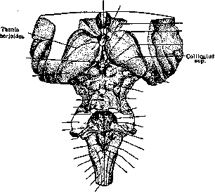
Habenula Pulvinar Lingula cerebelli Floor of the fourth ventricle Brachium conjunctivum Brachium pontis Corpus restiforme Recessus lat. ventriculi IV Taenia ventriculi IV Tuberculum cinereum Tuberculum cuneatum Clava Funiculus lat. Fasciculus cuneatus (Burdachi) Fasciculus gracilis (Golli)1 Corpus geniculatum mediale Colliculus inf. N. trochlearis Eminentia medialis Sulcus limitans rhombencephali Colliculus facialis Tuberculum acusticum et striae medullares Sulcus medianus fossae rhomboideae Ala cinerea Calamus scriptorius Obex Figure 2. Brainstem, its posterior surface. Floor of the fourth ventricle (third ventricle shown in the figure as a slit above corpus pineale).
and inflammatory changes. Primary hemorrhages into the ventricular cavities are very rare; more often, hemorrhages rupture from the surrounding tissue; clinically, this rupture manifests as a new apoplectic stroke, spread of paralysis to all limbs, muscle rigidity, convulsions, disorders of respiration and cardiac activity, drop in temperature, presence of blood in the cerebrospinal fluid. Death within 24 hours with signs of paralysis of the heart and respiration.-Neoplasms in the ventricles develop either in the ventricle itself from plexus choroideus or ependyma or grow into the ventricles from the surrounding brain tissue. The symptomatology of tumors developing in the lateral ventricles does not differ from the symptomatology of brain tumors (see); the clinical picture changes depending on their location. Small neoplasms in the cavity of the fourth ventricle may not be diagnosed during life - they are found at autopsy; the most common are cysticerci. Among the symptoms noted are disorders of the cranial nerves from VIII to XII, deafness, disorders of respiration, cardiac activity, swallowing, speech (dysarthria), sometimes glycosuria, diabetes, vasomotor disorders; less commonly, paresis or paralysis of the limbs, cerebellar ataxia, and finally, general cerebral symptoms - headaches, choked discs, blindness, dizziness, vomiting, etc. All these phenomena have an intermittent character, appearing periodically. Characteristic is the position of the head in such a patient (according to Bruns): he holds it motionless, because the slightest movement, turning to the side again causes dizziness, vomiting and other phenomena. With localization of the tumor in the third ventricle, drowsiness and mental disorders in the form of dementia paralytica are observed especially often, and sometimes as the only symptom, then polyuria, ophthalmoplegia and other symptoms appear, depending on the involvement of thalami optici, corpori quadrigeminum, pedunculi cerebri, floor of the third ventricle, etc.-Inflammatory processes in the ventricles in the form of ependymitis are observed in various inflammatory and toxic processes in the brain, e.g., in encephalitis, meningitis, syphilis, tuberculosis, alcoholism, etc., and are accompanied by hydrocephalus, the symptoms of which are observed in this case (see Hydrocephalus). For examining the condition of V. c., ventriculography (see) and encephalography (see) are used.
Related articles
Mentioned in
Cite this page
“Ventriculi Cerebri.” Soviet Medical Encyclopedia. English translation of Bolshaya Meditsinskaya Entsiklopediya, 1st ed. (Moscow, 1928–1936), ed. N. A. Semashko. https://sovietmedicalencyclopedia.pages.dev/article/ventriculi-cerebri/