Lumbar Puncture
Historical document, translated for reference. It reflects medical knowledge of the 1920s–30s and is not medical advice.
Summary
This article from the 1928–1936 Soviet medical encyclopedia details the technique, indications, and complications of lumbar puncture, a procedure used to obtain cerebrospinal fluid for diagnosis and therapy.
Encyclopedia article (1928–1936)
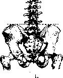
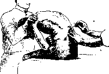
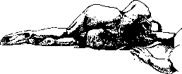
LUMBAR PUNCTURE (punctio lumbalis, lumbar or lumbar puncture) is performed to obtain cerebrospinal fluid from the spinal canal. According to Quincke, lumbar puncture is performed between L1 and L5. According to Tuffier, the puncture should be made between L4 and L5, and according to Chipault, between L5 and S1. Lumbar puncture was first performed by Quincke in 1890 on a 2-year-old child suffering from hydrocephalus. Even earlier, Corning (1885) developed the question of the action of injections of medicinal substances on the spinal cord. In the future, lumbar puncture became widely used for diagnosis and therapy in nervous system diseases, for spinal anesthesia according to Bier, and in recent times for encephalography. The puncture is made into the subdural sac (cavum subarachnoidale), which at this level contains only the filaments of the cauda equina, freely floating in the cerebrospinal fluid. In the lumbar part of the spine, the cavum subarachnoidale is expanded in the form of the cisterna terminalis over the course from L1 to S1. At this section of the spine, puncture can be performed in any intervertebral space without risking injury to nervous elements. - Most doctors orient themselves on L4 via the so-called "Tuffier's line" or the line of Jacobi, performing the puncture above or below it. This line is drawn between the highest points of the iliac parts or is determined by stretching a sterile towel over the iliac crests. The intersection of Tuffier's line with the line of spinous processes is the tip of the spinous process of L4 (Fig. 1). The puncture should be made with the following instruments: steel sharp needles 7–8 cm long with a short bevel or thin trocars of the same length (diameter 1–1.3 mm) and an ordinary Record syringe (5.0–10.0). There are special Quincke needles, Bier (Fig. 2), Kriging, Tomaszewski, and others. Lumbar puncture is performed in a hospital and, like any operation, strictly aseptically. - The puncture is performed in a sitting or lying position. The patient is seated either across the table with legs hanging over the stool, or with legs extended along the table (Fig. 3). The latter position is recommended (Borszeki, Sellheim, Furbringer, etc.) to avoid lordosis of the spine at the moment of puncture (cases of needle breakage). One can also seat the patient on a chair, facing the backrest. The patient's forearms rest on the thighs. The spine and neck are bent forward in an arc ad maximum. It is better if the head and shoulders are fixed by an assistant from the front. This position gives a clear prominence of the spinous processes, their separation, and better orientation relative to the puncture site, especially since the nerve roots of the dural sac gather into two bundles, diverging to the sides, which favors the introduction of the needle. Lumbar puncture is also performed in a lying position (Fig. 4), especially in severe patients and in infants: the patient turns to the side, legs are bent at the knee and hip joints, the torso is bent forward ad maximum, and the head is brought to the chest. - Technique of lumbar puncture. Marking the spinous process (L4) with the index finger of the left hand, an energetic puncture of the skin is made below it, in the interspinous space (according to Quincke, stepping back from the midline by 1/2 cm). In a sitting position, the needle should go almost horizontally, with a slight upward slope, while in a lying position, the needle is at an angle of 70° to the torso. The needle passes through the skin, subcutaneous tissue, lumbar aponeurosis, intervertebral yellow ligaments, and the dura and arachnoid mater. The depth of puncture varies depending on the patient's build and muscle development, on average from 4 to 7 cm in adults and from 2 to 5 cm in children. When passing the dura mater, a crackling sensation is felt, sometimes audible at a distance. After the sensation that the needle is in the subarachnoid space is felt, the stylet is removed. The entry of the needle into the cisterna terminalis is determined by the appearance of clear, yellowish fluid, which flows in a stream or in frequent drops. The needle should not be advanced further after this. The admixture of blood indicates puncture of the veins of the epidural space or an incorrectly taken direction. In this case, the needle should be withdrawn and the puncture repeated. The fluid is released in an amount not exceeding 10.0 ml in adults and 5.0 ml in children (only in meningitis from 20.0 to 40.0 ml). During puncture, the pressure under which the cerebrospinal fluid flows out is determined, its color, and then a series of analyses are performed (on protein, RW and various other physical, chemical, bacteriological, and cytological studies). The normal pressure under which the fluid flows out fluctuates from 125–150 mm of fluid. Pressure is measured by the Quincke method, using a Claude manometer measuring pressure in millimeters of water column. All movements of the body after lumbar puncture must be performed slowly and carefully. After puncture, the patient is placed horizontally, initially on the abdomen to avoid spontaneous leakage of fluid, and then turned onto the back; the patient's head is better placed lower; in this position he remains 1–2 days depending on the general condition. Lumbar puncture is applied in spinal anesthesia (see); in addition, it is a diagnostic means in nervous system diseases. Through lumbar puncture, air is blown into the brain cavities (see Encephalography), lipiodol. Lumbar puncture serves as a therapeutic measure for lowering intracranial pressure (in hydrocephalus, in skull injuries, brain tumors), for removing part of the infected fluid (in meningitis, encephalitis), and finally, lumbar puncture is used for the introduction of various medications (urotropin, pilocarpin, anti-meningococcal serum, etc.). Contraindications to lumbar puncture are the presence of neoplasms in the brain, especially in the posterior cranial fossa, because low pressure allows the tumor to move and press on important vital centers; lumbar puncture also presents a danger in cases where low pressure can cause a recurrent hemorrhage (in strokes), rupture of vessels (in uremia), etc. Lumbar puncture cannot be performed in the presence of an infectious focus nearby - bedsores, furuncle, carbuncle, etc. One avoids performing lumbar puncture in persons with heart diseases and with a general decrease in nutrition. Performing lumbar puncture ambulatorily is contraindicated. Subsequent complications of lumbar puncture: headaches, back pain, convulsions, dizziness, numbness, crawling ants, rise in temperature, nausea, vomiting, phenomena of the so-called "meningism" or "aseptic meningitis". The latter is explained by damage to the membranes by the needle (irritation) with subsequent leakage of fluid into the cavum epidurale (Baruch) (a scar forms according to Bungart through 8–10 days). Unfavorable consequences are often observed in psychopathic and hysterical subjects in the form of vomiting, a sensation of compression of the head, and phenomena of irritation of the lower extremities. Cases of paresis and paralysis, and even death after punctures, have been described. The latter is observed mainly in brain tumors. The frequency of complications according to various authors ranges from 10% to 30%. Treatment of complications: horizontal position in bed with the foot end raised, pituitrin, pilocarpin, infusion of NaCl, intravenous administration of a 40% solution of urotropin, etc. A variety of lumbar puncture is the suboccipital puncture according to Ayer and Eskuhen (see Myelography), performed between the occipital bone and the atlas; the patient sits with the head tilted forward; the needle is introduced to a depth of 4–5 cm. This method is unsafe for the patient due to the proximity of the medulla oblongata and is applied only for direct indications towards it (for introducing lipiodol into the subarachnoid space).
Fig.
patient's forearms rest on the thighs. The spine and neck are bent forward in an arc ad maximum. It is better if the head and shoulders are fixed by an assistant from the front. This position gives a clear prominence of the spinous processes, their separation, and better orientation relative to the puncture site, especially since the nerve roots of the dural sac gather into two bundles, diverging to the sides, which favors the introduction of the needle. Lumbar puncture is also performed in a lying position (Fig. 4), especially in severe patients and in infants: the patient turns to the side, legs are bent at the knee and hip joints, the torso is bent forward ad maximum, and the head is brought to the chest. - Technique of lumbar puncture. Marking the spinous process (L4) with the index finger of the left hand, an energetic puncture of the skin is made below it, in the interspinous space (according to Quincke, stepping back from the midline by 1/2 cm). In a sitting position, the needle should go almost horizontally, with a slight upward slope, while in a lying position, the needle is at an angle of 70° to the torso. The needle passes through the skin, subcutaneous tissue, lumbar aponeurosis, intervertebral yellow ligaments, and the dura and arachnoid mater. The depth of puncture varies depending on the patient's build and muscle development, on average from 4 to 7 cm in adults and from 2 to 5 cm in children. When passing the dura mater, a crackling sensation is felt, sometimes audible at a distance. After the sensation that the needle is in the subarachnoid space is felt, the stylet is removed. The entry of the needle into the cisterna terminalis is determined by the appearance of clear, yellowish fluid, which flows in a stream or in frequent drops. The needle should not be advanced further after this. The admixture of blood indicates puncture of the veins of the epidural space or an incorrectly taken direction. In this case, the needle should be withdrawn and the puncture repeated. The fluid is released in an amount not exceeding 10.0 ml in adults and 5.0 ml in children (only in meningitis from 20.0 to 40.0 ml). During puncture, the pressure under which the cerebrospinal fluid flows out is determined, its color, and then a series of analyses are performed (on protein, RW and various other physical, chemical, bacteriological, and cytological studies). The normal pressure under which the fluid flows out fluctuates from 125–150 mm of fluid. Pressure is measured by the Quincke method, using a Claude manometer measuring pressure in millimeters of water column. All movements of the body after lumbar puncture must be performed slowly and carefully. After puncture, the patient is placed horizontally, initially on the abdomen to avoid spontaneous leakage of fluid, and then turned onto the back; the patient's head is better placed lower; in this position he remains 1–2 days depending on the general condition. Lumbar puncture is applied in spinal anesthesia (see); in addition, it is a diagnostic means in nervous system diseases. Through lumbar puncture, air is blown into the brain cavities (see Encephalography), lipiodol. Lumbar puncture serves as a therapeutic measure for lowering intracranial pressure (in hydrocephalus, in skull injuries, brain tumors), for removing part of the infected fluid (in meningitis, encephalitis), and finally, lumbar puncture is used for the introduction of various medications (urotropin, pilocarpin, anti-meningococcal serum, etc.). Contraindications to lumbar puncture are the presence of neoplasms in the brain, especially in the posterior cranial fossa, because low pressure allows the tumor to move and press on important vital centers; lumbar puncture also presents a danger in cases where low pressure can cause a recurrent hemorrhage (in strokes), rupture of vessels (in uremia), etc. Lumbar puncture cannot be performed in the presence of an infectious focus nearby - bedsores, furuncle, carbuncle, etc. One avoids performing lumbar puncture in persons with heart diseases and with a general decrease in nutrition. Performing lumbar puncture ambulatorily is contraindicated. Subsequent complications of lumbar puncture: headaches, back pain, convulsions, dizziness, numbness, crawling ants, rise in temperature, nausea, vomiting, phenomena of the so-called "meningism" or "aseptic meningitis". The latter is explained by damage to the membranes by the needle (irritation) with subsequent leakage of fluid into the cavum epidurale (Baruch) (a scar forms according to Bungart through 8–10 days). Unfavorable consequences are often observed in psychopathic and hysterical subjects in the form of vomiting, a sensation of compression of the head, and phenomena of irritation of the lower extremities. Cases of paresis and paralysis, and even death after punctures, have been described. The latter is observed mainly in brain tumors. The frequency of complications according to various authors ranges from 10% to 30%. Treatment of complications: horizontal position in bed with the foot end raised, pituitrin, pilocarpin, infusion of NaCl, intravenous administration of a 40% solution of urotropin, etc. A variety of lumbar puncture is the suboccipital puncture according to Ayer and Eskuhen (see Myelography), performed between the occipital bone and the atlas; the patient sits with the head tilted forward; the needle is introduced to a depth of 4–5 cm. This method is unsafe for the patient due to the proximity of the medulla oblongata and is applied only for direct indications towards it (for introducing lipiodol into the subarachnoid space).
Fig.
Figure 3.
In a sitting position, the needle should go almost horizontally, with a slight upward slope, while in a lying position, the needle is at an angle of 70° to the torso. The needle passes through the skin, subcutaneous tissue, lumbar aponeurosis, intervertebral yellow ligaments, and the dura and arachnoid mater. The depth of puncture varies depending on the patient's build and muscle development, on average from 4 to 7 cm in adults and from 2 to 5 cm in children. When passing the dura mater, a crackling sensation is felt, sometimes audible at a distance. After the sensation that the needle is in the subarachnoid space is felt, the stylet is removed. The entry of the needle into the cisterna terminalis is determined by the appearance of clear, yellowish fluid, which flows in a stream or in frequent drops. The needle should not be advanced further after this. The admixture of blood indicates puncture of the veins of the epidural space or an incorrectly taken direction. In this case, the needle should be withdrawn and the puncture repeated. The fluid is released in an amount not exceeding 10.0 ml in adults and 5.0 ml in children (only in meningitis from 20.0 to 40.0 ml). During puncture, the pressure under which the cerebrospinal fluid flows out is determined, its color, and then a series of analyses are performed (on protein, RW and various other physical, chemical, bacteriological, and cytological studies). The normal pressure under which the fluid flows out fluctuates from 125–150 mm of fluid. Pressure is measured by the Quincke method, using a Claude manometer measuring pressure in millimeters of water column. All movements of the body after lumbar puncture must be performed slowly and carefully. After puncture, the patient is placed horizontally, initially on the abdomen to avoid spontaneous leakage of fluid, and then turned onto the back; the patient's head is better placed lower; in this position he remains 1–2 days depending on the general condition. Lumbar puncture is applied in spinal anesthesia (see); in addition, it is a diagnostic means in nervous system diseases. Through lumbar puncture, air is blown into the brain cavities (see Encephalography), lipiodol. Lumbar puncture serves as a therapeutic measure for lowering intracranial pressure (in hydrocephalus, in skull injuries, brain tumors), for removing part of the infected fluid (in meningitis, encephalitis), and finally, lumbar puncture is used for the introduction of various medications (urotropin, pilocarpin, anti-meningococcal serum, etc.). Contraindications to lumbar puncture are the presence of neoplasms in the brain, especially in the posterior cranial fossa, because low pressure allows the tumor to move and press on important vital centers; lumbar puncture also presents a danger in cases where low pressure can cause a recurrent hemorrhage (in strokes), rupture of vessels (in uremia), etc. Lumbar puncture cannot be performed in the presence of an infectious focus nearby - bedsores, furuncle, carbuncle, etc. One avoids performing lumbar puncture in persons with heart diseases and with a general decrease in nutrition. Performing lumbar puncture ambulatorily is contraindicated. Subsequent complications of lumbar puncture: headaches, back pain, convulsions, dizziness, numbness, crawling ants, rise in temperature, nausea, vomiting, phenomena of the so-called "meningism" or "aseptic meningitis". The latter is explained by damage to the membranes by the needle (irritation) with subsequent leakage of fluid into the cavum epidurale (Baruch) (a scar forms according to Bungart through 8–10 days). Unfavorable consequences are often observed in psychopathic and hysterical subjects in the form of vomiting, a sensation of compression of the head, and phenomena of irritation of the lower extremities. Cases of paresis and paralysis, and even death after punctures, have been described. The latter is observed mainly in brain tumors. The frequency of complications according to various authors ranges from 10% to 30%. Treatment of complications: horizontal position in bed with the foot end raised, pituitrin, pilocarpin, infusion of NaCl, intravenous administration of a 40% solution of urotropin, etc. A variety of lumbar puncture is the suboccipital puncture according to Ayer and Eskuhen (see Myelography), performed between the occipital bone and the atlas; the patient sits with the head tilted forward; the needle is introduced to a depth of 4–5 cm. This method is unsafe for the patient due to the proximity of the medulla oblongata and is applied only for direct indications towards it (for introducing lipiodol into the subarachnoid space).
Fig.
Figure 4.
The depth of puncture varies depending on the patient's build and muscle development, on average from 4 to 7 cm in adults and from 2 to 5 cm in children. When passing the dura mater, a crackling sensation is felt, sometimes audible at a distance. After the sensation that the needle is in the subarachnoid space is felt, the stylet is removed. The entry of the needle into the cisterna terminalis is determined by the appearance of clear, yellowish fluid, which flows in a stream or in frequent drops. The needle should not be advanced further after this. The admixture of blood indicates puncture of the veins of the epidural space or an incorrectly taken direction. In this case, the needle should be withdrawn and the puncture repeated. The fluid is released in an amount not exceeding 10.0 ml in adults and 5.0 ml in children (only in meningitis from 20.0 to 40.0 ml). During puncture, the pressure under which the cerebrospinal fluid flows out is determined, its color, and then a series of analyses are performed (on protein, RW and various other physical, chemical, bacteriological, and cytological studies). The normal pressure under which the fluid flows out fluctuates from 125–150 mm of fluid. Pressure is measured by the Quincke method, using a Claude manometer measuring pressure in millimeters of water column. All movements of the body after lumbar puncture must be performed slowly and carefully. After puncture, the patient is placed horizontally, initially on the abdomen to avoid spontaneous leakage of fluid, and then turned onto the back; the patient's head is better placed lower; in this position he remains 1–2 days depending on the general condition. Lumbar puncture is applied in spinal anesthesia (see); in addition, it is a diagnostic means in nervous system diseases. Through lumbar puncture, air is blown into the brain cavities (see Encephalography), lipiodol. Lumbar puncture serves as a therapeutic measure for lowering intracranial pressure (in hydrocephalus, in skull injuries, brain tumors), for removing part of the infected fluid (in meningitis, encephalitis), and finally, lumbar puncture is used for the introduction of various medications (urotropin, pilocarpin, anti-meningococcal serum, etc.). Contraindications to lumbar puncture are the presence of neoplasms in the brain, especially in the posterior cranial fossa, because low pressure allows the tumor to move and press on important vital centers; lumbar puncture also presents a danger in cases where low pressure can cause a recurrent hemorrhage (in strokes), rupture of vessels (in uremia), etc. Lumbar puncture cannot be performed in the presence of an infectious focus nearby - bedsores, furuncle, carbuncle, etc. One avoids performing lumbar puncture in persons with heart diseases and with a general decrease in nutrition. Performing lumbar puncture ambulatorily is contraindicated. Subsequent complications of lumbar puncture: headaches, back pain, convulsions, dizziness, numbness, crawling ants, rise in temperature, nausea, vomiting, phenomena of the so-called "meningism" or "aseptic meningitis". The latter is explained by damage to the membranes by the needle (irritation) with subsequent leakage of fluid into the cavum epidurale (Baruch) (a scar forms according to Bungart through 8–10 days). Unfavorable consequences are often observed in psychopathic and hysterical subjects in the form of vomiting, a sensation of compression of the head, and phenomena of irritation of the lower extremities. Cases of paresis and paralysis, and even death after punctures, have been described. The latter is observed mainly in brain tumors. The frequency of complications according to various authors ranges from 10% to 30%. Treatment of complications: horizontal position in bed with the foot end raised, pituitrin, pilocarpin, infusion of NaCl, intravenous administration of a 40% solution of urotropin, etc. A variety of lumbar puncture is the suboccipital puncture according to Ayer and Eskuhen (see Myelography), performed between the occipital bone and the atlas; the patient sits with the head tilted forward; the needle is introduced to a depth of 4–5 cm. This method is unsafe for the patient due to the proximity of the medulla oblongata and is applied only for direct indications towards it (for introducing lipiodol into the subarachnoid space).
Related articles
Mentioned in
Cite this page
“Lumbar Puncture.” Soviet Medical Encyclopedia. English translation of Bolshaya Meditsinskaya Entsiklopediya, 1st ed. (Moscow, 1928–1936), ed. N. A. Semashko. https://sovietmedicalencyclopedia.pages.dev/article/lumbar-puncture/