Myelography
Historical document, translated for reference. It reflects medical knowledge of the 1920s–30s and is not medical advice.
Summary
This article from the 1928–1936 Great Medical Encyclopedia describes myelography, detailing the introduction of contrast agents such as lipiodol and iodipin into the spinal canal to diagnose spinal cord tumors and blockages. It covers historical methods, technical procedures, radiographic findings, and potential complications including pain and aseptic meningitis.
Encyclopedia article (1928–1936)
MYELOGRAPHY (from Greek myelos - marrow and grapho - I write), the introduction of contrast agents into the cerebrospinal canal to determine its patency and to identify the level, extent, and nature of an obstructive process. In 1912, Krause first proposed injecting 10–20 cm3 of a 5% collargol solution before surgery in cases of spinal cord tumors. Later, solutions of sodium bromide and strontium bromide were proposed, but these did not come into use. In 1919, Dandy introduced air into the lumbar region of the cerebrospinal canal and made attempts to obtain contrast X-ray images. The air, rising upward, was arrested at the lower boundary of the tumor, and entered the ventricles with some delay. Dandy's method did not provide a clear picture of the obstruction and did not become widespread. In 1921, Sicard and Forestier in France introduced lipiodol for myelography; from that moment, myelography gained wide dissemination. Lipiodol and iodipin are most frequently used as contrast agents. Both lipiodol and iodipin are sterile and possess bactericidal properties.* Saponification of the injected oil occurs only partially, and absorption proceeds extremely slowly. Before performing myelography, the patient's bowel must be well cleansed. The contrast agent is introduced via a suboccipital puncture. The occipital region is shaved before the puncture, and the field is prepared accordingly. Lipiodol or iodipin, pre-warmed to make it less viscous, is drawn into a syringe in an amount of 1–2 cm3. Air bubbles are removed so that their presence does not delay the descent of the contrast mass or disturb the homogeneity of the shadow. The puncture is performed either with the patient in a sitting position or lying on their side with the head end of the table slightly elevated. The preparation is injected slowly. When withdrawing the needle, one should avoid letting drops of oil retained in the needle get into the soft tissues, as these drops can produce a false picture of retention on the radiograph. After the injection, the patient is left in a sitting position or, if the puncture was performed while lying down, the upper half of the torso is elevated. Injection can be performed in any interspinous space, but puncture in the cervical region and the upper half of the thoracic region is technically more difficult. In addition to contrast agents whose specific gravity is higher than that of the cerebrospinal fluid, ascending lipiodol (lipiodol ascendens), whose specific gravity is lower than that of the cerebrospinal fluid, is also used for myelography. Introduced via ordinary lumbar puncture, it rises upward and stops at the lower boundary of the obstruction. For the same purpose, ordinary lipiodol and iodipin can also be used by placing the patient in the Trendelenburg position after the lumbar puncture. X-ray examination is performed after 5–10 minutes, then after 3–4 hours, and after 1–2 days, depending on the retention and the speed of descent of the contrast agent. Before radiography, fluoroscopy should be performed preliminarily to establish the location of the contrast agent, and only then should the image be taken. The patient lies on a table tilted at an angle of 30°, or the image is taken in a vertical position. After the introduction of the contrast agent into the cerebellomedullary cistern, the latter appears on the anteroposterior image as a rhombus, and on the lateral image as a triangle, the upper corner of which reaches the foramen of Magendie, and the lower corner passes at the level of the lower edge of the atlas arch into the subarachnoid space of the spinal canal. Retention of the contrast agent in the cistern occurs with high-lying obstructions. In the absence of obstructions, the contrast agent flows down rather quickly, lingering briefly at the level of DIV, where there is a physiological narrowing, and after a few minutes collects completely in the blind end of the dural sac. Often, in the absence of any obstructions, the descent of the contrast agent occurs slowly; it leaves shadows in the form of bands, drops, or streaks along the course of the roots; the descent process may end within 24–48 hours. Having descended into the end of the sac, the contrast agent acquires the shape of a bullet or beet with the wide end facing upward. The contours of this shadow can vary greatly; in places along the course of the roots of the cauda equina, the contrast agent settles in separate drops and diverging streaks. The contours of the blind sac are projected at the level of LV and SI. The contrast agent can penetrate along the course of the roots and slowly descend down to the intervertebral ganglia and even further. Remaining mobile at first, it shifts with changes in position, but in about 2 weeks its mobility disappears, and it can remain in one place for years without noticeable change. Complete resorption of either iodipin or lipiodol apparently does not occur. Ascending lipiodol in the absence of obstructions rapidly rises upward and can be detected in the form of small drops in the basal cisterns, in the ventricles, and on the surface of the brain. Retention of the contrast agent can be false or true. In true retention, it is projected on the radiograph as a compact mass or in a pulverized form. The duration of retention varies from short-term to permanent. In diffuse processes in the subarachnoid space with the formation of adhesions, the contrast agent, having fragmented, is retained at various levels, giving scattered shadows in the form of drops or stalactites. Dissemination, slow movement of the contrast agent in separate drops and streaks, indicates the presence of arachnoidal adhesions. Sometimes in inflammatory processes, the contrast agent descends in two streams along the sides of the spinal cord or is retained in the form of a brush; less often, almost complete retention is obtained. In the latter case, differential diagnosis from a tumor can become very difficult. In spinal cord tumors, the contrast agent may be retained completely or partially, for a long time or only temporarily. With complete retention, a continuous shadow is obtained above the tumor, which covers the tumor like a cap or sits astride it. Characteristically, the lower edge of the shadow is concave, repeating the spherical contour of the upper pole of the tumor. However, one cannot assume that this concavity depends on the location of the contrast agent directly on the tumor; identical contour outlines can be observed in extradural and intramedullary tumors. Apparently, the shape of the lower boundary is determined by the nature of the slit-like space formed above the section of the spinal cord compressed by the tumor. Neither the shape of the shadow, nor the outlines of its edge, nor the nature of the descent of the contrast agent can provide an exact criterion for differential diagnosis between extra- and intramedullary tumors. The rapid passage of the contrast agent cannot completely exclude the presence of a tumor. In small, especially intramedullary tumors, the contrast agent may descend completely, and its short-term retention may be overlooked. In such cases, with increasing compression symptoms, repeated myelography is indicated. The height of retention in tumors corresponds to the border of the neoplasm, but it should be kept in mind that arachnoiditis phenomena often develop around the tumor, and the shadow on the radiograph may appear 1–3 segments higher than the true border of the tumor. Myelography with ascending lipiodol to determine the lower border is used less frequently. In multiple tumors or when a long tumor is suspected, a combined administration of ascending and descending contrast agent above and below the tumor is possible. At present, there are hundreds of cases of myelography without any harmful consequences. Nevertheless, the method is not free from complications. The very process of suboccipital puncture carries certain dangers of damage to the medulla oblongata, and isolated cases have been observed where myelography resulted in respiratory arrest, vomiting, etc. When the contrast agent descends into the affected segments, lightning-fast shooting pains may appear. Very frequently in the first days after myelography, headaches are noted; in half of the cases, an increase in temperature is observed, sometimes reaching 39°; there are pains in the lower back, in the legs, paresthesias, a feeling of tightness, and difficulty in urination. In many cases, myelography worsened compression symptoms, increasing paralysis, worsening sensory disturbances, and aggravating disorders of the pelvic organs. Such complications have given rise to a desire to remove the introduced contrast agent. Severe aseptic meningitides have been observed after myelography; several cases of death are known. Ascending lipiodol is tolerated worse than descending, causing a more violent reaction. The cerebrospinal fluid responds to myelography with an increase in protein content, the appearance of globulins, and an increase in formed elements (up to 500). * The specific gravity of both preparations is higher than the specific gravity of the cerebrospinal fluid; therefore, when introduced into the cistern, they sink downward.
During surgery at the site of the arrest, edema, clouding of the membranes, and encapsulation of the contrast medium in cavities were found. At autopsy, the development of chronic productive peripachymeningitis throughout the entire extent of the contrast medium arrest and the presence of oleogranulomas were observed. Experimentally, the irritating effect of iodized oil manifested as cellular infiltration of the pia mater, proliferation of connective tissue, and the formation of cystic cavities. With the introduction of myelography, the number of early-diagnosed and successfully operated spinal cord tumors increased. True, the clinical signs of a spinal cord tumor are so indicative that they allow an accurate topical diagnosis to be made without myelography, often based solely on neurological symptoms; nevertheless, the great auxiliary role of myelography in confirming the presence of a neoplasm and in specifying the level of the lesion cannot be denied. Therefore, in cases of spinal cord tumors, especially those subject to surgical treatment, myelography should conclude the clinical examination. During operations, droplets of the introduced lipiodol or iodipinal are easily removed. In tumors originating from the spine, myelography appears superfluous in the majority of cases. In chronic inflammatory processes and unclear spinal cord lesions, where it is difficult to establish the nature of the process from clinical data, myelography can significantly facilitate diagnosis, but due to the possibility of exacerbating pain, indications for myelography here must be set with extreme caution. In spinal tuberculosis, in some cases of intraspinal abscesses, myelography can reveal the relationship inside the spinal canal and resolve the question of the feasibility of surgical intervention. In some cases of old spinal fractures, myelography can resolve the question of whether the compression phenomena depend on the presence of scars and whether surgery is advisable. Contraindications to the use of myelography include Basedow's disease, iodine idiosyncrasy, and high fever. Taking into account the possibility of complications, the method cannot be considered completely indifferent, and the use of myelography must be performed according to strict indications after a comprehensive clinical examination.
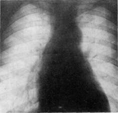
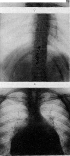
Fig. 1 and 2. Lipiodol arrest in spinal cord tumor. Fig. 1. Lipiodol descended to the end of the dural sac.
Figure 4. Typical lipiodol arrest in the form of a capsule in arachnoiditis at the level of the lower thoracic and upper lumbar vertebrae. Figure 5. Typical picture of mitral valve stenosis. Fig. 6. Stenosis with an insufficient mitral valve; marked enlargement of the right ventricle, left atrium, and left ventricle. Literature: Myelography. Heart defects. 1930; Bregman L. und Szpilman P., Zur Lipiodoldiagnose bei Rückenmarkskrankheiten, Deutsche Zeitschrift für Nervenheilkunde, B. CIII, 1928; Craig W., Use and abuse of iodized oil in the diagnosis of lesions of spinal cord, Surgery, gynecology and obstetrics, v. XLIX, 1929; Peiper H., Die Myelographie im Dienste der Diagnostik von Erkrankungen des Rückenmarks, Ergebnisse d. med. Strahlenforschung, B. II, 1926; Sgalitzer M., Myelographie mit sinkendem und aufsteigendem Jodol, Acta radiologica, B. IX, 1928; Sicard J. et Forestier J., Diagnostic et thérapeutique par le lipiodol, P., 1928. S. Bryusova. MYELOSES (myelosis), the process of development anywhere of myeloid tissue or its specific elements—myelocytes, myeloblasts, erythroblasts. In the case of obvious predominance or exclusive development of any of these elements, one speaks of myelocytic myelosis (myelocytosis), myeloblastic myelosis (myeloblastosis), and erythroblastic myelosis (erythroblastosis). Physiological, or typical, myelosis always occurs within the red bone marrow, serving as the main anatomical substrate of hematopoiesis (see). Under pathological conditions, an expansion of the territorial base of myelosis is often observed, and in some cases this expansion proceeds only within the bone marrow, the yellow (fatty) parts of which gradually turn into grayish-red, for example in leukemia, aleukemia (so-called leukemic and aleukemic myeloses; see Leukemia), and in certain chronic anemias (e.g., myeloblastosis in malignant anemia). Not infrequently, myelosis extends beyond the bounds of the actual myeloid tissue, i.e., the bone marrow (extramedullary, or atypical, myelosis), encompassing the most diverse organs. Most frequently, such extramedullary myelosis is observed in the spleen, lymph glands, and liver, especially in acute infectious diseases (acute infectious myelosis), in many blood diseases [leukemia (which is why myeloid leukemia has been proposed to be called leukemic myelosis), malignant anemia, anaemia splenica infantum, and others]. - Macroscopically, in myeloses of organs, one often notes some degree of, sometimes completely insignificant, enlargement of the organ (especially the spleen, lymph glands); on cross-section—significant juiciness, brain-like appearance, and frequently a distinct gray or grayish-green shade of the tissue; however, parallel phenomena of hyperemia often mask these gray shades, and recognition is made with certainty only microscopically. Myelosis can also develop outside organs, since all connective tissue cells (lymphocytes, fibroblasts, "resting wandering cells," etc.), as well as elements of vessel walls, endothelium, and adventitial cells, can undergo myeloid transformation. Myelosis is also observed in adipose tissue, which is accompanied by the atrophy of the latter. - In the recognition of myelosis under the microscope, of particular importance is the detection of the enzyme oxidase (see Blood, research methodology) in the protoplasm of myeloid cells, and practical diagnosis of myelosis often practically reduces to finding cells containing the specified enzyme. This method, however, does not always yield clear results, since the youngest of the myeloid elements, e.g., myeloblasts, may not give the oxidase reaction. On the other hand, it should be kept in mind that mature polymorphonuclear leukocytes and eosinophils always give a positive oxidase reaction, which in themselves do not indicate the presence of myelosis. Diffuse and nodular tumor-like myeloses of the bone marrow are commonly called myelomas.
I. Davydovsky.
Related articles
Mentioned in
Cite this page
“Myelography.” Soviet Medical Encyclopedia. English translation of Bolshaya Meditsinskaya Entsiklopediya, 1st ed. (Moscow, 1928–1936), ed. N. A. Semashko. https://sovietmedicalencyclopedia.pages.dev/article/myelography/