Myeloma
Historical document, translated for reference. It reflects medical knowledge of the 1920s–30s and is not medical advice.
Summary
This article from the 1928–1936 Great Medical Encyclopedia discusses myeloma, a tumor originating from bone marrow elements, detailing its historical background, pathological anatomy, clinical forms, and histological classification.
Encyclopedia article (1928–1936)
MYELOMA, myeloma, a tumor of bone marrow elements, one of the types of myeloblastic tumors. The term myeloma was first proposed in 1873 by Rustizky (Rustizky) to designate a tumor developing multiply (myeloma multiplex) in the bone marrow, which, according to Rustizky's opinion, stood closer in its histological character and course to hyperplasias than to true tumors. In 1889, Kahler (Kahler) detailed the clinical symptomatology of myeloma, which served as the basis from the clinical standpoint to frequently designate myeloma as "Kahler's disease." The name "Rustizky-Kahler disease" is also encountered. Soon after the first anatomical-pathological description of myeloma made by Rustizky, the indications given by him regarding the distinction of myelomas from true bone marrow tumors were forgotten, and the name myeloma began to be applied to various multiple tumors of the bone marrow; thanks to this, the term "myeloma" acquired as it were a collective meaning. Subsequently, thanks to the works of Paltauf, Pappenheim, Schmidt, Lubarsch, Hirschfeld, Fraenkel, Wallgren (Paltauf, Pappenheim, Schmidt, Lubarsch, Hirschfeld, Frankel, Wallgren) and others, the concept of "myeloma" was refined, and only a systemic lesion of the bone marrow, manifested in the development of multiple focal proliferations of bone marrow cells within it, began to be classified as myeloma. These proliferations do not exhibit true blastomatous growth and do not give metastases. However, to the present time, researchers still do not have complete agreement on the essence of myeloma, as well as on some details concerning the histological structure and course of myeloma (see below). Myeloma belongs to rare diseases, encountered in mature and especially in advanced ages; myeloma at the age of up to 15 years represents a great rarity. Men fall ill with myeloma significantly more often than women. Among bones, spongy bones are predominantly affected, specifically the bones of the skull, pelvis, ribs, sternum, and vertebrae; somewhat less frequently the lesion affects the long bones of the extremities and much less frequently the small bones of the hands and feet. There are cases of myeloma when the disease affects almost all bones of the skeleton, and on the other hand, cases can be observed in which myeloma nodes develop only in certain groups of bones, for example, in the bones of the skull or pelvis or in the ribs, sternum, and vertebrae. In appearance, bones may present no changes, or rounded or spindle-shaped swellings and thickenings may be noticeable in them. Bones in the affected areas are soft, easily bend, break, and are cut with a knife; sometimes this softness is also observed in bones in places where there are no myeloma nodes, which is explained by disuse osteoporosis. On cross-sections and cuts of bones in the bone marrow, rounded nodes are discovered, either few and rarely located or, conversely, in very large numbers, almost merging with one another. The size of the nodes is variable, but usually does not exceed the size of a chicken egg; the consistency is soft, but often somewhat denser than red bone marrow. The color of the nodes can present great variety; in the majority of cases, they have an uneven grayish-red color with lighter grayish or yellowish areas alternating with dark red foci; sometimes it is a yellowish- or pinkish-white color, similar to the color of a sarcomatous neoplasm; in some cases, the appearance of myeloma nodes is similar to the appearance of the surrounding red bone marrow. In general, myeloma nodes as a rule are more or less sharply contoured and demarcated from the surrounding bone marrow tissue; only when there are hemorrhages simultaneously involving the tissue of the myeloma node and the surrounding bone marrow does the boundary of the node appear unclear. Much rarer are those cases of myeloma when the process is expressed not in the formation of the above-described nodes, but in a diffuse proliferation involving the entire bone marrow of the given bones: this type of myeloma provides grounds, alongside the nodular form of myeloma, to recognize the diffuse form of myeloma, or diffuse myelomatosis. It must be pointed out that this form can present significant difficulties for diagnosis not only at the patient's bedside, but also at autopsy; due to the absence of nodes and the frequent external similarity of the myelomatous proliferation in such diffuse forms with the appearance of ordinary red bone marrow, the diagnosis is often made only after microscopic examination. The bone tissue within the limits of myeloma undergoes gradual resorption, and in the nodular form it is significantly more pronounced than in the diffuse form. Within the limits of the myeloma nodes, the trabeculae of the spongy bone disappear, and the compact cortical layer undergoes gradual thinning; in places, this may lead to the complete destruction of the cortical layer of the bone, and then the myeloma grows under the periosteum and lifts it up, producing a rounded soft node on the bone at such a place. Bone defects in the areas of myeloma nodes are clearly visible on macerated bone preparations, with the most demonstrative picture being given by the macerated cranial vault, in which through-holes with a delicately-laced network of trabeculae of the preserved bone substance are visible at the site of the myeloma nodes (see separate table, fig. 10). Beyond the periosteum, the myeloma proliferation usually does not go and does not give infiltrative growth into the soft tissues surrounding the bone. In rare cases in myeloma, bone destruction was not observed, but rather, conversely, osteosclerosis. As for the bone marrow adjacent to the myeloma nodes, in spongy bones it represents ordinary red bone marrow, in which hyperemia, hemorrhages, and small necroses can be observed in the immediate vicinity of the nodes; upon the development of myeloma nodes in the long tubular bones of the extremities, the nodes may be found among yellow, fatty bone marrow, or there is a picture of myeloid transformation of it to one degree or another, probably in connection with accompanying anemia. Microscopic examination of myeloma reveals that the nodes or diffuse proliferation consist of cellular mass located in a stroma, which has the appearance of a wide-meshed network rich in small vessels made of thin bundles of connective tissue fibers; in places, thin strands of stroma run parallel to each other, and then the myeloma cells are arranged as if in rows; in the tissue of proliferations, areas of necrosis and foci of hemorrhage can be observed. The cellular elements of myeloma generally correspond to one or another type of cells that make up the bone marrow; thus, at the basis of myeloma lies a multicentric proliferation of the bone marrow with the predominant manifestation of one or another type of its cells. In connection with this, many distinguish several types of myelomas: 1) in the majority of cases, myeloma nodes consist of cells similar in character to myelocytes (see); such types of myeloma are called myelocytomas; 2) in other cases, in which the cells resemble myeloblasts (see), they speak of myeloblastomas; 3) isolated cases have been described in which myeloma cells revealed significant similarity with erythroblasts; in particular, the protoplasm of some of the cells contained hemoglobin; this type of myeloma, named erythroblastoma, is controversial, since the belonging of the cells making up the nodes to erythroblasts arouses some doubt; 4) further, there are cases in which myeloma nodes consisted of cells resembling leukocytes; some give them the name leukocytomas; 5) in some cases, myeloma turned out to be structured according to the type of lymphadenoid tissue with cells similar to lymphocytes; such cases are classified as lymphocytomas; 6) finally, not a few cases have been published in which the elements making up the myeloma nodes were very similar to plasma cells; this relatively infrequent type of myelomas is called plasmacytoma, plasmoma, plasmocellular myeloma. Plasmacytoma is not accepted by everyone as a type of myeloma; some classify it as a completely different process—lymphogranulomatosis; the majority, however, considers multiple plasmacytoma of the bone marrow as one of the types of myeloma. The fact that the cells of plasmacytomas are not fully similar to ordinary plasma cells suggests their myeloid origin. An acidification tumor from bone marrow myeloplaxes (megakaryocytes) has also been described under the name "myeloplaxoma"; however, in all probability, this is a polymorphic sarcoma of the bone marrow. This division of myeloma into six types is not currently generally accepted. In the very recent time, Wallgren's point of view has been becoming more and more widespread; he pointed out the impossibility in many cases of myeloma to determine with precision to which category of cells the elements of myeloma belong. Therefore, Wallgren, and later Heilmann, expressed themselves in favor of not subdividing myeloma into various types on the basis of cellular morphology alone.
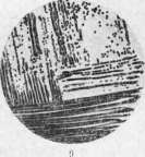
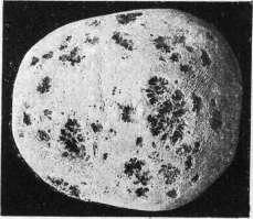
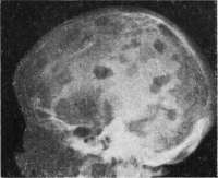
Figure 1. Micrococci. Fig. 2. Lactic acid bacteria (Streptococcus lacticus) in milk as diplococci. Figure 3. Lactic acid bacteria (Streptococcus cremoris) as streptococci. Fig. 4. Lactic acid bacilli (Bacterium casei E). Fig. 5. Preparation from the milk of a cow suffering from inflammation of the udder: leukocytes and bacteria causing udder inflammation - Streptococcus mastitidis. Figures 6, 7, and 8. Plate cultures. Serial inoculation of material with a single spatula on a series of plates. Fig. 9. Inoculation of material with a loop in streaks on a single plate. Figure 10. Myeloma of the skull. Figure 11. Radiograph of the skull in myeloma. M. :–». To the articles: Myeloma, Micrococci, Microorganisms, Lactic acid bacteria. 8S of cell types, which probably represent merely various stages in the development of one and the same indifferent bone marrow cell, there is no need whatsoever; the cells of myeloma should simply be called "myeloma cells" (German: Myelomzellen). Much controversy surrounds the questions of whether myeloma can be characterized by the sprouting into the periosteum and soft tissues surrounding the bone, as well as the formation of metastases. All of the above characterization, based on the vast majority of myeloma cases, is the characterization of typical myeloma; but cases of myeloma with sprouting into the periosteum and soft tissues, as well as the formation of metastases in the liver, lymph nodes, spleen, and kidneys, are occasionally described. Many authors, who believe that true myelomas never sprout into the periosteum and do not produce metastases, view the inclusion of these cases in myeloma negatively; they believe that these cases belong not to myeloma, but to true malignant tumors of the bone marrow; some have even proposed designating such cases with the term "myelosarcoma," implying a neoplasm analogous to the lymphosarcoma of lymphadenoid tissue. Others believe that all myelomas belong to true malignant tumors and as such can infiltrate tissue and give rise to metastases; some even say that metastases are usually not found in myeloma simply because the patient dies before they could form. Most correctly, however, it should be considered that typical myeloma does not produce destructive sprouting of soft tissues and metastases in the sense of cell transfer; what is described as sprouting into the periosteum and the soft tissues closest to it is in most cases not a manifestation of the infiltrative growth of the tumor, but that cellular infiltration which is an expression of appositional growth and takes place, for example, also in the capsule of lymph nodes and in the neighboring adipose tissue in aleukemic hyperplasias of lymphadenoid tissue. As for the metastases in the liver, lymph nodes, spleen, and kidneys observed in some cases of myeloma, it can now be considered established that the described metastases are not such, but represent the local development of myeloma nodes from bone marrow tissue that appeared in the indicated organs by way of so-called myeloid metaplasia; the formation of these extra-bone-marrow myeloma nodes in such cases merely confirms the systemic nature of the disease. The finding of foci of extramedullary hematopoiesis in the liver, lymph nodes, and spleen in myeloma is not very rare. Along with this, in some cases one cannot exclude the possibility of the transition of myeloma into a truly malignant bone marrow tumor of the myelogenous sarcoma type. Among rare findings in myeloma, mention should be made of the formation in myeloma nodes of protein crystals, apparently relating to the Bence-Jones protein body, and the presence of clumps and crystals of amyloid among the myeloma tissue. In rare cases, myeloma was accompanied by generalized amyloidosis. The formation of calcareous metastases in various internal organs as a result of the destruction of bone tissue has been described in myeloma more than once. The question of the pathogenetic essence of myeloma is still not definitively resolved. It remains not entirely clear whether myeloma belongs to true tumors—blastomas—or not. On the one hand, growth in the form of nodes with a tint of limitless tumor growth, structure from cells of a single type, the delimitation of myeloma nodes from the adjacent bone marrow, and passive changes in the latter speak, according to Helly, for the blastomatous character of the process; on the other hand, however, the diffuse forms of myeloma, the leukemic alteration of the blood in individual cases of myeloma, the absence of true metastases, the topography of myelomatous proliferations in the bone marrow and sometimes in organs with myeloid metaplasia bring myeloma very close to leukemic and aleukemic myeloses, that is, to those hyperplasias of bone marrow (myeloid) tissue that occur with and without leukemic changes in the blood. Based on the above-described properties, the essence of myeloma is quite often defined as a process standing on the border between aleukemic hyperplasias of the bone marrow and true tumors, its blastomas. However, it is more correct to align with the more definite opinion of such researchers as Hart, Frenkel, Pappenheim, and others, and to consider that myeloma is not a true tumor, but represents a nodular or diffuse hyperplasia of bone marrow tissue of the aleukemic myelosis type with mild anoplasia of the proliferating tissue and with its expansive growth. The clinical picture of myeloma boils down to a characteristic skeletal change accompanied by gradually increasing weakness, anemia, and exhaustion. On the part of the bones, there are pains in various places, the formation of multiple tumor-like swellings or expansions of the bones, of which those that protrude multiply on the head when the cranial vault is affected are especially characteristic; spontaneous bone fractures (sometimes healing very well), spinal curvature often with symptoms of spinal cord compression, and sometimes shortening of the trunk. X-ray examination reveals multiple foci of bone tissue rarefaction (see separate table, figure 11). On the part of the blood, in most cases, apart from phenomena of secondary anemia of one degree or another, no changes are discovered; but sometimes leukocytosis is observed with such a strong shift of the formula to the left that it indicates blood myelocytosis; cases of the presence of plasma cells in the blood in plasmacytomas have been described; very rarely, the picture of myeloid leukemia was present in myeloma. In many cases of myeloma, the presence of the Bence-Jones body is discovered in the urine (see Bence-Jones protein body). The course of myeloma is chronic; the disease progresses over months and years and, as a rule, leads the patient to death. Remissions of the disease in the form of a decrease in the volume of tumor-like proliferations are temporary and are associated with the appearance of necroses in the myeloma nodes, or sometimes depend on the loss of fluids by the organism (diarrhea, profuse sweating, etc.).
Related articles
Mentioned in
Cite this page
“Myeloma.” Soviet Medical Encyclopedia. English translation of Bolshaya Meditsinskaya Entsiklopediya, 1st ed. (Moscow, 1928–1936), ed. N. A. Semashko. https://sovietmedicalencyclopedia.pages.dev/article/myeloma/