Periosteum
Historical document, translated for reference. It reflects medical knowledge of the 1920s–30s and is not medical advice.
Summary
The periosteum is a fibrous membrane covering most of the outer surface of bones, serving to connect them with surrounding tissues. It plays a crucial role in bone growth, healing, and nutrition, with different structural characteristics in growing versus mature bones.
Encyclopedia article (1928–1936)
Periosteum (periosteum, периост), a fibrous membrane of yellowish-white or pink color, covering almost everywhere the outer surface of bones and facilitating their connection with surrounding soft tissues. Parts of bones free from the periosteum are those covered with cartilage, as well as such areas of bones as the rough lines of long bones (lineae asperae), tuberosities (tuberositates), the anterior surface of the patella, etc. The thickness of the periosteum varies: in places where it connects with muscle tendons or fascia, it appears thickened and mostly has a whitish, shiny appearance, while in places of direct connection with muscles, it appears as a thin membrane. The periosteum in adults is 2-8 times thicker than in newborns. The connection of the periosteum with the bone occurs through vessels branching in the periosteum and "entering the cortical layer of the bone, through connective tissue accompanying the vessels, as well as with the help of fibers or bundles of them, starting in the fibrous layer of the periosteum (tunica fibro-elastica) and entering the bone tissue as a component part of it (Sharpey's, or perforating fibers). In old people, the periosteum may attach to the bones very loosely, and then free spaces may form between the periosteum and the bone. In some places, a close fusion of the periosteum with the overlying mucous membrane is noted. The periosteum is rich in blood vessels, which through the openings of Haversian, Volkmann's and other channels penetrate into the bone or leave it. The largest vessels are found in the outer layers of the periosteum. Lymphatic vessels of the periosteum have been the subject of several studies; true lymphatic vessels with their own walls have been found only in the most superficial layers of the periosteum. The periosteum is abundantly supplied with nerves, forming numerous plexuses. Nerve fibers either branch in the periosteum or penetrate into the bone accompanying the vessels. In these plexuses, both medullated and non-medullated fibers can be found. Numerous Vater-Pacini corpuscles have also been found in the periosteum. The dura mater is especially abundantly supplied with nerves, equal in structure and significance to the periosteum (see Dura mater). In terms of its histological structure, it is necessary to note the difference between the periosteum of young growing bones and the periosteum of bones with completed growth. The periosteum in young individuals consists of three well-distinguishable layers: 1) outer (adventitia), 2) middle (t. fibro-elastica) and 3) inner, osteoblastic layer. The adventitia consists of intersecting bundles of poor in cells and relatively dense connective tissue, which becomes loose in the vicinity of muscles and passes into their interstitial tissue. Elastic fibers, located rather disorderly, are found especially in the outer parts of the layer; in some places there is also adipose tissue. The adventitia is particularly rich in blood vessels and nerves. In some places, for example on the bones of the cranial vault, the adventitia is absent; the periosteum here merges without a sharp boundary with the galea aponeurotica.
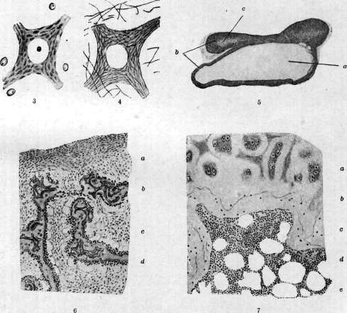
Figure 1-2. Comparative picture of chromatolysis of a nerve cell when staining chromophilic substance and neurofibrils. Figure 3-4. Comparative picture of a nerve cell of the spinal cord when staining chromophilic substance and neurofibrils. Figure 5. Cyst of the adrenal gland (a); b - cortical substance; c - medullary substance. Figure 6. Periosteum in an active stage from the diaphysis of the femur of an embryo: a-periosteum; b - unossified cartilage; c-periosteum between bone trabeculae; d - bone trabeculae from cubical osteoblasts. Figure 7. Periosteum in a resting state from the epiphysis of the femur of a middle-aged human: a-cartilage; b-bone; c - osteoblasts located parallel to the bone; d - bone marrow; e - fat cells. (Figures 1-4 - according to Rakhmanov, figures 6 and 7 - according to Kaufmann.)
See article Periosteum, Adrenal glands, Nerve cells. The osteoblastic layer (proliferative Virchow, cambial Billroth) consists of two parts: a) outer-loose fibrous and b) inner-cellular (osteoblastic in the proper sense of the word). This layer is 4-5 times thinner than the previous one; elastic fibers in it are found only in the connective tissue accompanying the vessels. The inner part of this layer consists of cells (osteoblasts), which in embryos are arranged in rows resembling rows of epithelial cells; in young bones this arrangement in rows is already lost (see separate table, figures 6 and 7). Osteoblasts are large cells, on average 20-30 μ, usually cylindrical or cubic in shape, more rarely osteoblasts have the appearance of elongated cells with processes and large nuclei of round or oval shape. The typical osteoblastic layer, rich in osteoblasts, exists in embryonic life and at least in still growing bone. In adults, typical osteoblasts generally do not exist in the periosteum or are found very rarely (Heitz-Boyer, 1918; Vereshchinsky, 1924; Riess, 1924; Leriche and Policard, 1924). Originally rich in cells, the osteoblastic layer gradually turns into fibrous tissue, consisting of bundles of fibers and poor in cellular elements. Cells scattered among these fibers of connective tissue, in their structure, approach fibroblasts or polyblasts; occasionally individual cells rich in protoplasm and resembling osteoblasts in structure are found. The number and size of elastic fibers in this former osteoblastic layer increase, and the difference between it and the fibro-elastic layer disappears. Thus, the periosteum of adults should be correctly divided into 2 layers, namely: 1) outer layer (adventitia) and 2) containing somewhat more cells than the outer layer, inner (designated by authors as the "formative layer" or as fibro-elastica). The periosteum is the tissue through which vessels pass and in which branch, feeding the bone; thus, damage to its integrity leads to a disorder of bone nutrition. In the growth of the bone in the embryo and especially in the young organism, the periosteum takes a significant part, with the formation of bone from the periosteum occurring in two ways: 1) in the form of multiplication of periosteal cells (osteoblasts) and formation of osteoid tissue and 2) in the form of direct transformation (metaplasia) of periosteal connective tissue into bone without cell multiplication (figures 1 and 2). The periosteum has bone-forming function in the embryonic period and in the period of bone growth. In the adult organism, under normal conditions, the periosteum does not have osteogenic properties (Maksimov, Weidenreich), but under pathological conditions (with mechanical injuries, with bone fractures, etc.) its embryonic properties in the form of ability to form new bone tissue seem to return. The periosteum plays an important role in the healing of bone fractures, manifested by the growth of its elements and the formation of the so-called external callus (callus externus). Pathology of the periosteum. In so-called osteogenesis imperfecta (see), a congenital insufficiency of the bone-forming function of the periosteum is noted with normal activity of osteoblasts and proper growth of the epiphyseal cartilage. This is a congenital dysplasia, "weakness" of the periosteum. Traumatic injuries (bruises) of the periosteum lead to more or less significant hemorrhages in it itself and mainly under it (hematomas of the periosteum). Often at the bruised place, periostitis ossificans traumatica (see Periostitis) develops in the periosteum, as a result of which the bone thickens, but this thickening is mostly of a temporary nature. In inflammatory processes of the periosteum-see Periostitis. Tuberculosis of the periosteum can be primary or secondary at
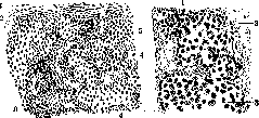
Figure 1.
Figure 2.
Figure 1. Formation of osteoid beams from the periosteum (from a fracture of 2 weeks): 1-periosteum (outer layer); 2-periosteum (inner layer); 3-cartilage tissue; 4-bone marrow tissue; 5-osteoid beams. Figure 2. Formation of cartilage and bone from bone marrow osteoblasts: 1-osteoblasts; 2-bone marrow tissue; 3-cartilage tissue. In the transition of the process from the bone. Tuberculous granulation tissue develops in the inner layer of the periosteum and destroys it.-Periosteal gummas are elastic thickenings, which on cross-section have the appearance of a jelly-like mass; they develop in the inner layer of the periosteum. Periosteal gummas are most commonly observed on the bones of the cranial vault, sternum, tibia, and clavicle.-Actinomycosis affects the periosteum usually when the process spreads from the surrounding soft tissues; the periosteum turns into granulation tissue and disintegrates.-Tumors of the periosteum most often originate from its inner layer. Among benign tumors of the P., it is necessary to note fibromas, which most often originate from the bones limiting the oral and nasal cavities, as well as the bones of the base of the skull and its vault. Fibromas of the P. are often rich in blood vessels, so that on cross-section they sometimes resemble cavernous tissue (telangiectatic fibromas). They occur almost exclusively in adolescence, mostly in males. Cases of neurofibromas originating from the P. have been described (see Neurofibromatosis), as well as very rare cases of myxomas and lipomas of the P. Regarding tumors of the gums, which according to some authors originate from the P., see Epulis. The P. can be the source of the formation of exostoses, which most often develop on the vault of the skull, on the walls of the facial cavities, and on the jaws. The P. is the source of the formation of so-called periosteal or peripheral sarcomas; they are most often located at the ends of long bones, but can be in any part of the bones. Histologically, these are round-cell, spindle-cell, or mixed forms of sarcoma; sometimes giant-cell forms can also occur. These sarcomas, starting in a limited area, can circularly encompass the bone and in a short time develop into a large tumor. Metastases to the lungs, pleura, lymph glands, etc., are common. Treatment is surgical; the nature of the surgical intervention depends on the size of the tumor, involvement of surrounding tissues, etc.-Until recently, the periosteum was attributed an important role in the engraftment of bone transplants and the formation of new bone tissue. Thus, according to old studies by Ollier (ОШег), the engraftment of bone tissue without the P. was considered impossible. Over the last 2 decades, based on research by a number of authors, it has been clarified that the P. does not play such a significant role in bone transplants as was previously thought; at present, the opinion is beginning to prevail that the formation of new bone tissue occurs from surviving bone cells, from elements of the bone marrow, from the periosteum, endosteum, and finally from the young connective tissue surrounding the transplant.-The endosteum is close to the periosteum in genetic, morphological, and physiological aspects, representing the internal connective tissue lining of the marrow spaces and Haversian canals.
Related articles
Mentioned in
Cite this page
“Periosteum.” Soviet Medical Encyclopedia. English translation of Bolshaya Meditsinskaya Entsiklopediya, 1st ed. (Moscow, 1928–1936), ed. N. A. Semashko. https://sovietmedicalencyclopedia.pages.dev/article/periosteum/