Adrenal Glands
Historical document, translated for reference. It reflects medical knowledge of the 1920s–30s and is not medical advice.
Summary
The adrenal glands are paired endocrine organs located in the upper part of the abdomen on the upper inner surface of the kidneys. This article describes their anatomy, blood supply, lymphatic drainage, and nerve connections.
Encyclopedia article (1928–1936)
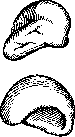
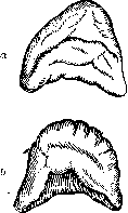
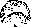
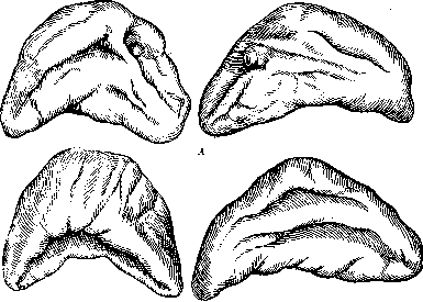
ADRENAL GLANDS (glandulae suprarenales, epinephra, hypernephra, paraganglia), paired endocrine organs located in the upper posterior part of the abdominal cavity on the upper inner surface of the kidneys. Their discovery and first description are attributed to the anatomist Eustachius of the 16th century, but the attention of physicians was first drawn to them by the work of Addison in 1855; he pointed out the importance of the adrenal glands as organs necessary for life. The disease associated with damage to the adrenal glands is named after him. Anatomy. In adults, the adrenal glands have a rounded-triangular or crescent shape (the shape of a 'thick comma' according to Testut). This shape is not established immediately: up to the 3rd month of intrauterine life, the adrenal glands have the shape of a lentil; from the 3rd month until birth, their shape is triangular or conical, flattened from front to back and with the base directed backward (fig. 1-3). In childhood, the thickness of the adrenal glands is greater than in adults (10-11 mm compared to 7 mm). Three surfaces are distinguished in the adrenal glands (anterior-lateral, posterior-median, and inferior or base), the upper pole, and lateral and median edges. The surfaces, which are smooth at the beginning of development, later become wrinkled; the most significant of these folds on the anterior surface of the adrenal gland forms the so-called hilus of the adrenal gland (hilus gl. suprarenalis), into which several arterial branches enter and from which the central vein (vena suprarenalis, s. v. centralis gl. suprarenalis) and lymphatic vessels exit.
Figure 1. Adrenal glands of a human fetus at 5.5 cm (A) and 10 cm (B): a-from the front; b-from the back; 1-right; 2-left. to the adrenal glands by the work of Addison (Thomas Addison) in 1855; he pointed out the significance of the adrenal glands as organs necessary for life. The disease associated with damage to the adrenal glands is named after him. Anatomy. In adults, the adrenal glands have a rounded-triangular or crescent shape (the shape of a 'thick comma' according to Testut). This shape is not established immediately: up to the 3rd month of intrauterine life, the adrenal glands have the shape of a lentil; from the 3rd month until birth, their shape is triangular or conical, flattened from front to back and with the base directed backward (fig. 1-3). In childhood, the thickness of the adrenal glands is greater than in adults (10-11 mm compared to 7 mm). Three surfaces are distinguished in the adrenal glands (anterior-lateral, posterior-median, and inferior or base), the upper pole, and lateral and median edges. The surfaces, which are smooth at the beginning of development, later become wrinkled; the most significant of these folds on the anterior surface of the adrenal gland forms the so-called hilus of the adrenal gland (hilus gl. suprarenalis), into which several arterial branches enter and from which the central vein (vena suprarenalis, s. v. centralis gl. suprarenalis) and lymphatic vessels exit.
Figure 2. Adrenal glands of a human fetus at 17 cm (A) and a newborn (B): a-from the front; b-from the back; 1-right; 2-left. The average size of the adrenal glands:
Figure 4. Various positions of the adrenal gland (diagram): 1-posterior edge of the liver; 2-right kidney; 3-renal vein; 4-inferior vena cava; dashed line-high position of the adrenal gland; dotted line-medium; solid line-low. (From Testut.)
Figure 3. Adrenal glands of an adult: A-from the front; B-from the back; 1-right; 2-left. height 30 mm, width 25 mm, thickness 7-8 mm; average weight 12 g (Testut); in adults
The volume and weight of the adrenal gland is on average equal to 1/25 of the volume and weight of the kidney (Latarjet). The color of the adrenal glands is dark yellowish, the consistency is quite soft. There are many variants in the shape of the adrenal glands: ovoid, triangular, and quadrangular adrenal glands are found. Adjacent to the kidneys, the adrenal glands occupy one of 3 positions (fig. 4): high, when the adrenal gland lies on the upper pole of the kidney at the level of DX-XI; low, when the adrenal gland is located immediately above the renal vessels at the inner edge of the kidney at the level from DXI to LIII, and medium-between the first two. Whatever position the adrenal glands occupy, they are always placed in the renal bed (fascia renalis), occupying its upper-inner part and being surrounded by a fatty capsule (capsula adiposa). The adrenal glands are connected by loose connective tissue with the kidneys at the point of contact. The layer between the adrenal glands and the kidneys in adults consists of fatty tissue, the thickness of which individually varies (from 0.5 to 3 cm); in embryos and newborns, it is completely absent (G. Ivanov). In any case, this connection is very weak, so that when the kidney is displaced, the adrenal glands always remain in their place; in addition, the adrenal glands are firmly held by wide fibrous bands extending from the capsule of the adrenal gland to neighboring organs and tissues; such ligaments are distinguished in three for each adrenal gland: 1) a ligament extending from the anterior surface of the right adrenal gland to the inner edge of the inferior vena cava (in the left adrenal gland, a similar ligament goes to the aorta); 2) the adrenoliver ligament, extending to the inferior surface of the liver; 3) the adrenodiaphragmatic ligament, the strongest of all adrenal ligaments, extending from the upper angle of the adrenal gland to the dome of the diaphragm. These ligaments are not true ligaments, but only connective tissue, particularly strong around the vessels of the adrenal gland (Latarjet). Topography of the adrenal glands. With their posterior surface, both adrenal glands lie on the lateral part of the spine at an angle of 45° to it, on the pillars or lumbar part of the diaphragm. The left adrenal gland lies lower than the right, descending to LIII, and usually occupies a low position; medium and especially high positions are extremely rare for it (S. et M. Gerard). Between it and the posterior wall of the abdominal cavity pass the truncus sympathicus and the endings of the n. splanchnici majoris et minoris and the vena lumbalis ascendens. With a high position, the left adrenal gland lies at the level of the XII rib; in this case, when approaching it from behind, the pleural cavity is encountered, namely the sinus costodiaphragmaticus. The anterior surface of the left adrenal gland is partially covered by the posterior leaf of the parietal peritoneum; in front of the adrenal gland above lie the inferior surface of the left lobe of the liver and the posterior edge of the spleen (not always), in the middle-the stomach, below-the tail of the pancreas (not
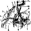
Figure 5. Right adrenal gland (1) from the front: 2-its hilus; 3-the part in contact with the liver, always; across the adrenal gland covered with peritoneum; 4-the spleen is also covered with peritoneum; 5-right kidney; 6 and 9-12-duodenum; 7-pancreas; 8-central vessels. With its base, the left adrenal gland, in its high position, lies on the upper pole of the kidney, in the low position (which is more common) - on the renal vessels. The median, convex edge of the adrenal gland touches the ganglion semilunare sin. and is 1-2 cm away from the aorta. The shortest distance of the left adrenal gland from the midline of the body is on average 1.8 cm (Promptov).-The right adrenal gland lies somewhat higher than the left and usually occupies a medium position; low position is rare for it, and high position is very rare (Gerard). Between the right adrenal gland and the posterior wall of the abdominal cavity, in addition to the elements listed in the description of the left adrenal gland, there passes an anastomosis between the n. phrenicus dext. and the plexus Solaris (Latarjet). The anterior surface of the right adrenal gland is covered in its medial part by the v. cava inferior, in the lower medial angle by the duodenum, and in the remaining part by the liver, which covers the adrenal gland; its middle part is separated by peritoneum (fig. 5). With its base, the right adrenal gland lies on the kidney, descending with its lower medial angle in the low position to the renal vessels. The median edge of the adrenal gland lies in close proximity to the ganglion semilunare and the v. cava inf., i.e., it is 7.5 cm away from it between it and the spine. The shortest distance of the adrenal gland to the midline of the body is on average 1.5-2 cm (Promptov).-With their upper poles, both adrenal glands are directed obliquely upward, outward, and slightly forward. Blood vessels of the adrenal glands. The adrenal glands are very abundantly supplied with blood, which they receive through 4 groups of arteries: 1) art. suprarenalis sup. (a branch of art. phrenicae inf.), 2) art. suprarenalis media (a branch of aortae abd.), 3) art. suprarenalis inf. (a branch of arteriae renalis) and 4) art. capsulae adiposae renis (a branch of aortae abd. or art. renalis). The arteries, after giving numerous anastomoses with each other, branch in the substance of the adrenal gland.-The veins of the adrenal gland, in most cases, unite into a large vein (vena suprarenalis, s. v. centralis gl. suprarenalis), emerging from the anterior surface of the adrenal gland; it empties on the right into the v. cava inf., on the left-into the v. renalis sin. Almost constantly present in the left adrenal gland is the vena reno-capsulo-diaphragmatica-an anastomosis between the v. centralis gl. supraren., v. phrenica inf. and v. renalis (Albarran). This vein is a collateral pathway between the vena renalis and the parietal veins of the abdominal cavity.-Lymphatic vessels of the adrenal gland, upon emerging from the depth of the adrenal gland, 1-9 collect on the inner edge of the adrenal gland, from where they go to the lymphatic glands; they are divided into anterior and posterior. In the right adrenal gland, the anterior vessels, numbering 4-5, are very voluminous, go obliquely downward and to the middle, cross in front of the right renal vessels and the v. cava inf., and empty into 2-3 paraaortic lymphatic glands immediately below the renal vessels; the posterior lymphatic vessels, numbering 2-3, are very short, go to the lymphatic glands on the posterior surface of the vena cava inf., between it and the pillars of the diaphragm (fig. 6). Lymphatic vessels of the left adrenal gland: the anterior ones, numbering 4-6, go almost vertically downward and flow into the paraaortic lymphatic glands immediately below the left renal vessels; the posterior lymphatic pathways (4-5) go differently: part of them empties into the left paraaortic glands immediately below the renal vessels, another part goes upward, passes through the slit between the inner and lateral pillars of the diaphragm into the thoracic cavity, where it empties into the posterior mediastinal glands at the level of DIX between the aorta and the vertebra.-NervesThe adrenal glands receive nerves from two sources: some go to the posterior surface of the adrenal gland from the n. splanchnicus major, others-to the anterior surface from the plexus Solaris (fig. 7).
Figure 6. Regional lymphatic glands and vessels of the adrenal glands.
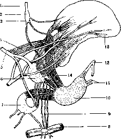
Figure 7. Endings of sympathetic nerves. Nerves of the kidneys and adrenal gland: 1 and 2-right n. phrenicus; 3-a branch to the adrenal gland; 4-nerves from the n. splanchnicus major (5) and from the adrenal gland node (6); 7-ending of the n. splanchnicus major, in the additional node; 8-right renal vein; 9-adrenal-kidney anastomoses; 10-semilunar node; 11-branches connecting the node with a similar one on the other side; 12-ending of the n. vagi dextri; 13-right adrenal gland; 14-nerves from the semilunar node.





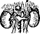
A Sirtokin. Comparative anatomical data. The adrenal glands (suprarenal glands) in lower vertebrates are represented by organs of two kinds: interrenal glands and adrenal glands (glandulae interrenales and glandulae suprarenales, s. adrenales). The chromaffin glands are fundamentally metameric formations, showing in their phylogenetic development a tendency to fuse with each other. In sharks, the adrenal glands consist of metamerically arranged bodies connected with the sympathetic nervous system; the interrenal glands form paired or unpaired masses located closer to the tail, and their metameric arrangement is indistinguishable (figure 8: interrenal (5) and suprarenal (2) organs of sharks; 1-kidneys). In bony fishes, the adrenal glands are included in the walls of the cardinal veins, while the interrenal glands lie on the kidneys, sometimes penetrating their parenchyma. In amphibians, both kinds of suprarenal glands come into contact with each other and are covered by a common capsule, forming the adrenal glands, which in tailed amphibians lie along the medial edge of the kidneys, differing from them by a golden-yellow coloration, while in tailless amphibians they are located on the ventral surface of the kidneys also in the form of yellow angularly curved strips. In reptiles and birds, the adrenal system penetrates into the interrenal masses, forming a close, irregular interweaving of strands; the adrenal glands in the form of elongated, so-called golden-yellow bodies are located near the sex glands. Finally, in mammals, the adrenal glands are located along the upper edge of the kidneys, with the penetration of the adrenal system into the interrenal substance being completed and the first being transformed into the medulla, and the second into the cortex of the adrenal gland. Embryological data. The adrenal glands originate from two sources: first, from the mesoderm of the abdominal cavity, which gives rise to the interrenal system - the cortical substance, and second, from the rudiments of sympathetic ganglia, at the expense of which the adrenal medulla develops (fig. 9). The cortical substance appears in human embryos in the 1st month of intrauterine life (5 mm in length) between the epithelial rudiment of the genital tubercle and the root of the mesentery in the form of proliferations of the peritoneal mesothelium, which then separate from the mesothelium and lie on either side of the abdominal aorta. In lower vertebrates, the rudiments of the cortical substance develop in a metameric order; in humans, the area of the rudiment is limited to a narrow strip corresponding to the upper part of the Wolffian body (Soulie). Significantly later, in the second half of the 2nd month (in a 19 mm embryo), the rudiments of the medulla appear, which, according to the opinion of most authors, originate from the trunk of the sympathetic nervous system. This separation occurs over a considerable extent to the pelvic region (Kohn), but only a part of the cells (sympatogonia) enters the composition of the adrenal glands, the others form the so-called paraganglia and the organ of Zuckerkandl (see). In addition, the medulla is formed by those chromaffin cells that migrate here from the cranial poles of the abdominal aortic paraganglia (Ivanov, M. Wrete). The formation of the adrenal glands occurs as follows (Poll): the rudiment of the cortical substance is surrounded by a mesenchymatous capsule, to which sympathogonia approach along with blood vessels; then they begin to penetrate into the depth and form branching strands; only gradually does the medulla occupy a central position. In the 3rd month, the adrenal glands are located on the cranial and partly on the ventral surface of the kidneys and have a relatively large size; by the end of the 3rd month, they shift further in the cranial direction. Histological differentiation begins approximately at the same time, earlier in the cortical substance, which divides into three zones; somewhat later in the medulla, the elements of which differentiate into ganglion and chromaffin cells. The size of the adrenal glands in the fetus is relatively very large due to the strong development of the cortical layer; after birth, there is a rapid decrease in the size of the adrenal glands due to the degeneration of the inner layers of the cortical layer.
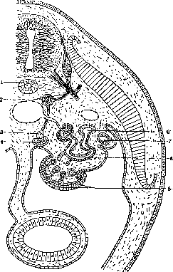
Figure 9. Diagram of the development of the adrenal gland: 1- notochord; 2- sympathetic ganglion of the trunk; 3- rudiment of the medulla; 4- rudiment of the cortical layer; 5- genital tubercle; 6- canals of the primary kidney; 7- Müllerian duct; 8- Wolffian duct. (According to Corning.)
Histology. In a cross-section of the human and mammalian adrenal gland, two kinds of substance can be distinguished with the naked eye [see separate table (pp. 15-16), fig. 9]: cortical (substantia corticalis) and medullary (substantia medullaris). The former is yellowish-white, turning to brown in the deeper layers, is denser, and tears easily in the direction perpendicular to the surface, with a thickness of 0.28-1.12 mm (Kölliker). The medulla is grayish-pink, sometimes red from the large blood content, less dense; in thickness at the edges 0.35-0.75 mm, in the middle 2-3 mm. The cortical substance (fig. 10) is covered on the
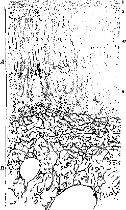
Figure 10. Cross-section of the adrenal gland (low magnification): A-cortical substance; B-medullary substance; 1-capsule; 2-zona glomerulosa; 2'-zona fascicularis; 3-zona reticularis. (According to Ebner.)
by a fibrous capsule, in the superficial layers of which are found fat lobules and blood vessels; in its depth it is denser, and from it connective tissue septa extend into the organ, forming the stroma of the cortical substance. The capsule contains smooth muscle fibers. The parenchyma of the cortical substance of the A. consists of cords of epithelial cells (cylinders of Kölliker), arranged perpendicular to the surface of the organ. According to the character and arrangement of the cords, three zones are distinguished in the cortical substance, running parallel to the surface (Arnold): 1) the outer glomerular zone (zona glomerulosa), 2) the middle, fascicular zone (zona fascicularis) and 3) the inner, reticular zone (zona reticularis). However, not all agree with such a division: it is pointed out that in humans z. glomerulosa is indistinct and there is no basis for its isolation (Ebner); others, on the contrary, paying attention to the character of the cells, distinguish 4 zones, isolating the spongy zone from the fascicular zone (Guieysse), and even 5 zones, dividing the fascicular zone into 3 (Bernard and Bigard). The glomerular zone is named so because the epithelial cords under the capsule are more or less strongly curved, sometimes bulb-like at the ends, and on sections appear separated from the fascicular zone by layers of connective tissue. The cells in this zone are small in size, in many animals having a narrow cylindrical shape; they contain small grains or droplets of fat and lipoids. Many authors, following Gottschau, consider the glomerular zone a collection of young, embryonic elements which, gradually maturing, pass into the following layers; cell divisions have been repeatedly described in it.
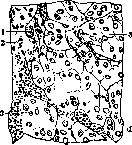
The fascicular zone is characterized by the fact that the epithelial cords in it are arranged in parallel, slightly curved rows; between them, closely adjacent, run blood capillaries. The cells here are larger, cubic or polyhedral in shape, 9-20 μ in diameter, and contain a round nucleus. The uneven distribution of protoplasmic inclusions gave some authors reason to distinguish different types of cells in the fascicular zone (figure 11): vacuolated cells - spongiocytes (adelomorphic cells of Bogomolets), dark cells (principal cells of Bogomolets), siderophilic cells, microcytes, etc.
The reticular, or mesh, zone is characterized by the fact that the epithelial cords curve to the sides and anastomose, forming networks with wide intervals; accordingly, the course of the capillaries also changes. The cells of the reticular zone are smaller in size (8-18 μ), darker, and starting from the 20th year of life, contain grains and clumps of brown-yellow pigment. Its origin is not established: most authors consider it a result of the transformation of inclusions of the fascicular zone or an expression of the atrophic state of the cells. According to this, the reticular zone is usually considered a zone of involution in which cells that have migrated from the fascicular zone die. According to another view (Bernard and Bigard), the production of pigment has a special functional significance, analogous to the accumulation of lipoids. The most characteristic feature of the cells of the cortical substance is their content of a large amount of fat substances, located in the protoplasm in the form of droplets or grains. For the most part they belong to lipoids and in particular to compounds of cholesterol (see Lipoids); in addition, with special stains, the presence of free fatty acids (mostly in the reticular zone) and small amounts of neutral fat can be detected. The distribution of lipoids in the cortex of the A. is uneven. The largest amount of them is found in the fascicular zone. In the glomerular zone, their quantity is significantly less, and sometimes cells are noted that do not contain lipoid inclusions. In the reticular zone, there are also fewer lipoids than in the fascicular zone. The droplets of fat substances here are very small. The content of lipoids in the cortex of the A. varies greatly in animals of the same species (Kolosov), which makes observations on changes in lipoids in the cortex of the A. under the influence of experimental effects very difficult and repeatedly led to errors.
In the protoplasm of the cells of the cortex of the A., organelles have been found: centrioles, the Golgi reticular apparatus, chondria, and various inclusions: siderophilic substance (gives a reaction to iron), oxyphilic substance (Ciaccio) and the above-mentioned pigment of the reticular zone. The medullary substance (fig. 12) consists 1) of connective tissue stroma with a large number of elastic fibers and cells; 2) of blood vessels, among which wide veins stand out, and 3) of cellular cords arranged in the form of networks around the vessels. A characteristic feature of the medullary substance is the combination in it of two kinds of elements: sympathetic ganglion cells and a special kind of epithelial cells - chromaffin cells (Kopp) or phaeochromic cells (Poll). These cells have a polyhedral, often cylindrical shape, 8-18 μ in diameter, and contain grains that stain brown-yellow after treatment with chrome salts (Henle's reaction); they also get a greenish-blue color from the action of ferric chloride (Vulpian's reaction), and black when impregnated with silver according to the Cajal method. Most authors assume that the grains in the cells of the medullary substance represent adrenaline. In the very wide blood capillaries of the medullary substance, among clotted blood plasma, one can find accumulations of a substance that gives a color reaction with ferric chloride (dissolved grains of adrenaline). Attempts to cause shifts in the morphology of the cells of the medullary substance by irritation of the secretory nerves (n. splanchnicus major) did not give sufficiently convincing results.
The stroma is formed by so-called reticular fibers (Gitterfasern): they surround the blood vessels and pass to the capillaries, enveloping them in a dense delicate network. From the capillaries, the reticular fibers pass to the cords of epithelial cells of the cortex and medulla, forming around them the finest, small-meshed sheaths. The blood vessel system of the cortical and medullary substances are closely connected. From numerous arterial branches branching in the capsule, capillaries go into the cortical substance and run radially between the epithelial cords; they pour in a large part into the veins of the medullary substance. The medullary substance receives blood from special arterial branches piercing the cortical substance; they then pass into the winding capillaries of a sinusoidal character, which flow into the central vein.
The nerves of the A. Numerous nerve trunks, originating mainly from the solar plexus and belonging to the sympathetic nerve (greater and lesser splanchnic nerves) (in the origin of these trunks, according to some authors, the vagus nerve participates), enter the capsule where they are located in connective tissue; the A. contains numerous nerve ganglia with typical sympathetic cells. Further, nerve trunks, following the course of the blood vessels, enter the cortical substance, giving off lateral branches to the cortex, and enter the medullary substance. The number of nerve fibers in the medullary substance is extremely large. The nerve fibers form endings on the cells of the medullary substance in the form of small loops or rings (Pines, Kolosov). The number of nerve cells in the medullary substance varies greatly in different animals and even in animals of the same species. The manner of termination of their processes is not clarified. On the basis of data on the development of the A., it must be assumed that the nerve cells of the cortex and medulla of the A. are connected with sympathetic nerve fibers.
V. Karpov. Pathological Anatomy. Developmental Defects. Cases of agenesis of both adrenal glands (in viable individuals otherwise normal), repeatedly described in the old medical literature (before 1860), are completely inconsistent with the currently proven absolute necessity for life of this organ and are obviously the result of error or insufficient accuracy of observation. Probably in these cases it was a matter of congenital ectopia of the adrenal glands, which is usually bilateral and in which the adrenal glands, located either under the fibrous capsule of the kidneys or even in the connective tissue around the renal vessels, can easily be overlooked. Even the absence of one adrenal gland represents an exceptional rarity (in the entire literature 9 more or less reliable cases; in 8 of them the right adrenal gland was absent). More often significant degrees of underdevelopment of this organ are observed, which are always combined with major defects in the development of the central nervous system (anencephaly, acrania). The essence of this combination has not yet been clarified. Histological studies in relation to the adrenal glands have established only that 1) underdevelopment here mainly concerns the cortical layer, while the medulla may be present even in a relatively excessive amount, and 2) that underdevelopment is exclusively quantitative, while the degree of maturity of the organ is usually greater than that of a normal fetus of corresponding age (see Anencephaly). To very rare defects belong the fusion of both adrenal glands into one unpaired organ (similar to a horseshoe kidney), as well as unilateral or bilateral doubling of them. On the contrary, so-called accessory adrenal glands are extremely common (especially in early childhood). The latter usually consist only of cortical layer cells and represent isolated nodules ranging from microscopic size to cherry-sized, which may either be located in the adrenal gland itself (in the depth of its tissue or in the capsule) or be scattered in the adjacent connective tissue, as well as inside or near certain abdominal organs (kidneys, a. et v. suprarenalis, v. cava, plexus Solaris, colon transversum, spleen, right lobe of the liver, pancreas, v. spermatica interna, m. ilio-psoas, spermatic cord, testis and its appendix, broad ligaments, tube, ovary). Such nodules are formed either by detachment from the already formed interrenal organ (the cortical part of the future adrenal gland) during the ingrowth of germ cells of the medullary layer (sympatogonia) or even earlier, during the period of development and fusion of individual mesodermal rudiments from which this organ is formed and which at that time are in the closest proximity to the primary kidney, forming sex glands and adjacent vessels. In newborns such accessory cortical nodules can be found in one or another of the indicated places in almost 100%, and during the nearest years of life most of them usually atrophy and disappear. Postmortem changes, circulatory disorders, degeneration, necrosis. On the dissecting table changes in the adrenal glands are very often determined, regarding which the question remains unresolved to what extent they are antemortem and to what extent, on the other hand, postmortem processes play a role in their origin. This includes above all the so-called softening of the adrenal glands. It is expressed by the formation of a slit-like cavity between the cortical and medullary substance, mainly in the place of the innermost, most pigmented layer of the zona reticularis, so that in markedly pronounced cases there is as it were sequestration of the entire medullary substance. Where the latter is absent, e.g. at the medial and lateral edges of the adrenal gland or in the child's organ in which the medullary layer is generally poorly developed, such softening produces a central impression. At the basis of this phenomenon, formerly attributed entirely to postmortem changes (or according to some to mechanical injuries during removal and cutting of the organ), lies according to the currently generally accepted view antemortem disorders in the form of severe hyperemia, edema, hemorrhages, cellular degeneration, etc. Being most strongly expressed in the reticular layer due to its looseness and greater vulnerability, they in turn give rise to the extremely rapid development here of autolytic postmortem or possibly atonal softening. Accordingly the described picture is most often found in infectious diseases accompanied by severe circulatory disorders in the adrenal glands, as well as in stagnant conditions, especially in old age in arteriosclerotic subjects. To the same category of phenomena arising from the combined action of antemortem and postmortem processes probably also belong the often noted in the adrenal glands, sometimes extensive, necrotic foci without signs of reaction in the surrounding tissue (so-called pseudonecroses). Circulatory disorders. Hemorrhages into the adrenal glands (see separate table fig. 7) are usually observed in connection with severe venous congestion and can be very diverse in size, ranging from small extravasates to complete hemorrhagic infarction of the organ or to the formation of large hematomas converting part or even the entire adrenal gland into a blood cyst [see separate table (art. 119-120), fig. 5]. Since such hemorrhages are very often bilateral, in extensive sizes they can lead to death due to acute insufficiency of the adrenal glands. The main cause of hemorrhages is most often various traumatic and infectious-toxic influences. Among the first, first place is occupied by birth trauma of newborns (especially in pelvic positions), the effect of which on this organ is increased by the physiologically occurring at the moment of birth extremely severe hyperemia and loosening of the inner zone of the cortical layer. Considerable importance as an etiological factor may also have revival by the Schulze method. As for infectious-toxic influences, there is hardly any acute contagious disease in which in more or less severe cases hemorrhages into the adrenal glands would not have to be encountered. They are more often observed in diphtheria, then in various forms of sepsis, typhoid fever, dysentery and other diseases, especially in elderly subjects. Very often a moment favorable to their occurrence is one or another preliminary vascular disorder, such as: severe hyperemia, toxic or stagnant thrombosis of veins, bacterial emboli, etc. Among other pathological conditions giving rise to hemorrhages into the adrenal glands, mention should be made of severe and prolonged venous congestion (for example in heart defects), extensive burns, leukemia and various forms of hemorrhagic diathesis. Among degenerative-atrophic processes, disorders of fat metabolism are most often encountered, expressed mainly in a decrease in the total amount of lipoids of the cortical layer (together with a decrease in the size of individual lipid droplets in the cells), as well as in a change in their chemical composition in the sense of disappearance of cholesterol compounds with relative or even absolute increase of phosphatides. Often the matter goes almost to complete disappearance of lipoids from the organ. Usually simultaneously with this, but sometimes also completely independently, other metabolic disorders arise, especially the so-called vacuolar degeneration (see), also very often encountered in the adrenal glands as in various pathological conditions, and incidentally also in their physiological involution. It is observed in both the cortical and medullary layers and is expressed by the appearance in the cells of either large vacuoles occupying often the entire cell body, or small droplets penetrating the cell and giving its protoplasm on preparations a fine-meshed appearance. In the cortical substance vacuoles sometimes arise inside lipid droplets, which gradually take on a ring-shaped or half-moon form, and then disappear completely. Due to the often occurring complete melting and death of individual cellular elements or small cell groups and accumulation of edema fluid in their place, small cavities may arise in the cortical substance, very similar to glandular ones due to the fact that the surrounding cells form as it were a continuous epithelial lining along their walls. However, such cavities have nothing in common with true glandular follicles. In the medullary substance along with the formation of vacuoles one can sometimes notice the appearance in the protoplasm of cells of peculiar, weakly staining hyaline-like droplets of various sizes. Sometimes in the medullary substance a weakening or even complete loss by cells of the ability to give reactions to adrenaline (see Adrenaline) is observed, in particular the commonly used in histological practice so-called chrome reaction of Henle. With the greatest certainty about this reaction one can judge when fixing for a day fresh pieces of the adrenal gland, taken no later than 3 hours after death, in a mixture of 90 parts of a 3.5% solution of bichromate of potassium and 10 parts of commercial formalin. Disappearance of the reaction indicates a decrease or cessation of the production of the specific hormone by the cells and is often accompanied by their vacuolar degeneration.
The main cause of all the mentioned metabolic disorders is primarily various acute infectious and toxic effects (diphtheria, scarlet fever, typhoid, paratyphoid, cholera, pneumonia, various forms of sepsis, extensive burns, etc.), to a lesser degree some chronic intoxications (many forms of tuberculosis, pernicious anemia, some leukemias, some tumors, sometimes diabetes). In severe forms of acute infections (especially diphtheria and scarlet fever), limited foci of necrosis, undoubtedly of antemortem origin with a clearly expressed reactive zone, are sometimes found in the cortical layer. The deposition of amyloid in the adrenal glands also deserves mention, which occurs in every case of general amyloidosis (see Amyloid degeneration) and sometimes can reach great intensity. It arises mainly around the capillaries of the cortical layer, especially the zona fasciculata, and as it develops, causes atrophy and death of the corresponding parenchymal cells. Hypertrophic processes can affect the entire organ as a whole (e.g., in vicarious hypertrophy of one adrenal gland due to the death of the other), as well as its individual part. In the cortex, changes of a hypertrophic nature are almost always closely related to fat metabolism, developing in accordance with an increase in its lipid content. This is manifested not only by an increase in their quantity in the usual places of deposition (zona fasciculata), but also by the involvement of cells of the zonae glomerulosae et reticularis, which are normally poor in lipids, in their accumulation. Physiologically, such a condition is observed as a transient phenomenon during pregnancy and in the postpartum period; pathologically—in arteriosclerosis, shrunken kidney, long circulatory disorders (heart defects) and some others. Tuberculosis, pernicious anemia, leukemias, tumors, diabetes can occur with both increased and decreased lipid content. Isolated hypertrophies of the medullary substance are little studied and difficult to determine. Better known are hypertrophies of other parts of the chromaffin system (see Paraganglia), which sometimes occur when the medullary part of the adrenal gland is destroyed by some destructive process. The picture of acute inflammation can often be seen in the adrenal glands in severe forms of various acute infectious diseases, especially of a septic nature. It consists of alterative and vascular changes, which are complemented by the appearance of infiltrates (from neutrophils, round plasma cells, monocytes, etc.), located mainly in the reticular zone. Rarely, in the outcome of such inflammations, sclerosis of the organ with atrophy of the parenchyma can apparently develop. Among the chronic inflammatory processes in the adrenal glands, tuberculosis and syphilis (see Hypernephritis) are of the greatest importance, especially the first, which forms the anatomical basis for the vast majority of cases of so-called Addison's disease (see). Primary tumors of the adrenal glands are naturally divided into 2 separate groups: tumors of the cortical layer and tumors of the medullary substance. Representatives of the first are the so-called cortical adenomas [see separate table (pp. 103-104), Figure 4], otherwise called strumae suprarenales (Virchow), or adrenal hypernephromas (Birch-Hirschfeld). These are limited, mostly located under the capsule (rarely in the cortex itself or even in the medullary substance), usually encapsulated accumulations of cortical cells, often arranged in parallel, radial or interlacing rows similar to the zona fasciculata or reticularis. The material for them is apparently given partly by developmental abnormalities (in the form of the additional cortical nodules described above), and partly by the processes of restructuring and regeneration that arise after various destructive changes so often observed in the adrenal glands in all sorts of infections. Some facts (such as the greatest frequency and multiplicity of adenomas in such conditions of the body that are accompanied by an increased content of lipids in the adrenal glands, an increase in their number with age, etc.) suggest that it is not always possible to draw a line here between true tumor nodes and so-called nodular hyperplasia. On the other hand, these formations sometimes show unmistakable signs of blastomatous growth, can reach very large sizes, destroy the organ itself, and even (especially in childhood) give atypical metastasizing malignant forms. Such tumors often exert a specific hormonal influence on the body, accompanied by either premature sexual maturity or hermaphroditism, hirsutism, etc., in short—a number of signs constituting the so-called adrenogenital syndrome (see Hypernephroma, Hirsutism). From the elements of the medullary substance, tumors of different maturity but also different differentiation can arise. Since the embryonic form of cells of the medullary layer is common with the same form of cells of the sympathetic nervous system, it is natural that immature tumors of this part of the adrenal gland have the character of tumors from nervous tissue (sympathogoniomas, sympathoblastomas); while cells of mature tumors can differentiate in two directions and give either nervous (ganglioneuromas) or chromaffin (paragangliomas, pheochromocytomas) tissue (see Ganglioneuroma). In addition to the specific tumors mentioned, lipomas, hemangiomas and lymphangiomas are rarely found in the adrenal glands. Among metastatic ones, carcinomas are most common.
M. Skvortsov. Normal and Pathological Physiology. Complete removal of both adrenal glands leads to death in most animals regardless of whether the adrenal glands are removed simultaneously or over a long period of time one after the other. Only in rats, and according to some authors in rabbits, especially with two-stage removal, survival is sometimes achieved (in rats 50%, in rabbits 15-20%; Biedl). The latter is attributed to the presence of accessory adrenal glands. The survival period after removal of adrenal glands varies greatly in different animal species; it also depends on the surgical technique (longer survival after two-stage operation), age (young animals die later than old ones), metabolic state (winter frogs die later than summer ones), and interaction with other endocrine glands (see below). Feeding with food rich in nitrogen shortens the survival period. The latter equals* with simultaneous removal in dogs-4-6 days, cats- about 40 hours, rabbits-24-36 hours, guinea pigs- about 15 hours, frogs-8-12 days. Phenomena after removal of adrenal glands: the first 1-3 days animals (dogs) appear as in normal condition, then loss of appetite, lethargy, weakness, drowsiness develop; the animal often lies motionless with hind legs extended. During work, signs of fatigue appear very quickly, finally complete adynamia (see) develops. Excitability of motor nerves decreases (in frogs) to 1/« of normal. Shortly before death, muscle twitching is often observed, rarely convulsions. Sensitivity remains fully preserved. Vomiting (in dogs) and diarrhea are often observed. Blood pressure often drops (to half normal and below), however not immediately and not in the first hours after surgery, but much later. Excitability of vasomotor and respiratory centers decreases; temperature may be slightly elevated at first, then drops. Death occurs from paralysis of respiration, the heart survives for several more minutes. Blood studies have given contradictory results; many note thickening of the blood, erythrocytosis. The amount of cholesterol and phosphatides in the blood often increases. Increased pigmentation of the skin, as in Addison's disease, is not observed in animals after removal of adrenal glands, however pieces of skin from such animals darken more during storage (especially at 56° and even in formalin) than those removed from normal animals. General metabolism, heat production decreases. The amount of sugar in the blood (inconstant and mainly before death) and glycogen in the liver decreases. Resistance to poisons (veratrine, atropine, diphtheria toxin, tetanus toxin, histamine, adrenaline, cobra venom, choline, morphine) and to the action of infectious pathogens is sharply reduced. Blood and organ extracts from animals after removal of adrenal glands are quite poisonous. With longer survival of the animal after epinephrectomy, all phenomena are weaker, but adynamia is quite pronounced; thermoregulatory ability is reduced, body weight decreases, hair falls out and becomes brittle, stomatitis is observed. In the liver, fat deposition is observed, sometimes hemorrhages, the amount of glycogen may not be reduced; lymph nodes and thyroid gland increase in volume; in the intestine often - edema of the walls, hemorrhages, necrosis and ulcers on the mucosa; in the cerebral cortex and medulla oblongata, as well as in sympathetic ganglia - degenerative changes in nerve cells. When removing adrenal glands in young animals in case of long survival, retardation of development and growth is observed (especially of the body, while bones of extremities may even be longer than normal). Unilateral removal of adrenal glands is well tolerated by animals, but a sharp hypertrophy of the remaining adrenal gland (especially in young animals, mainly due to the cortical layer) and accessory interrenal organs occurs. Only the cortical substance of the adrenal glands is vital for life. Removal of one medullary substance (for example by cauterization with subsequent histological control) is tolerated by animals without severe disturbances, probably due to the presence of the remaining chromaffin tissue. Transplantation of cortical substance after removal of adrenal glands significantly prolongs the survival period. Transplantation of an entire adrenal gland is equivalent to transplantation of one cortical substance, since the medullary substance in the transplant dies quickly, while elements of the cortex survive much longer and possibly, in some animals, take root completely. Injections of adrenaline do not preserve life or improve the condition of animals after removal of adrenal glands. After removal of one medullary layer, only some instability of vascular tone and blood pressure, as well as thermoregulation and blood sugar level, is observed. The exclusion of the secretory function of the medullary layer is also achieved by denervation of one adrenal gland after removal of the other: animals continue to live, but die if the remaining adrenal gland is removed. Thus, the cortex of one adrenal gland (and even part of it) is sufficient to preserve life. Experiments by Biedl also speak in favor of the vital importance of the cortical substance - removal of the interrenal organ in selachians (lying separately from the adrenal system) leads to the death of animals. The adrenal glands apparently play an important role in the phenomena of avitaminosis. Thus, in experimental B-avitaminosis in pigeons, the adrenal glands increase, which is associated with disturbance of cholesterol metabolism. In C-avitaminosis in guinea pigs, disappearance of lipoids from the adrenal cortex and atrophy of its cells are found. The function of the medullary substance is mainly the production of adrenaline. The question of the existence of constant secretion of adrenaline in normal conditions and its significance remains controversial (see Adrenaline, Adrenalinemia). Increased excretion of adrenaline from the adrenal glands and decreased content of it in the medullary substance occurs under many conditions, for example when stimulating the vagus nerve, central stimulation of the sympathetic nervous system (pricking the bottom of the fourth ventricle), strong sensory stimuli, muscular work, cold, fright, asphyxia, anemia of the brain, action of many poisons (chloroform, morphine, nicotine), action of some hormones (insulin, thyroxine). Some find a decrease in adrenaline content in the medullary substance in severe acute infections, which some associate with excitation of the sympathetic nervous system in experimental infections, since after cutting the vagus nerve, decrease in adrenaline content in the adrenal glands does not occur or is observed only before death. Resistance of the animal to cocci after removal of one adrenal gland and denervation of the other does not decrease. Enhanced excretion of adrenaline-like substances from an isolated adrenal gland is observed under the influence of bacteria and their toxins (Nikolaev), as well as many other substances (tyramine, nicotine, morphine, etc.). - The significance of the hypofunction of the adrenal glands, resp. decreased content of adrenaline in them, in the pathogenesis of cardiovascular disordersg recognized by some, especially in acute infections, is not precisely established.-Functional relationships of internal secretion of the medullary substance of the adrenal glands and other endocrine organs are as follows: adrenaline is an antagonist of insulin (see), thyroxine stimulates the secretion of adrenaline, the hormone of the anterior pituitary enhances the action of adrenaline (Kepinov); phenomena of parathyroid tetany decrease after removal of adrenal glands and increase under the influence of adrenaline. After removal of the adrenal glands, enlargement and hyperemia of the thyroid gland often occur. Extracts of sex glands increase the secretion of adrenaline, and the latter increases the excretion of the secretion of the seminal gland. During pregnancy, hypertrophy of the cortical substance and to a lesser degree of the medullary substance of the adrenal glands is sometimes observed. The following processes are associated with the function of the adrenal cortex: 1. Neutralization of endogenous and exogenous poisons; the following is presented as proof: a) blood after removal of adrenal glands becomes very poisonous; b) resistance to poisons under the same conditions decreases; c) in many infections and intoxications, marked changes are observed in the adrenal glands, especially in the cortex; d) suspensions of adrenal cortex weaken the poisonous properties of many substances (nicotine, strychnine, atropine, cobra venom, diphtheria toxin) in vitro and in vivo; e) the cortical substance is very rich in cholesterol, which neutralizes many poisons (saponin, digitonin, tetanus toxin, curare, etc.). Poisonous substances that are neutralized by the adrenal glands are similar, it is thoughtg 146 to substances formed during muscle fatigue; accordingly, there are observations that during intense muscular work (as in infections) lipoids disappear from the adrenal cortex and that there is a certain correlation between the mass of musculature and the development of the adrenal cortex. 2. The adrenal glands were attributed to the production of lipoids (also partly related to neutralization of poisons); the view of the adrenal glands as organs producing lipoids is now rejected; the following speaks against it: a) secretory poisons do not cause in the adrenal cortex changes specific to increased secretion of the gland; b) stimulation of the vagus nerve does not cause an increase in lipoid content in the blood of the adrenal vein and a decrease in content in the adrenal cortex; c) removal of adrenal glands causes not a decrease, but rather an increase in lipoid content in the blood; d) feeding animals with cholesterol leads to a sharp increase in lipoid content and especially cholesterol in the adrenal cortex (Anichkov, Khalatov, Krylov).
Both of the latter arguments suggest that lipoids are not produced in the cortex of the adrenal glands (Chauffard and his school), but only accumulate by being absorbed from the blood (Landau, Aschoff, Mc Nee). However, it must be admitted that some chemical transformations of the absorbed lipoids in the cortex of the adrenal glands and their return to the blood are possible. The severe symptoms of adrenal insufficiency in the absence of lipoids in the cortex without any changes in the medulla indicate the important role of lipoids in the function of the adrenal glands. Whether the cortex has a regulatory role in cholesterol metabolism (Krylov) is very debatable; in any case, it can participate in this process along with other organs (liver, pancreas, reticulo-endothelial system). In general, there is no complete parallelism between the cholesterol content in the cortex of the adrenal glands and the degree of cholesterolemia. The increase in the amount of lipoids in the cortex during pregnancy may be associated with the lipoidemia of pregnancy, but also with other factors (see below). 3. The formation of choline (see) is also considered one of the functions of the cortex of the adrenal glands (Lohmann, Ciaccio, Goldzieher), based mainly on its increased excretion from the adrenal glands under the influence of pilocarpine and its transition (or some muscarine-like substances) into the washing fluid passed through the cortex of the adrenal glands (Shkavera and Kuznetsov). Choline is formed in the cortex of the adrenal glands apparently from lipoids and acts as an antagonist to adrenaline. Hence the attempts to link the function of the cortex with the parasympathetic nervous system and to oppose it to the function of the medulla, associated with the sympathetic nervous system, as well as attempts to explain some symptoms of Addison's disease by a decrease in parasympathetic tone (especially intestinal phenomena). In any case, it is now considered certain that choline is not a specific hormone of the adrenal cortex, determining its place in the endocrine system. 4. The formation of pigment in the zona reticularis, which apparently has no relation to the skin pigment or to the production of adrenaline and probably originates from the lipoids of the cortical layer; when the latter increase in quantity, the amount of pigment also often increases. 5. The supposed participation of the cortex in the production of adrenaline, assumed by some (Hartmann, Castaldi), probably depends on the presence of individual islands of medulla in the cortex or on the rapid postmorten impregnation of the cortex with adrenaline diffusing from the medulla. 6. Extracts of the adrenal cortex, free from adrenaline, delay the onset of fatigue during muscular work. 7. The same extracts have a pronounced accelerating effect on growth (experiments on tadpoles-Herwerden, Adler and on guinea pigs-Castaldi, McKinley, Fischer); feeding pregnant guinea pigs with extracts of the adrenal cortex increases the weight of the fetuses. These data are consistent with clinical observations: in increased function of the adrenal cortex (tumors of the cortex) in young age, accelerated growth is observed, in decreased function-delayed growth. The particularly accelerating effect of adrenal cortex extracts on sexual maturation is especially pronounced, which is also confirmed by clinical observations (symptoms of hirsutism in tumors of the adrenal cortex). The specific hormone of the adrenal cortex has not yet been isolated. Stephan isolated from the cortex a substance (Rindenhormon) that caused an increase in the number of red blood cells and recommended it for the treatment of pernicious anemia. Szent-Gyorgyi obtained from the adrenal cortex a strongly reducing crystalline substance close to glucuronic acid. Correlations of the cortical substance of the adrenal glands have been reliably established with the following endocrine organs: 1. With the thyroid gland: partial removal of the adrenal glands increases the function of the thyroid gland; feeding with the substance of the adrenal cortex gives some improvement in Basedow's disease; the weight of the adrenal glands decreases in hyperfunction of the thyroid gland (the amount of lipoids in the cortex also decreases) and increases in hypofunction (the amount of lipoids increases). 2. With the pancreas: in its atrophy, hypertrophy of the cortical substance of the adrenal glands is found; injections of insulin lead to the disappearance of lipoids from the cortex. 3. With the sex glands, in favor of which speaks: a) the commonality of embryonic development from the epithelium of the whole; b) the morphological similarity of the cells of the cortex with the lutein cells of the ovary; c) the parallelism in the fluctuation of the lipoid content in the cortex of the adrenal glands and in the interstitial cells of the sex glands; d) the longer survival of pregnant animals after removal of the adrenal glands; e) an increase in the adrenal glands and the amount of lipoids in them during estrus and during pregnancy; the latter is connected by some with the detoxifying function of the cortex with respect to endogenous poisons, others-with hypercholesterolemia, constantly accompanying pregnancy, thirdly-with the manifestation by the adrenal cortex of a function similar to the function of the corpus luteum; f) hypertrophy of the adrenal cortex when placental substance is introduced; in connection with this, the probably larger size of the adrenal glands in fetuses; g) after castration and in the climacteric, hypertrophy of the adrenal cortex is observed, which is connected with the compensatory function of the cortex with respect to the sex glands or with the hypercholesterolemia occurring after castration; finally, changes in the sex organs and secondary sexual characteristics in hyperfunction of the adrenal cortex (see the clinical part). 4. With the mammary gland: the increase in the adrenal glands during pregnancy persists in the lactation period; administration of preparations of the mammary gland causes hypertrophy of the adrenal glands and an increase in lipoids in the cortex; in hypertrophy and tumors of the cortex, accompanied by virilization of female individuals, the mammary glands often do not atrophy. 5. With the adrenal system: in favor of such a connection speak the close anatomical relationships between the interrenal and adrenal systems, the possibility of the occurrence of Addison's disease with isolated lesions of both the cortex of the adrenal glands and the medulla, the antagonistic nature of adrenaline and choline. What is the functional connection of the adrenal system with the interrenal system is unclear: some speak of the production of proadrenaline in the cortex, others-of the penetration of adrenaline into the cortex and its effect on the production of the cortex's secretion, thirdly-of the 'protective role' of the cortex (detoxification of poisons) with respect to the very sensitive medulla, fourthly-of the activation of adrenaline by the hormone of the cortex. The latter concept explains to a large extent the occurrence of Addison's disease with isolated lesions of both the cortex and the medulla, as well as the beneficial effect in Addison's disease from preparations of the cortex together with adrenaline (Rowntree) and the absence of effect when treating each of these substances separately.
N. Apichkov. Clinic. Insufficient function of the Adrenal Glands (hypofunction) is observed in congenital hypoplasia of them and is accompanied by reduced resistance of such people to infections. (On hypofunction of the Adrenal Glands-see also Addison's disease.) Disorders of growth, possibly related to hypofunction of the Adrenal Glands, manifest in particularly severe form in rare cases of so-called progeria (see) or "nanisme type senile": growth is sharply retarded, cachexia is observed, the skin is thin, all in folds, hair is absent, patients have an aged appearance. In the Adrenal Glands (also in other endocrine glands) sclerosis is found in this condition. Some mental disorders are attempted to be associated with insufficient function of the Adrenal Glands. As for increased function of the Adrenal Glands (hyperfunction of them), there are no convincing proofs in favor of its existence, but such phenomena as arteriosclerosis, essential hypertension, primarily shrunken kidney, epilepsy, are by some associated with hyperfunction of the Adrenal Glands (medulla). Tumors of the Adrenal Glands are not easily diagnosed, and they are most often confused with tumors of neighboring organs-kidneys and liver. The appearance of secondary sexual characteristics not inherent to the given sex, especially in women, may suggest a tumor of the Adrenal Glands: appearance of hair all over the body, especially on the face, change of voice, development of musculature, delay of menstruation, etc. In men, atrophy of the testes and appearance of a female habitus may occur. Cases of premature sexual development have been described in tumors of the cortical layer of the Adrenal Glands. After surgical removal of such tumors, symptoms of hirsutism often disappear. As for the symptomatology of tumors of the medullary layer of the Adrenal Glands, two types described by Hutchison (1907) and Pepper (1901) come to the forefront. The Hutchison syndrome occurs in children from three months to nine years and consists in the appearance of ecchymoses of the eyelids, unilateral exophthalmos, development of a tumor of the orbit, swelling of the parotid and submandibular lymph nodes. The primary tumor of the Adrenal Glands remains inaccessible to palpation and is discovered at autopsy; at this time, multiple metastases are found in the kidneys and bones. The Pepper syndrome consists of a rapidly growing tumor of the abdominal cavity in a child-diffuse nodular infiltration of the liver and Adrenal Glands and metastases in other organs (Gibson and Vereshchagin). The general symptomatology and diagnosis of tumors of the adrenal glands was developed by Israel (1905). Surgery of the Adrenal Glands. Surgical approaches to the adrenal gland or to a tumor affecting it are proposed as follows: lumbar, transperitoneal, and extraperitoneal abdominal. Of all three approaches, Kummell prefers the lumbar with resection of the XII rib, although he also considers resection of the rib to be unnecessary. The kidney is displaced into the wound as much as possible in its fatty capsule, behind it the Adrenal Gland is pulled out, its vessels are carefully ligated, and then the organ itself is easily removed. Removal of a tumor of the Adrenal Gland is significantly more difficult with its large size. In such cases, the retroperitoneal lumbar approach is less successful, giving less space than the transperitoneal one. In the latter, the incision is made either along the median line from the xiphoid process to the navel with the addition of a transverse incision or obliquely, according to Kronlein, from the xiphoid process to the midaxillary line. Further, after opening the abdominal cavity, the posterior leaf of the peritoneum is dissected over the tumor, and the latter is separated much more easily than with the lumbar incision (Korte). Promptov tested on cadavers the transperitoneal method of Bruening, the extraperitoneal method of Melnikov, and the combined method of Oppel and came to the conclusion that the best is the transperitoneal method with an incision along the median line and with an additional transverse incision to the anterior axillary line. By this method, V. S. Levit operated on 9 patients; the advantage of the approach, the possibility of operating all the time under visual control, are completely obvious. In recent years, interest in operations on the Adrenal Glands has increased in connection with proposals to perform adrenalectomy for various diseases. Thus, according to the proposal of Fischer (H. Fischer), the Adrenal Glands can be removed to reduce in the body poisons causing convulsive seizures; Bruning removed the Adrenal Glands (left) in genuine epilepsy, and noted at this time cessation or reduction in the number of seizures; however, Kummell in 11 operations performed for mild and severe cases of epilepsy obtained no positive results. Amburyants reported on extirpation of the Adrenal Glands in 2 cases and found no improvement. Levit (1924) performed extirpation of the Adrenal Glands in 10 cases of epilepsy without positive results. Recently, Galata proposed to remove the Adrenal Glands in primary hypertension; in one case he obtained a good effect. The observations of Egorov (from the clinic of Oppel) confirm what has been said: in patients with spontaneous gangrene, blood pressure after extirpation of the Adrenal Glands falls. Steffen proposed to remove the Adrenal Glands in polycythemia. Oppel proposed to remove the Adrenal Glands in so-called spontaneous gangrene, in particular in its variety, named by him gangraena arteriotiea suprarenalis. The basis of Oppel's theory is the hypothesis of the pathogenetic role of hyperadrenalinemia in this condition and hence as a method of treatment of hyperadrenalinemia, and therefore of gangrene-adrenalectomy. However, Oppel's theory did not receive universal recognition and met with sharp criticism. Indications for adrenalectomy (besides cysts and tumors of the Adrenal Glands themselves) are still insufficiently developed. Even less developed seems to be the transplantation of the Adrenal Glands, excluding cases of Addison's disease: in the latter, heteroplastic transplantation of the Adrenal Glands in a number of cases gave positive results. Homoplastic transplantations of the Adrenal Glands would undoubtedly give more lasting results in Addison's disease. Material for this can serve the Adrenal Glands, taken during nephrectomy or removed for the above indications. Favorable results of homoplastic transplantation of the Adrenal Glands were reported by Leschke, Reinhardt, etc., and the duration of the cases they followed was 6 months and 1 year; all symptoms of the disease disappeared, cachexia passed, menstruation was restored. However, unsuccessful homoplastic transplantations of the Adrenal Glands in Addison's disease have also been described (Curschmann). In so-called spontaneous gangrene (in opposition to the teaching of Oppel and his school), transplantation of the adrenal gland has also been proposed and performed by I. P. Dmitriev with success in 13 cases (heteroplasty). The theoretical basis, laid by the author under his proposal, is based mainly on the fact that in spontaneous gangrene hyperfunction of the Adrenal Glands and hyperadrenalinemia are not proven, and with the same right the author considers hypofunction or dysfunction of the Adrenal Glands permissible. Transplantation eliminates these defects. -Taking into account the close correlation between the cortical layer of the Adrenal Glands and the sex glands, Oppel proposes to do transplantation in hyperfunction of the testes, accompanied by hypofunction of the Adrenal Glands (Ganeshina). Stephan established that there is a certain antagonism between the spleen and the Adrenal Glands, and from his observations he concluded that in polyglobulia there is hyperfunction of the Adrenal Glands, and in essential pernicious anemia-hypofunction. Stephan, and after him Arinkin, proposed to do in pernicious anemia transplantation of the Adrenal Glands and described a favorable effect from the operation: transplantation of the Adrenal Glands temporarily inhibits the function of the spleen, disintegration of red blood cells decreases, the amount of Hb increases (Arinkin).
Related articles
Mentioned in
Cite this page
“Adrenal Glands.” Soviet Medical Encyclopedia. English translation of Bolshaya Meditsinskaya Entsiklopediya, 1st ed. (Moscow, 1928–1936), ed. N. A. Semashko. https://sovietmedicalencyclopedia.pages.dev/article/adrenal-glands/