Amyloid Degeneration
Historical document, translated for reference. It reflects medical knowledge of the 1920s–30s and is not medical advice.
Summary
Amyloid degeneration, or more precisely amyloidosis, is a disorder of protein metabolism in which a special homogeneous substance called amyloid is deposited in tissue spaces and along blood vessels. The article describes the chemical structure of amyloid and its infiltration of the liver.
Encyclopedia article (1928–1936)
AMYLOID DEGENERATION, or, more precisely, amyloidosis, is such a disorder of protein metabolism in which in the tissue spaces and along the course of blood vessels is deposited and precipitated a special homogeneous substance—amyloid, according to its chemical structure
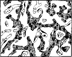
Amyloid infiltration of the liver: the amyloid substance is located between the liver cords and capillaries, while the former are displaced and partially atrophied from pressure (according to E. Kaulmann).
defined as the main protein. Well-developed cases of A. p. are easily determinable by the naked eye, since the affected organs greatly increase in weight (by 3-5 times), become extremely dense and glassy. The spleen is predominantly affected, then the liver, kidneys, adrenal glands, intestines, and lymph glands, but in milder degrees A. p. can also occur in all other organs; most rarely - in striated muscle (tongue), brain and spinal cord, nerves, nerve nodes, bone marrow, pancreas and lungs. In the spleen, amyloid substance is deposited either only in the follicles, and then it has a 'sago' appearance, or diffusely throughout the organ; in the latter type of cases, depending on the blood filling, a picture of either 'lard-like' or 'ham-like' spleen is obtained. In the liver, amyloid is located in the walls of arteries, periportal veins and along capillaries, compressing, sometimes to complete destruction, the liver parenchyma (see figure). In the kidneys, the glomeruli are selectively affected, as well as the vessels and capillaries of the cortex and medulla, more rarely - the tubule membrane and the connective tissue stroma of the medullary rays. Often A. of the kidneys is accompanied by hyaline-drop degeneration and fatty degeneration of the tubular epithelium with subsequent fatty degeneration of the stroma as well. In the intestines, A. p. of the villi and submucosa can lead to the formation of ulcers. The heart is affected in one-third of cases of general A., with the amyloid masses located between the muscle fibers and are only detectable microscopically; amyloid of the heart, visible to the naked eye, is very rare. As for the localization of amyloid in the vessels themselves, it is most often found here in the middle layers of small arteries and veins and under the endothelium of capillaries, markedly thickening the wall, narrowing the lumen, but almost never leading to its complete closure. Amyloid is always deposited intercellularly; in connective tissue - between its fibers or on them. The question of the causes of the predominant localization of A. p. in individual organs is still not sufficiently clarified. Some authors in this case attribute importance to the nature of fluid flow or the functional state of the organ, while others believe that amyloid should precipitate most strongly in the organ where protein breakdown occurs most intensely, i.e., protein metabolism, as for example in the spleen. General A. is a secondary phenomenon. It complicates prolonged chronic diseases: tuberculosis, osteomyelitis, syphilis, abscesses, bronchiectasis, chronic suppurations, malaria, nephritis, pyelitis, intestinal diseases, leukemias, tumors (cancer and sarcoma), fecal, urinary and biliary fistulas, actinomycosis and many others. Etiologically, the largest number of cases of widespread A. p. occurs in tuberculosis (lungs, intestines, bones)--about 65%, followed by: about 15%-chronic suppurations, in 8%-ulcerated and non-ulcerated tumors, in 5%-chronic kidney diseases, in 1%-syphilis, in 6%-other diseases (dysenteric ulcers, leukemia; data of Lubarsch). Extremely rarely, general amyloidosis occurs spontaneously, without an apparent cause (Nisten).-Besides general amyloidosis, local amyloidosis is also observed--primary A.: in the cartilaginous part of synchondroses, in the connective membrane of the eye, in the mucous membrane of the pharynx, larynx, bronchi, in the walls of the bladder and seminal vesicles, with amyloid sometimes lying here in the form of large tumor-like masses. The cause of local amyloidosis is unknown. [The name 'amyloid' was introduced by Virchow, who, based on microchemical reactions of amyloid, considered it to be a starchy substance (amylum-starch)]. Soon, however, this view was refuted, and the protein nature of amyloid was established, with it initially being assumed that it is a complex compound of protein with chondroitin-sulfuric acid. At present it is known that the latter does not enter into the composition of amyloid, and that amyloid is a high-molecular, protein-like protein, whose elementary analysis determines 50.26% C, 7.29% H, 14.79% N, 3.39% CN and S in traces. The high content of diamino acids gives amyloid the character of a basic protein. As a protein substance, it has positive reactions: biuret, xanthoprotein, Millon's, Molisch's, Hopkins' and with PbSO4-Hansen's. It differs from other proteins in its extreme resistance to acids, alkalis, pepsin-hydrochloric acid and autolysis. It dissolves only in barium water. As a basic protein, it is well stained with acid dyes, especially Congo red, which allows microscopic determination of its minimal amounts (see table to art. 287-288, fig. 3). The possibility of even intravital staining of amyloid with Congo red and Trypan blue has been proven. From hyalin, which is close to it in type, amyloid differs by the following microchemical reactions: 1) reaction with I (amyloid parts of a fresh organ acquire a dark reddish-brown color-see table to art. 287-288, fig. 2); 2) reaction with I and H2SO4 (areas of amyloid treated after iodine with 10% sulfuric acid acquire a dark blue or dirty green color); 3) reaction with methyl violet and gentian violet dyes (exclusively for microscopic sections); amyloid is stained cherry-red, non-amyloid areas-blue. These reactions, however, are soon transient and inconstant. Apparently, they relate to different complex groups, more or less loosely attached to the protein core of amyloid; therefore, the absence of one or another reaction indicates that amyloid of different origins apparently has an unequal composition. When these groups are cleaved off, amyloid may lose the ability to give color reactions altogether, while remaining optically unchanged. Klebs designated such morphologically complete but chemically imperfect amyloid 'achromatic' (achroamyloid). As early as 1904 it was established that the basis of amyloidosis is not tissue degeneration, but the deposition of amyloid substance in the tissue slits from the carried fluid. At the same time, some authors interpreted such precipitation of protein substance as a process of enzymatic coagulation; others, however, considered the circulation of a preformed protein body in the blood and the inability of the tissue to excrete the sulfuric acids accumulated in it, under the influence of which the protein substance is precipitated, as necessary for the deposition of amyloid. Kuczynski transferred the question of the genesis of amyloid to the plane of physical chemistry, i.e., the process of amorphous or crystalline precipitation of amyloid is considered by him as the process of precipitation of protein from a saturated colloidal solution under the influence of a change in the H-ion concentration of the medium, with H-ions of chondroitin-sulfuric acid, always found where there is amyloid, acting as electrolytes. The saturation of the blood plasma with products of bacterial and tissue (leukocytic) protein breakdown in cases of A. p. does indeed occur. Domagk, however, does not consider A. a purely passive process. He believes that the precipitation of amyloid is preceded by the breakdown on site of proteins brought with the blood flow, with the endothelium playing an active role in this respect. Then, under the influence of appropriate enzymes, the deposition of amyloid occurs in the form of a dense substance. A. can also be obtained experimentally, by creating suitable conditions in the experimental animal, which was first established by Kravkov. Thus, for example, amyloid is obtained in rabbits, chickens and dogs when bacterial cultures (staphylococcus, coli bacillus, etc.) are introduced under the skin with the formation of chronic suppuration. But the reproduction of aseptic suppuration (by injecting turpentine) also leads to widespread A. By overfeeding white mice with casein, chicken protein and milk, as well as by intramuscular injection of nutrose, A. can be obtained, in its localization, appearance and color reactions fully corresponding to human A. The same results are obtained when feeding the animal with silicic acid or by intravenous injection of it. Domagk observed the deposition of amyloid after intravenous injection of a culture of golden staphylococcus as early as 2 minutes after injection, whereas before him the earliest appearance of experimental amyloid was described only on the 7-10th day, and in humans it is reliably described only on the 10th week after the start of the disease causing A. p. The clinical significance of A. depends on the extent, intensity and localization of the process. Once started, A. progresses relentlessly and does not disappear even after eliminating the cause that caused it. In this sense, the experimentally established fact of the resorption of amyloid by plasma and giant cells, despite its enormous fundamental interest, has little practical significance. The duration of A. cannot be determined. It leads to exhaustion and, in cases of kidney damage, to albuminuria and edema. In any case, its course is chronic.
Related articles
Mentioned in
Cite this page
“Amyloid Degeneration.” Soviet Medical Encyclopedia. English translation of Bolshaya Meditsinskaya Entsiklopediya, 1st ed. (Moscow, 1928–1936), ed. N. A. Semashko. https://sovietmedicalencyclopedia.pages.dev/article/amyloid-degeneration/