Obliteration
Historical document, translated for reference. It reflects medical knowledge of the 1920s–30s and is not medical advice.
Summary
Obliteration refers to the closure or destruction of a cavity or lumen through tissue proliferation from the walls, typically involving connective tissue. This process can occur in various body systems including vessels, respiratory passages, intestines, ducts, and serous cavities, with consequences ranging from loss of patency to restricted organ mobility.
Encyclopedia article (1928–1936)
OBLITERATION (Latin obliteratio - destruction), a term used to denote the closure or destruction of a particular cavity or lumen through the proliferation of tissue originating from the walls of this cavity. This proliferation most often concerns connective tissue and represents either a manifestation of productive inflammatory processes or the so-called organization (see) of some dead material present in the cavity's lumen. More rarely, the proliferation causing obliteration of a cavity is related to tumor growth. Obliteration can be observed in the tubular systems of the body, such as vessels, respiratory passages, intestines, gland ducts, vagina, etc., as well as in serous cavities - pleura, pericardium, peritoneum; vaginal cavity of the testis, joints, etc. Obliteration of vessels is expressed in the complete closure of the vessel's lumen over a certain length of it and is most often either the result of organization of a thrombus formed in the vessel or a consequence of productive inflammation manifested by progressive proliferation of connective tissue in the inner lining of the vessel (e.g., in syphilitic lesions of arteries). Additionally, obliteration of vessels, especially arteries, occurs when blood flow through their lumen ceases, the lumen collapses, and subsequently becomes overgrown with connective tissue. This is what happens in the distal part of an artery ligated with a ligature. The physiological prototype of such obliteration is the obliteration of umbilical vessels and the ductus arteriosus in a child, occurring after birth. Obliteration of respiratory passages occurs as a consequence of various ulcerative processes of the bronchial mucous membrane, where in the area deprived of epithelial covering, proliferation of connective tissue occurs, eventually closing the bronchial lumen; or such proliferation is based on the organization of a fibrinous exudate present in the bronchial lumen. Obliteration of the intestinal lumen, excretory ducts of glands, bile ducts, vagina most often has an inflammatory origin and is associated with the processes of scarring of various ulcerative defects that resulted either from injuries (e.g., in the bile duct from a stone, in the vagina from a pessary) or from non-specific and specific inflammations. The persistent closure of the intestinal lumen, gland duct, or vagina as a result of obliteration is sometimes called atresia (see). Obliteration of serous cavities, also denoted by the term concretio, is expressed in the complete or partial overgrowth of a given cavity (pleura; pericardium, peritoneum, etc.) with connective tissue; in most of these cases, obliteration is a consequence of a former acute inflammation of the cavity with the exudation of fibrinous exudate and organization of fibrin after the acute period of inflammation has subsided (see Adhesive process). Sometimes in this case, masses of fibrin remain among the connective tissue, undergoing calcification (obsolescentio). In rarer cases, obliteration of a serous cavity is the result of chronic productive (e.g., tuberculous) inflammation. With respect to the processes causing obliteration, the adjective 'obliterating' (obliterans) is used; e.g., obliterating inflammation, obliterating endarteritis, endophlebitis, obliterating pericarditis. The consequences of obliteration are very diverse, but in general, in tubular systems they amount to the cessation of their patency, e.g., of vessels for blood, of respiratory passages for air, of gland ducts for corresponding secretions, etc., whereas obliteration of serous cavities leads to difficulties and limitations in the mobility of organs enclosed in these cavities (lungs, heart, intestines) or in the case of joint obliteration, movement of the limb in this joint. In turn, these immediate consequences of obliteration can have further results; e.g., obliteration of an artery - local anemia, atrophy or necrosis of tissue, obliteration of veins - stasis of blood, obliteration of glandular ducts - development of retention cysts, obliteration of the bile duct - retention of bile and jaundice, obliteration of the vagina - haematocolpos and haematometra. a. Abricosov. METABOLISM. Contents: I. History of the question..............
704 VI. Carbohydrate metabolism.............717 VII. Mineral metabolism............727 VIII. Water exchange ...............741 IX. Metabolism in children...........750 I. History of the question. Metabolism and energy. Under this term, which is a translation of the German 'Stoff- und Kraftwechsel,' is understood not only the direct material and energy exchange between the external environment and the living organism, which is such a characteristic feature of life, but also the entire complex of transformations of the substances and energy received in the body, which determines the vital activity of the organism. If, however, one wants to denote these two parts separately, the first is called general metabolism, the second intermediate. The French, English, and Americans use the term metabolism instead of 'metabolism and energy' (see). The dependence of life on the intake of substances into the body was undoubtedly recognized already in antiquity, however all the views put forward at that time are merely more or less ingenious intuitions. Aristotle believed that the body needs to receive food for threefold purposes: for growth, for the development of heat, and to cover its material losses. He believed that the development of heat occurs in the heart by boiling, that heat is carried by blood throughout the body, and that the task of breathing is to cool the body from the excess heat formed. The authority of Aristotle was so high not only in antiquity but also throughout the Middle Ages that during this entire period the doctrine of metabolism and nutrition represented only one or another paraphrase of his positions. A shift in this respect was brought about by the Renaissance, when speculation about nature gave way to its actual study. In 1613, Sanctorius (Sanctorius, cited after Voit) published his experiments on weighing food, drink, excreta (urine and feces), and the subject himself, establishing a significant deficit in the weight of expenditure against the weight of intake. Weighing further a fasting person, who did not lose either urine or feces, Sanctorius established a significant loss of weight. This invisible path of body losses was called by him insensible perspiration (perspiratio insensibilis) and, in his opinion, occurred through the lungs and skin. The result of this first attempt at quantitative research on metabolism undoubtedly had great significance, however further steps were impossible due to the complete lack of chemical knowledge and a very vague understanding of gas exchange.-Around the same time, the famous representative of the iatrochemical school van Helmont (van Helmont; 1577-1644) discovered that when wood burns, a gas is formed that cannot support combustion, and that the same substance is formed when acid acts on limestone, as well as during fermentation (brewing). He expressed the idea that metabolism in the body occurs through the fermentation of introduced substances acted upon by the body's juices; during this process, juices are formed that are used by the body and waste products are removed from it. Van Helmont already had the concept of chemical elements as indecomposable bodies, and from the four basic substances of Aristotle he denied the indecomposability of earth, fire, and air, but recognized it for water, to which he therefore attributed special significance in life processes, asserting for example that the fact of the growth of fish living in water proves the possibility of building animal tissue from water. The important significance of respiration and therefore of air for the life of higher animals was of course generally known since ancient times, however a clearer understanding of this became possible only after the works of Torricelli (1643), Guericke (1602-1686), and Boyle (1662), who proved the materiality of air. Guericke showed at the same time that in airless space neither the burning of a candle nor the life of warm-blooded animals is possible. In 1674, Mayow (John Mayow, 1645-1679) explained that air contains a substance, spiritus nitroaereus, which combines with metals, increasing their weight, turns venous blood into arterial blood, and reacts during combustion and respiration with combustible substances, as a result of which heat develops. This substance, besides air, is contained in saltpeter (from which its name originated). Mayow's thoughts, which contained the germ of Lavoisier's doctrine, did not receive recognition and were forgotten due to the triumph of Stahl's (1660-1734) phlogiston theory, which became a brake on the successful development of chemistry. The doctrine of metabolism, i.e., about the entire series of transformations in the body of substances received from outside, is the doctrine of the chemistry of life and as such is a branch of chemistry. It is therefore natural that it could be divided and developed in connection with and parallel to the successes of chemistry. Lavoisier, the great reformer of chemistry, is at the same time the founder of the doctrine of metabolism in the animal organism. A series of his studies on respiration and his joint work with Laplace on the question of animal heat laid the foundation for the continuous work of subsequent researchers on animal life. In 1789-97, Lavoisier together with Seguin published experiments on the influence of food intake, work, ambient temperature, and the composition of the atmosphere (oxygen content) on oxygen consumption. To connect the subject with the respiratory apparatus, they used a copper mask that hermetically sealed the face of the subject. The principles that formed the basis of the construction of later respiratory apparatus, including modern ones, were given by Lavoisier (see Gas exchange, methodology). The basic idea of Lavoisier's works: 'respiration is a special kind of combustion of substances in the animal body, constituting the main source of animal heat' contained in itself a program, the development of which took up the entire 19th century and which passed into the 20th century still far from completed. Lavoisier's successors had to solve a whole series of questions regarding the substances subject to combustion in the body, the quantity and quality of the material being burned in various states of the organism, the places and very processes of combustion, etc. The second half of the doctrine of metabolism consists of questions about the exchange of energy between the external environment and the animal body. The basic position of this doctrine, the law of conservation of energy, arose and developed half a century later than the works of Lavoisier and Laplace on examples from the inorganic world; however, the basis for the applicability of this law to phenomena of animal life - the fact of the development of heat as a result of the combustion of substances in the animal body - was still given by Lavoisier and Laplace. Lavoisier considered the affinity of oxygen for insufficiently saturated body substances as the cause of combustion in the body, whereas regarding the place of combustion he did not express himself so definitely in favor of the blood of the lungs, as his followers began to assert. In any case, in the time of Lavoisier and for almost 8/4 of a century after him, the blood was considered the main place of oxidative processes. After the works of Lavoisier it became possible to compare material intake and expenditure, but only by weight, and not by composition, because there were no reliable methods for analyzing complex organic substances of food and excretions. For quite a significant period of time, researchers of metabolism limited themselves to establishing the so-called balance equations, i.e., comparing the weight quantities of elements (C,H,0,N) of food and excretions. It was assumed that everything taken in during the day should be excreted during the same time, and therefore the difference in the obtained quantities of elements was attributed to loss by respiration and the skin without any experimental verification of this. Parallel to these studies went the study of the composition of foodstuffs, food substances, substances excreted by the body and formed in it. Outstanding chemists and physiologists of that time took part in this work: Chevreul, Dumas; 1800-1884), Boussingault; 1802-1887), Prout; 1785-1850), Magendie, Wöhler, Liebig, and others, who laid the foundation of organic and biological chemistry. An outstanding place in this constellation undoubtedly belongs to Liebig. He not only gave guidance for the analysis of organic substances and in a series of works (see literature) collected and systematized all the scientific material of organic and biological chemistry, but with remarkable insight and breadth of mind foresaw the paths of further research and practical application of scientific data. Knowledge of the composition of organs and excreta gave him the opportunity to assert that in the organism there is a gradual transformation of complex compounds into simpler and simpler ones up to the final products excreted from the body, and that on the basis of the quantity and quality of these products conclusions can be drawn about the quantity and quality of substances broken down in the body. He asserted that the nitrogen of urine should be considered as a measure of the breakdown of protein substances in the body. He saw the task of physiology in studying the course of transformations of substances in the body and its changes under the most diverse conditions. Liebig's theoretical views on the significance of the transformation of substances in the animal organism are extremely interesting: the destruction of the organized constituent parts of working organs is the basis of all manifestations of the physiological activity of the organism, and in particular therefore also the basis of the mechanical work performed by muscles.
Therefore, following the prevailing opinion at the time that organized substance consists almost exclusively of protein, Liebig recognized only the proteins in food as true nutrients, which were supposed to serve for the restoration of tissues destroyed during work, i.e., for plastic processes (hence the name 'plastic substances'). When tissues are destroyed, which according to Liebig occurs under the influence of oxygen, nitrogen-containing waste products and part of the exhaled carbon dioxide are formed. He considered the combustion of protein, besides the 'organized' protein, to be completely excluded. On the contrary, fats, carbohydrates, and other non-nitrogenous substances, in his opinion, do not participate in the construction of body tissues and are oxidized by oxygen in the first place. The oxidation of these substances serves exclusively for warming the body, and therefore these substances received the name 'respiratory substances' and were not by Liebig considered true food substances participating in obliteration. Protein substances in food could be oxidized for the purpose of heat production only in case of insufficiency of respiratory substances. Great Medical Encyclopedia, Vol. XXI. In 1842, an article by J. R. Mayer appeared in Liebig's 'Annals' formulating the law of conservation of energy, and in 1848, his treatise 'On Organic Movement in Its Connection with Metabolism' was published as a separate book. Sharing the common view that oxidation occurs in the blood and under the action of oxygen, Mayer connects the processes of oxidation with the work of muscles as follows: 'part of the heat arising from the oxidative processes occurring in the capillaries of muscles becomes during muscle activity latent or expended, and this expenditure is proportional to the mechanical effect produced.' Thus, the organized substance of the muscle is merely a transformer of energy, being itself relatively stable: 'one cannot deny the wearing out of organs, but this is a separate issue; and in steam engines, wearing out occurs daily and hourly, but one cannot put on the same level the materials needed for repairs with the expenditure of coal.' On the one hand, Mayer's theory established a close connection between metabolism and energy exchange, and on the other, it substantially limited the basic provisions of Liebig's theory: the extreme destructibility of organized living substance and the exclusively plastic significance of food proteins. [The fact that the life of animals (dogs) cannot be maintained on food devoid of proteins was established by Magendie in 1836.] Thanks to Liebig's authority, his theory was in the forefront. From it followed a whole series of questions that became accessible to experimentation thanks to Liebig himself. He gave researchers the method for quantitative determination of urea, i.e., the main product of protein breakdown in the body, and Dumas-the method for determining the total nitrogen in food and excretions, and so it became possible to clarify: 1) whether the nitrogen of food is completely excreted in urine and feces, 2) what is the effect of different amounts of food proteins, and 3) how the breakdown of body proteins proceeds during muscular work. Respiratory experiments by Lavoisier gave him the basis to assert that nitrogen does not participate in respiration, however, a series of researchers of nitrogen metabolism, headed by Boussingault, could not establish in their experiments the equality of food nitrogen and nitrogen in excretions, from which arose the doctrine of the so-called nitrogen deficit in urine and feces and its excretion through the lungs and skin. The famous study of gas exchange in various animals, conducted (1849) by Regnault and Reiset (see Gas Exchange) on a very large scale, gave some basis to the doctrine of nitrogen deficit, because in many experiments a change in the amount of nitrogen in the apparatus was noted, although small and moreover fluctuating sometimes in the + direction, sometimes in the -. Classical in their strict methodology experiments by Bidder and C. Schmidt in Dorpat (Bidder, Schmidt; 1852) on dogs and cats showed however the correctness of Liebig's thought that all nitrogenous breakdown products are excreted in urine and feces, and this was then confirmed by Bischoff in 1857 and especially by C. Voit and Gruber (Gruber, 1857) in experiments establishing nitrogen equilibrium with its verification by sulfur equilibrium. In more recent times, the respiratory experiments of Krogh (Krogh) have finally, it seems, settled this question in accordance with Lavoisier and Liebig. Investigating the effect of different amounts of food protein on the breakdown of protein in the body, Bidder and Schmidt established with certainty the increased breakdown of protein when its content in food increases, all other conditions being equal, and there was also an increase in heat production. This obviously spoke against Liebig's doctrine, according to which only organized protein should break down and therefore in these experiments it should break down on an increased scale only to allow the newly incoming protein to replace the old one. Therefore, a correction was introduced that the animal's need for protein is determined by the breakdown of protein during starvation, and if during feeding more protein is given than the specified 'typical minimum', then the excess protein is oxidized by the blood oxygen, like non-nitrogenous substances, and therefore there is 'excessive consumption' (Luxusconsumption) of this protein as unnecessary for body plasticity. However, Bischoff rightly pointed out the improbability of the existence of two causes for protein destruction and the unjustifiability of oxygen coming into action as soon as the delivery of protein crosses a certain boundary. Subsequently, the Munich school, headed by Voit and Pettenkofer, using its respiratory apparatus (see Gas Exchange), proved that during muscular activity the breakdown of protein does not increase at all or increases only insignificantly, while the breakdown of non-nitrogenous substances (judging by the excretion of carbon) increases to a considerable degree. On the other hand, numerous studies by Voit showed the close dependence of protein breakdown on its intake and the possibility of establishing the organism in a short time in a state of nitrogen equilibrium with very different amounts of food nitrogen. Voit further showed that an excess of protein protects non-nitrogenous substances from breakdown, and an excess of carbohydrates in turn spares proteins, so that under these conditions one can bring the organism into nitrogen equilibrium with an amount of food protein less than the amount of protein breaking down during the animal's starvation. All these facts are in contradiction with Liebig's theory and are easily reconciled with Mayer's theory. Voit's doctrine distinguishes between 'organized' protein and 'circulating' protein in the body fluids. The first possesses considerable stability and is destroyed only in the process of wearing out, while the latter is easily destroyed, coming into contact with cellular elements. It remained little understood, as Liebig and Pflüger (Pflüger) emphasized, how Voit explained the transition of circulating protein into organized protein and back (during starvation). The Munich school and in particular Voit is indebted to the doctrine of metabolism for the extremely systematic development of the chapter on protein metabolism under various nutritional conditions, during starvation, at rest and during work, as well as the doctrine of nutrition. By the beginning of the last quarter of the 19th century, the doctrine of obliteration, extremely enriched with factual material, in its theoretical part continued to remain in the sphere of ideas of Lavoisier and partly of Liebig. The cause of the destruction of substances in the body was recognized as the oxidative capacity of inhaled oxygen, the place of oxidation-blood and tissue fluid, the substrate of oxidation-food substances and only during starvation the dissolving organized substance. The latter was recognized as stable, its wearing out as insignificant and replenishable from food proteins, since the organized substance itself was considered 'living protein'. In 1875 appeared the famous critical-theoretical article by Pflüger 'On Physiological Combustion in Living Organisms,' which completely overturned previous ideas about the cause and place of oxidative processes in the body. Not the influx of oxygen determines, according to Pflüger, the breakdown of substances in the body, but, conversely, the intensity of metabolism determines the greater or lesser uptake of oxygen, and the place of oxidative processes is not the blood, but the cellular elements of the body, i.e., organized living substance. Pflüger's opinion was supported by brilliant experiments from his laboratory, which showed, first, that frogs whose circulatory system had been washed with a physiological solution of salt and filled with it ('salt frogs') for a long time continued to secrete a normal amount of carbonic acid, and second, that normal frogs placed in an atmosphere of pure nitrogen could exist for about 20 hours and excreted during this time a significant amount of CO2. The experiment by Georg Liebig, proving the excretion of CO2 by frog muscles cut out of the body, and numerous subsequent studies of tissue respiration (see Respiration, tissue respiration) made it indisputable the position of Pflüger that 'the causes of the destruction of substances lie in the cells.' This same thought is extremely vividly expressed by C.
by Bernard in the following words of his treatise 'On the phenomena of life common to animals and plants': 'If a muscle contracts, will and sensation manifest themselves, thought arises or a gland secretes, then the substance of muscles, nerves, brain and gland is disorganized, destroyed and consumed'. At first glance one might think that scientific thought had returned to the view of Liebig, however this would be true only to a certain extent, because the new concept encompassed the functions of all organs of the body and developed a different viewpoint on the composition of organized living substance. The latter was no longer only 'living protein' but, on the basis of the study of its decay products, was recognized as being 'composed of various substances, including proteins' (Voit), and specific in its structure and composition for different organs. These ideas received further development in the direction that 'living organized substance' is currently not considered as a homogeneous 'substance' but as an extremely complex physico-chemical system, specific in each individual case in its structure and composition, in the course and character of the chemical processes occurring in it, and in the products that arise during this process. A common property of these systems, inherent in their structure and composition, is their destructibility and ability to regenerate and, as a consequence, the exchange of substances between cellular elements and their surrounding internal environment of the body—blood and tissue fluid, the same for all cells but specifically utilized by each of their kinds. From this point of view, the specific action of medicinal substances and the specificity of hormones, vitamins, etc., become partly understandable. Now that it has become possible to study the conditions of tissue culture (see) outside the organism, that it has become possible through microsurgery (see) to directly affect individual parts of cells, and through microchemical methods to study certain chemical processes in the cell, the study of O. v. now makes further very important advances, especially in the area of understanding the various intermediate stages of the transformation of substances in tissues. The thought of Lavoisier that respiration is a special kind of combustion, which is the source of animal heat, already connected the exchange of substances with the exchange of energy, however the principle of the transformation of energy was not known to Lavoisier and therefore he could not seek in the process of combustion the source of energy for the mechanical work of animals. J. R. Mayer, who formulated the law of conservation of energy, pointed to its applicability to the animal organism, which, according to Mayer, had to be considered as an energy transformer. However, experimental proof of this was first given (1891-94) by Rubner in calorimetric studies on dogs, and then (1897-1899) by Atwater on humans. There are two external sources of energy for the animal body: the destruction in the body of food substances or body substances (during starvation) with the release of potential chemical energy from them, and the totality of all effects from the external world on the sense organs or generally on the surface of the body. The magnitude of the intake from the first source can easily be determined from the heat of combustion of substances, determined calorimetrically, but the external effects on the sense organs in their entirety cannot be measured, and therefore this deficiency in the experimental solution of the question is inevitable, but quantitatively it is undoubtedly extremely small. As for the expenditure of energy, it in the resting and starving animal consists of heat losses (directly as heat or by evaporation), from the heat of combustion of excreted urine and feces, as well as from the internal work of blood circulation, respiratory movements, intestinal movements, etc., which in turn pass almost entirely into heat (the external work of respiratory movements, for example, the expulsion of air from the lungs, is obviously extremely insignificant). Thus, under the specified conditions, both the intake and expenditure of energy from the animal side can be expressed in heat units, with negligible deficiencies on both sides, and compared with each other. It goes without saying that the taking of food or the performance of mechanical work, without introducing anything new in principle, only somewhat complicate the compilation of the energy balance. The question of whether it is permissible to equate to each other in terms of thermal effects two processes of combustion as different in conditions as burning in a calorimetric bomb and oxidation in the organism, was resolved affirmatively (in 1838) by the Petersburg academician Hess in the form of the following proposition: 'the amount of heat accompanying a given chemical process depends only on the initial and final state of the reacting bodies and does not depend on the intermediate stages of transformation between them'. Therefore, since carbohydrates and fats give in the calorimetric bomb and in the organism the same products CO2 and H2O, their heats of combustion in both cases are the same, while for protein substances, which burn in the organism only to urea, uric acid, etc., a correction must be made. This correction was determined by Rubner, who established the following standard heats of combustion of food substances in the body: 1 g of carbohydrates-4.1 calories, 1 g of fat-9.3 and 1 g of protein-4.1. Rubner's experiments, and later Atwater's, showed the extreme closeness of the figures (to fractions of a percent) of the income and expenditure parts of the balance, which, on the one hand, confirmed the correctness of establishing the income from the heats of combustion, and on the other hand, made it possible to recognize the expenditure of energy as a characteristic summary of the breakdown of substances in the body. Proceeding from the energetic concept of nutrition, Rubner conducted a series of observations on the replaceability of food substances with each other and came to the establishment of the so-called law of isodynamicism (see) of food substances, according to which under certain conditions of insufficient nutrition, substances destroyed in the body replace each other in heat-equivalent quantities. From this it was concluded that the organism in each given case has a certain 'need for energy', which it can cover with different substances. For protein, however, an exception had to be made: 'the impossibility of feeding the organism exclusively with fats and carbohydrates and completely eliminating the last traces (3-4%) of protein decomposition depends only on the fact that for certain chemical processes, such as for the replacement of destroyed tissue parts, protein is necessary, because animals are unable to synthesize this substance' (Rubner). Considering the organism as an energy transformer, Rubner naturally came to the conclusion that in the greater or lesser scale of this transformation, i.e., the exchange of energy, one should seek the criterion for the intensity of life processes. However, since the exchange of energy is closely connected with the exchange of substances, Rubner proposes to speak of the general exchange of substances, even without adding the words 'and energy'; in the word 'general exchange' is already, in Rubner's opinion, an indication of the measurement of the exchange in a general unit, the energy unit, i.e., calories. Rubner's doctrine, revolving in the sphere of J. R. Mayer's ideas, rightly emphasizing the energetic aspect of the exchange, is nevertheless too one-sided and simplified in its depersonalization of food substances and its evaluation of them only by caloric value. The moment of simplicity undoubtedly played a major role in the popularization of Rubner's doctrine, especially among nutrition practitioners, while among physiologists it met with objections. The failure of experiments with feeding animals mixtures of as pure food substances as possible, calorifically quite sufficient, and all further doctrine of vitamins and one-sided nutrition clearly emphasize the material aspect of the exchange, without of course diminishing in any way the importance of energetics. If one recognizes that the basis of the life activity of 'living organized matter' is its destruction, its partial breakdown, and yet this substance undoubtedly possesses dynamic stability, this means that along with the processes of dissimilation, breakdown, processes of assimilation, restoration of losses occur in it at the expense of interaction with those chemical groups and bodies with which it can react and which it can assimilate. This possibility is determined, of course, by the composition and structure of the chemical groups and bodies, and not only by their energetic value. P. Methodology of research. The methods of research of the general O. v. and energy are based on the exact quantitative determination of the constituent parts of the intake and expenditure of solid, liquid and gaseous substances. Food substances to be used in the experiment must be stored, if they withstand storage, in quantities sufficient for the entire duration of the experiment. From the stocks, average samples must be taken, analyzed according to the task either for food substances (proteins, fats, carbohydrates, salts, fiber) according to the general rules of food analysis or for individual elements (C, H, N, S, P, etc.) according to the rules of elementary organic analysis. Of course, preliminary analysis makes sense only in the case when one or another food substance is given in food without culinary processing.
Therefore, in experiments with humans, in most cases it is necessary to analyze an aliquot part of all the food offered or to mix these parts or even analyze them separately, which of course requires a large expenditure of labor and time. The use of tables of the composition of foodstuffs is inadmissible due to the significant variation in the composition of market products. It is also extremely dangerous to use various kinds of coefficients such as N : C in urine, meat, etc., as evidenced by the experiments of Voit, which according to his calculations demonstrate the formation of fat from proteins, but according to the calculations of Pfleuger with other more accurate N : C coefficients in urine and meat—devoid of any evidentiary value. The duration of the research can of course be very different, but the element of the experiment in time is recognized as a day, with the last meal being no later than 12 hours before the start of the next day. At the beginning of each new day, after the voiding of urine (in humans voluntarily, in animals by catheter) and, if necessary, after defecation, weighing is performed. Special attention must be paid to the quantitative collection of urine and feces. Urine is usually collected daily, feces—* mostly for the entire period of the experiment. In humans, the collection of urine presents no difficulties with a conscious attitude on the part of the subject, in animals catheterization is used, but to avoid accidental loss of urine, animals are always placed in cages with an inclined or grated floor ensuring the drainage of urine into a receiver. In cages for small animals, a mesh of appropriate density is placed under the grated floor to separate urine from feces. The delimitation of feces belonging to the experimental period is achieved in dogs by giving them a small amount of bones at the beginning and before the end of the experiment, which form a well-formed plug in the intestine, and in humans by giving 300-500 cm3 of milk, which produces a characteristic light-colored milk stool, or charcoal tablets, charcoal suspension, berries, etc. The delimitation in humans is not always sufficiently sharp, but since the feces are usually collected for the entire experimental period, no less than several days, the error is not of great importance. Methods for the analysis of urine and feces—see Urine, analysis of urine and Excreta, chemical examination. The accounting of gaseous components of intake and expenditure is carried out using respiratory apparatus (see Gas exchange), which makes it possible to determine the amount of consumed 02 and exhaled CO2 in long-term and short-term experiments. The former are necessary where it is a matter of establishing the material and energy exchange, the latter are appropriate when establishing only the expenditure of energy. Analyses of air associated with respiratory experiments are conducted according to the rules of gas analysis (see), mostly by the Haldane method. Since in the study of the overall O. of substances and energy, if there are no special tasks, it is only a matter of the exchange of dynamogenic substances (proteins, fats and carbohydrates), it is only the quantitative determination of N and C in urine, inhaled oxygen and exhaled CO2 that is absolutely necessary. Then, from the N in urine, the amount of decomposed protein is calculated (g × 6.25), and from the 'net' respiratory coefficient, the share of participation in the expenditure of carbohydrates and fats is determined, using the Zuntz-Lusk table. Knowing the amount of dynamogenic substances decomposed in the body and their heat of combustion, it is easy to calculate the amount of energy released during this decomposition. The calorimetric experiments of Rubner and Atwater, as mentioned above, showed excellent agreement in the numbers of energy expenditure calculated in this way and established directly in the experiment. If it is intended to establish only the expenditure of energy, it is sufficient to determine the gas exchange or even only the amount of oxygen absorbed, the energy expenditure is then calculated from the thermal value of oxygen. Of course, the calculation of the overall exchange from the thermal value of oxygen is inadmissible if in the body not only proteins, fats and carbohydrates are burned, but also other substances, e.g. alcohol, or if in the body there is a conversion of carbohydrates to fat, as well as the formation of under-oxidized products, e.g. acetone bodies. Direct determinations of energy exchange in respiratory calorimeters, as requiring expensive and complex apparatus and a large expenditure of labor, are available to only a few laboratories. III. Basal metabolism. Overall O. of substances and energy. The characteristic of the intensity of the overall O. of substances and energy of the organism in different periods of its life is the so-called basal metabolism, i.e., that decomposition of substances in the body which takes place under the maximum possible limitation for a living organism of the activity of all its organs. Basal metabolism consequently encompasses the entire set of those transformations in the body which are necessary for maintaining the basic vital functions, but as a concept it is still a conditional quantity, although for a given organism at a certain
table 2. period of his life is sufficiently constant. Factors that determine it to some extent are weight, height, age and sex. Taking these factors into account, Harris and Benedict established empirical formulas for calculating the normal basal metabolism; for the male sex (over 1 year): 66.473 + 13.7516.kg + 5.0033.cm - 6.7550 . years; for the female sex (over 1 year): 655.0955 + 9.5634.kg + 1.8496. cm - 4.6756. years; for children under 1 year: boys - 22.1 + 31.05.kg + 1.16. cm, girls - 44.9 + 27.84. kg + 1.84. cm. Based on these formulas, derived purely empirically, Harris and Benedict compiled tables of basal metabolism, which proved very satisfactory and found wide application. According to these tables, the normal basal metabolism is determined as the sum of two numbers: the basic number by weight and the second number by height and age, e.g.: male 60kg, 163cm2)years. Basic number by weight - 892 cal., second number by age and weight - 647 cal., basal metabolism 1,359 cal. (see tables). Table 1. Basic number by weight. Kg For men For women Kg For men For women 1 167 1 387 562 1038 1 552 1 579 1 114 1 662 1 152 The weight of the body cannot of course be considered a determining factor for metabolism due to the different composition and structure of the body, therefore the calculation per 1 kg of weight, often used for comparison purposes, is very imperfect, but still acceptable if we are comparing organisms that are at least similar in external constitutional characteristics. Thus, organisms of similar height, weight, muscular development and nutrition usually have similar basal metabolism, i.e. possess a similar amount of "organized living substance", called for brevity "living protoplasm", the life activity of which determines the basal metabolism. Folin showed that with protein-free food, creatinine is excreted as a product of endogenous metabolism of substances. American researchers (Palmer, Means and Gamble) compared the excretion of creatinine with the observed heat production and found quite constant ratios: in men per 1mg of excreted creatinine - 0.98 cal., and in women - 1.26 cal. According to Burger, this creatinine coefficient is a good index of muscle mass. Table 5. Second number by age for girls from 0 to 12 months. Months 10 2 V. cal. -535 : -475 ! -420 -370 According to Rubner, the main factor determining metabolism is the surface of the body as the surface of its cooling, assuming that heat loss determines heat production. If this is true for warm-blooded animals and moreover under certain conditions to a certain extent acceptable, then as a principle it is undoubtedly incorrect. For practical purposes, relating the amount of metabolism to the body surface is just as acceptable as relating it to weight. The most satisfactory formula for determining body surface area in humans is recognized as the formula of D. and E. F. Dubois: surface in m2=kg0.426 x cm0.725 x 71.84, or more simply - surface in cm2 = 167.2√kg x √cm. This formula is very accurate. According to the same authors, the basal metabolism in large calories per 1 m2 of surface and per 1 hour varies with sex and age as follows: At present, basal metabolism is calculated either according to Benedict or according to Dubois, since the results are almost the same. The basal metabolism of an average constitution and nutrition person is taken as equal to 1 calorie per 1 kg of weight and hour, i.e. e.g. with 70 kg per day it will be 70 x 24 = 1,680 calories. However, during sleep metabolism decreases even more due to an even greater restriction of life activity and is considered equal to 0.9 per 1 kg per hour. In ordinary life, the so-called working addition is always added to the basal metabolism, caused by the activity of muscles, the intake and digestion of food, if physical heat regulation is insufficient, etc.
M. Shaternikov. Pathology of Basic Metabolism. One can note deviations in metabolism by tracing the extraordinary transformations of substances that occur in various diseases. But one can also consider the other side of metabolism and evaluate, from the most general point of view, the amount of substance broken down in the body, or, what is the same thing, determine the amount of heat formed during this process. Data concerning the measurement of this understood general metabolism do not characterize the details and changes in the course of individual reactions within the body. Up to the present time, clinical medicine has been interested mainly in changes in the so-called basic metabolism and to some extent in changes in the specific dynamic action of food. An increase in basic metabolism by itself is not accompanied by an increase in temperature, and the excess heat formed leaves the body by various paths. That increase in temperature observed in febrile disease is a consequence of disturbance of temperature regulation with simultaneous increase in metabolism. The increase in heat production in these cases can be considered a constant phenomenon, but in various stages and cases of the febrile process it is expressed differently. Being very strong at the beginning with a high and steep rise in temperature, it later becomes less. The increase in basic metabolism sometimes reaches 48%. The connection between increase in metabolism and temperature according to some authors corresponds to the rule derived for chemical reactions by van't Hoff ('a rise in temperature of 10° corresponds to an increase in the rate of reaction by 2-3 times'). The respiratory coefficient RQ in fever is increased only during the chill due to the destruction of glycogen during muscular movements. At other times it is rather below normal (participation of fat oxidation processes).-Malignant new growths and leukemia in many cases similarly increase the basic metabolism.-Anemia remains without any particularly characteristic influence on basic metabolism, just as polycythemia does too (sometimes metabolism is increased).-Diseases of the liver and kidneys are not associated with an increase in metabolism. However, with a significant increase in the spleen in hepatolienal syndromes, and also in hypertension, metabolism although not necessarily, may be increased.-Similarly, no characteristic changes in basic metabolism have been noted for diseases of the lungs and heart. If, then, the influence of diseases of various internal organs on basic metabolism is small, the same cannot be said about lesions of the glands of internal secretion. The greatest significance for the entire pathology of general metabolism is the thyroid gland. Removal of this organ lowers metabolism significantly (by 20%), myxedema sometimes even more (by 50%). Similarly, lowered metabolism figures can be found in ordinary goiter.' In contrast to this, the introduction of thyroidin by mouth or thyroxin under the skin strongly increases metabolism. In Basedow's disease a typical increase in basic metabolism is observed. These same analyses have made it possible to control the effect of medicines and operative intervention in diseases of the thyroid gland.-Research on basic metabolism in lesions of the hypophysis encounters all the difficulties that are met in the path of research on the pituitary gland. They consist in the fact that the pituitary gland, with its small size and superficial location, is a complex formation and moreover irritation of the entire area of the brain lying near this organ also gives changes in metabolism. In acromegaly most authors speak of an increase in gas exchange, whereas cases of dystrophia adiposo-genitalis are very often associated with a decrease in basic metabolism.-Diabetes insipidus is not accompanied by changes in basic metabolism. General metabolism in diabetes mellitus deserves special attention. For decades the question of general metabolism in diabetes mellitus was given great importance. There was talk sometimes of an increase, sometimes of a decrease in it. At the present time it is recognized that there are no strictly characteristic changes in basic metabolism in diabetes. Only in cases of very severe diabetes can one as a rule see an increase in metabolism. But these increases, which are subject to dietary influence, can hardly be attributed entirely to diabetes, since the patient is also exhausted, suffers from ketonuria, and therefore his disturbance of metabolism depends on a number of factors. Experimental diabetes, caused by removal of the pancreas or poisoning with floridzin, is accompanied by an increase in metabolism.-Even greater significance than in diabetes is given to general metabolism in obesity, since fat, considered as reserve material, ultimately comes from food substances not destroyed in metabolism. Conversely, with insufficient feeding the missing calories are extracted in the largest quantity from fat reserves, which are noticeably reduced. The calorie expenditure, depending to a large extent on muscular activity and fat consumption, also increases depending on the mobility and activity of the subject being examined. Thus the deposition and expenditure of fat are regulated by two factors little accessible to accounting: the activity of the subject observed and his food, which is usually determined in quantity by appetite. The question arises- can food eaten in excess also be broken down to end products of decomposition (apart from the participation of muscles). For the fact that in principle some change in the size of basic metabolism can occur, speak the studies of fasting people and observations during prolonged insufficient nutrition. In both cases the magnitude of basic metabolism (i.e. the amount of oxygen consumed) is noticeably reduced. From this it follows that one cannot speak of absolute stability of the basic metabolism figure, and if metabolism under the influence of nutrition can decrease, then probably it can also increase. The increase in metabolism from food is always noted for several hours. Fats and carbohydrates increase metabolism by a barely noticeable amount, protein nutrition acts much more strongly. Proteins were ascribed the specific dynamic action. It is possible however that not only the postprandial but also the morning basic metabolism in cases of prolonged overeating (and of protein, and fat, and carbohydrate) is established at higher figures of oxygen consumption. Such an ability, if it exists, would guarantee the organism against the transformation of excess nutrition into burdensome fat ballast. Not all researchers recognize the existence of such a 'luxury consumption' (Luxusconsumption). As long as in the doctrine of obesity overeating obesity and lack of movement obesity are recognized above all. In these cases basic metabolism shows no deviations from normal. In addition the existence of obesity connected with the state of internal secretion is also recognized. In these cases it is sometimes possible to establish normal appetite with decreased metabolism or with normal basic metabolism an insufficiently clearly expressed rise from the specific dynamic influence of nutrition.As for the path by which endocrine influence makes itself felt on O., according to some the secretions act directly on the oxidation processes in the body, others consider it possible that the glands of internal secretion through the central nervous system act on the entire behavior of the organism, which is closely connected with the work of the muscles, and in this way bring about an increase in metabolism. e. Fromgold. '87 Pathological anatomy of disturbances of O. of substances can manifest itself in various ways. First of all one can point out that a whole series of serious irregularities in O. of, for example, nitrogen, carbohydrates, salts, may not be accompanied by any morphological changes in organs and tissues. In other cases of general disturbance of one or another O. of pathological-anatomical changes are observed which are consequences of the improper O. of and manifest themselves in atrophic processes or in the form of one or other pathological deposits (see Arteriosclerosis, Atrophy, Starvation, Gaucher's disease, Diabetes mellitus, Cachexia, Niemann-Pick disease, Obesity, Gout). Local manifestations of disturbance of O. of in tissues are expressed in the appearance in the cells or intercellular spaces of various materials, physically or chemically foreign to this tissue, which is designated as degeneration or infiltration of tissue or deposition in the tissue (see below). IV. Protein metabolism. Nitrogen metabolism (syn. protein metabolism)- in the broad sense the aggregate of all phenomena in the body more or less connected with the metamorphosis and transport of nitrogenous compounds throughout the body under different conditions of nutrition and states of the body. These phenomena are divided into three groups: 1) digestion and absorption of nitrogenous compounds in the intestinal canal; 2) transformations of nitrogenous compounds and their breakdown products in the fluids and tissues of the body, (intermediate nitrogen metabolism); 3) excretion of nitrogenous compounds from the body through various excretory organs. In particular also under nitrogen metabolism is meant the establishment of nitrogen balance (see below). Nitrogenous products of digestion-amino acids and some polypeptides-on absorption penetrate through the intestinal mucosa into the capillary blood, then into the portal system of the liver and from there further into the general circulation of the blood, considerably increasing the content of amino acid nitrogen in it.'
At the same time, the amino acid nitrogen of the tissues absorbing free amino acids from the blood also increases; thus, in the experiment of Folin and Daines, after introducing 61 g of egg white into a loop of the small intestine of a cat, after 11/2 hours, an increase in 'residual' nitrogen (non-protein blood nitrogen minus urea nitrogen) was found in the portal vein blood from 14 to 22 mg%, and in the carotid artery blood from 12 to 22 mg% (i.e., by 57%, resp. 83%); 45 minutes after the introduction of glycine, the residual nitrogen in the portal vein increased by 433% (from 12 to 64 mg%), in the carotid artery by 140% (from 15 to 36 mg%), and in the muscles by 43% (from 223 to 319 mg%). In all these experiments, N of urea remained unchanged at the beginning. After a certain period of time, the amount of amino acid N in the blood and tissues decreased (in the former sooner than in the latter), while the amount of urea began to increase. Example: in a person after administration of 135 g of gelatin, amino acid N in the blood after 1 hour (in mg%) was 9.4, after 2 hours-11.4, after 3 hours-7.2, after 8 hours-6.8 (before taking gelatin - 5.5). After the same intervals, the urea content in the blood in mg% corresponded to 16.0-14.2-20.2-25.0 (before eating-17.3). Different tissues do not absorb amino acids equally, as seen from the following table: Table 1. Amino acid nitrogen and urea in a dog Before injection (mg%) After injection into the vein of extract of digested meat after after 11/2 hours 4 hours (mg%) (mg%) Amino acid nitrogen: in muscles .... in liver .... in kidneys .... Urea in blood .... 4.7 . 5 70 156 88 13.7 71 71 89 11 During periods of increased concentration of amino acids in the blood, some of them manage to pass into the urine. Between blood amino acids (Ac), adsorbed amino acids of tissues (At) and tissue protein (Pt), there must exist some reverse equilibrium, which can be symbolically expressed as Pt ⇌ At ⇌ Ac. The arrival of new batches of amino acids into the blood, and from it into the tissues during active digestion, leads to a disruption of this equilibrium, and according to the law of mass action, the amount of tissue protein must increase. However, amino acids and peptides adsorbed by tissues selectively go to the synthesis of more complex polypeptides, resp. protein molecules in these tissues; unused amino acids can return to the blood (with subsequent relative depletion of the blood with amino acids) and be adsorbed elsewhere. As a result of such redistribution of amino acids, some time after their absorption from the intestine, all tissues to some extent become 'saturated' with them, while carrying out the indicated protein synthesis; however, many amino acids remain unused and undergo deamination in the tissues (mainly in the liver), as a result of which NH3 and keto acids are obtained: the first in combination with CO2 gives urea (see), the second partially undergo further oxidation, reaching CO2 and H2O, with energy release, while partially they can be converted either into glucose and glycogen or into higher fatty acids and fats. The breakdown of amino acids is a very complex process, many details of which are still far from being clarified. Of great importance for understanding the phenomena involved is the observation of Embden (method-passing amino acids through a surviving liver), that α-amino acids give transformations not like their corresponding fatty acids, but like acids with one less carbon atom. For example, leucine (α-aminoisocaproic acid) gives acetone under experimental conditions, just like isovaleric acid (one C less), while the acid corresponding to leucine (isocaproic) does not have this property. From this it was concluded that amino acids during breakdown are converted into fatty acids with the loss of one carbon atom: R-CHNH2-COOH → R-COOH. Thus, in this breakdown, three processes take place: 1) loss of the NH2 group (see Deamination), 2) loss of CO2 from the COOH group-decarboxylation, 3) oxidation. The question of the order in which these processes proceed in different amino acids has not yet been fully resolved, however, a number of facts now allow establishing the general scheme of the course of these reactions. The usual research methods are: 1) study of the transformations of certain amino acids when solutions containing these amino acids are passed through the blood vessels of a surviving liver. 2) Feeding large amounts of certain amino acids, the transformation products of which do not have time to oxidize completely and are excreted in the urine as intermediate products. 3) Introduction (per os, intravenously, per rectum) of such derivatives of amino acids, which are foreign to the animal organism and in their transformations pass only the initial stages of changes during deamination or decarboxylation and are then excreted in the urine, where they are determined. 4) Introduction of certain amino acids into the organism of a diabetic or an animal with experimental diabetes (after pancreatectomy or poisoning with floridzin), as well as into the organism of an alkaptonuric (see Alkaptonuria) for the purpose of observing the transformations of the introduced substances (or their derivatives) into sugar (in diabetes) and into homogentisic acid (in alkaptonuria). Similar observations are also made in other diseases of metabolism, arising spontaneously or experimentally. All these methods have led to the view that the first stage of amino acid transformations is deamination. Against this, it would seem to speak the relatively strong bond between N and C in amino acids, however, under the influence of bacteria, as is known, this bond breaks easily [e.g. phenylalanine (C6H5-CH2.CHNH2.COOH) is converted into phenylpropionic acid (C6H5.CH2.CH2.COOH)]; similarly in the organism after abundant feeding of tyrosine (HO || \C6H4 \CH2.CHNH2.COOH/) it was possible to notice in the urine the appearance of p-oxy-phenyllactic acid (C6H4 \CH2.CHOH.COOH)* According to Dakin, the first steps in breaking the N and C bond probably go in such a way that first, according to the general rule, the β-carbon atom is oxidized, R', R', R' CH2', CHNH2', CNH2 I', '!', 'i', COOH', COOH'
SOON, after which a double bond appears between the α and β atoms with the subsequent rearrangement of the compound R - CH - C.NH2.COOH ^NH into the imino acid R-CH2.C-COOH and its hydrate R-CH2-C^COOH ^NH2. The probability of this assumption follows from the fact that the compound C(CH3)3 - CH. NH2. COOH (α-aminotrimethylpropionic acid), in which a double bond cannot appear between the α and β atoms, hardly burns in the body. Imino acids or their hydrates, being unstable bodies, easily lose ammonia, turning into keto acids: R. CO. COOH. Thus, the process of deamination is accompanied by oxidation (oxidative deamination). The previously accepted view that deamination proceeds by hydrolysis (R - CH.NH2.COOH + H2O = R-CHOH. .COOH + NH3) was based on such facts as the appearance of hydroxy acids in the urine (e.g., lactic acid) after abundant feeding with corresponding amino acids (e.g., alanine), as well as the fact that the fate of the amino acid and the corresponding hydroxy acid in a number of cases is the same; e.g., both leucine [(CH3)2-CH.CH2.CH(NH2).COOH] and the corresponding leucinic acid [(CH3)2-CH.CH2. CHOH. COOH] can be sources of acetone bodies in diabetes (see below). However, recent research has established the incorrectness of this view; on the contrary, a number of facts speak in favor of the fact that during deamination, keto acids mainly appear, while alcohol acids (hydroxy acids) are a secondary product (reduction of keto acids) or a product of an accompanying side process. The main facts forcing such a conclusion are the following: 1) the possibility of imitating the appearance of keto acids from amino acids in vitro (through oxidation by permanganate); 2) the appearance of keto acid in the urine when foreign amino acids are introduced into the body: for example, phenylglycine (C6H5CH. .NH2.COOH) gives phenylglyoxylic acid (C6H5.CO.COOH) with a small amount of benzoic (C6H5COOH-product of decarboxylation) and mandelic (C6H5. .CHOH.COOH-product of reduction) acids. In addition, the keto acid is a more reactive body than the corresponding hydroxy acid and is more capable of reverse transformation into an amino acid than the hydroxy acid; this gives more weight to the view that the main compound during deamination is the keto acid, i.e. RC^COOH-^ ->R-C-COOH-| -NH3. However, there are facts that do not fit into such a scheme. There are also reactions in which keto acids can arise not through the stage of imino acids; for example, phenylglycine (C6H5CH.NH2.COOH) can turn into phenylglyoxal (C6H5C^C^H) and then be oxidized into the corresponding keto acid-phenylglyoxylic (C6H5-CO-COOH). However, this course of the process cannot be considered typical. After the formation of the keto acid, decarboxylation occurs, resulting in an aldehyde R- "CH O and then the corresponding acid R - C^~OH. Thus, as a result, we have an acid, poorer by 1 carbon atom than the original amino acid, which agrees with Knoop's rule. The general scheme of these transformations is as follows: R
R
R1 /H
-H I
.. +H2O ! ,OH I \NH2 amino acid G=NH imino acid I XNH2COOH hydrate of imino acid
The amino acids that give keto acids upon deamination can be called ketoplastic amino acids, while the amino acids of the first group are aketoplastic. The introduction of aketoplastic compounds in diabetic acidosis, as a rule, reduces the formation of acetone bodies and therefore they are also antiketoplastic. 3) Of the remaining amino acids, tryptophan, lysine, glucosamine (histidine?) do not belong to either group, and therefore can be called aketo and aglycoplastic amino acids. In the midst of the absorption and deamination of amino acids (and the formation of urea), there is a peculiar phenomenon-enhanced roil
R
R -NH3 | -CO2 i +0 | -> C-O -> C=O -> C=O ketone acid \ fatty acid dehydration water entry The resulting fatty acids from the reactions above have in the body a fate common to these compounds (see fat and carbohydrate metabolism). However, a series of reactions that amino acids undergo during deamination and decarboxylation to some extent determine their further course of transformations in the body, both toward final combustion to CO2 and H2O, and partly toward synthetic reactions (transformation into carbohydrate and fat). As a general rule for the oxidation of fatty acids (see fat metabolism), the beta-carbon atom acquires an OH-group, and then with further oxidation, two carbon atoms are cleaved off. However, this rule may only apply to monobasic fatty acids of the fatty series with an unbranched chain and moreover a sufficiently long one (five-six C). The fatty "residues" of amino acids, with the exception of normal valeric acid from normal leucine, do not belong to this well-studied group, but for the most part have either shorter chains, or are branched, or represent hydroxy- and keto acids, dicarboxylic acids, etc. The systematic study of various amino acids with respect to the products obtained from them that are excreted in the urine in pathological cases (see above diabetic and alkaptonuric organisms) allows one to form some idea of the further transformations after deamination. All known amino acids that make up the proteins of the body and food can be divided into three groups: 1) amino acids that can be transformed into glucose - glucoplastic: glycine, alanine, serine, cystine, aspartic acid, glutamic acid, proline, arginine (histidine?). 2) Amino acids that do not give gluconeogenesis (in diabetic organisms, in normal organisms such gluconeogenesis can be achieved by a longer pathway) - aglucoplastic: valine, leucine, isoleucine, phenylalanine, tyrosine (histidine?). These amino acids in diabetic organisms instead of sugar give acetone bodies (see), therefore their deamination decarboxylation oxidation in tissues leads to an increase in heat production. An illustration can be the experiment of Lusk (Figure 1), from which it is seen that after the administration of protein food, the course of heat production and the course of N excretion in the urine (by the hour in a day) run completely parallel to each other. Such an influence of protein substances in food Administration of protein food 40Ca1 2.0g 1.5 'W hours22 0 3 в в 12 16 18 21 Figure 1. Lusk's experiment: columns at the top-heat production; dotted curve-excretion of N in the urine. was known for a long time, and M. Rubner called it the specific dynamic action. The cause of this phenomenon still constitutes a subject of discussion. In any case, all apparently agree that the penetration of protein digestion products into tissues produces in them (under normal conditions) a kind of "irritation", the result of which is a noticeable increase in heat production (resp. oxygen consumption and CO2 excretion). The specific dynamic action of proteins does not manifest itself 1) under conditions causing increased heat production (low temperature, work); 2) during increased synthesis of proteins in tissues, occurring at the expense of food proteins, e.g. during vigorous growth or after prolonged fasting. Nitrogen balance. Nitrogen equilibrium. With the same intake of nitrogen into the body day after day in the protein-amino acids of tissues-amino acids of blood system, such a dynamic equilibrium is established, in which the amount of decomposed (deaminated) nitrogenous compounds in tissues is equal (in nitrogen content) to the amount entering. This is externally expressed in the fact that the N balance (N food - N feces - N urine) equals 0. Such a state in the body is called nitrogen equilibrium. If at established nitrogen equilibrium the level of nitrogen "93 60g ' «DO 5 + Щ™-™.,-..' ш . 4 6 a 10 12 14 that N food N excretions positive balance N Fig 2.1-nitrogen equilibrium before the experiment; and III-first and second nitrogen equilibrium. food is raised and kept at that level for some time, then due to the influx of amino acids into tissues, the synthesis of protein in them will increase, but at the same time the accumulation of protein masses as labile substances simultaneously enhances the breakdown of protein molecules, and after some time (in a few days) a new nitrogen equilibrium is established at a new, elevated level of N entering the tissues. It must be emphasized that the factor establishing the equilibrium is ceteris paribus the level of N in food. With a decrease in dietary N, the relationships change in the opposite way, and as a result, nitrogen equilibrium is again obtained at a new, reduced level of nitrogen. An illustration of the above can be any experiment with the establishment of nitrogen balance. From Fig. 2 (Voit's experiment on a dog) it is seen that with the transition to a diet with an increased nitrogen content, the dog retained 26.4 g of nitrogen, resp. synthesized 165 g of protein; in five days after reducing the nitrogen in food it - lost 14.7 g of nitrogen, resp. lost from the body's reserves 91.9 g of protein. The establishment of nitrogen equilibrium in normal occurs after a short period of time; in convalescents and generally after fasting, partial or complete, the period with positive nitrogen balance is considerably prolonged, which depends on the previous depletion of tissues with protein. Positive N balances also occur during tissue growth (in young organisms, in pregnant women) and during periods of training in muscular work, which leads to hypertrophic changes in muscles. Thus, Caspari in a dog (18-day experiment) showed that with the same food (calorie content 2 088-2 099; N=25.11 g pro die) and the same daily work (about 600 cal.) approximately on the 6th day a significant retention of nitrogen began to be detected, which can be considered a consequence of protein synthesis in muscle tissue. However, with an established muscular system, under ordinary working conditions, no difference is found in protein consumption when comparing nitrogen metabolism during periods of work and periods of rest, as seen in the table 8 below. Table 8. Average numbers per 1 day From what has been said above, it follows that the more N there is in the body, the more is required in food to keep the body in nitrogen equilibrium; therefore it is quite clear that persons with well-developed musculature require a larger protein ration than persons of the same weight with less developed musculature. On the other hand, it is obvious that low protein regimes, when applied for a long time, lead to a decrease in protein reserves in tissues. In this connection, Kestner's experiment is very demonstrative, conducted during the last economic crisis in Germany on patients of the clinic not suffering from metabolic diseases. Table 9. Days Food balance ' urine feces N ! ' 7.3 3.6 1.8 I + 1.9 1 8.6 3.1 1.8 + 3.7 i 1 8.1 ! 6.4 1.5 + 0.2 ! ! 7.9 j 5.0 3.6 -0.7 ! 1 12.5 ! 4.0 1.4 + 7.1 1 13.9 ! 4.7 1.8 +7.4 I 13.2 I 4.3 3.2 +5.7 ! ! 8.0 3.0 2.3 +2.7 1 Marked with * are ! days of meat administration-110-120 g. | Food Periods N in g calories Rest (6 days) . Normal work (9 days) ..... Intense work (4 days) . . . . 5.9 6.0 5.9 2 300 3 000 3 200 A z o total ammonia i 4.77 0.35 4.40 0.38 3.94 0.42 creatinine i 0.605 0.60 0.56 From Table 9 it is seen that with a very low nitrogen content in food, usual for the selected patients (here only one of six cases is given), the nitrogen balance undergoes significant fluctuations with positive and negative signs due to the incomplete identity of food from day to day. When 110-120 g of meat was added to the ration, the influx of nitrogen increased significantly (by 60 - 70%), but the amount of protein decomposed in the body did not increase, as we might have expected, so that the N balance increased significantly in the positive direction. This indicates an increased protein synthesis in the tissues of the subjects, arising from the extreme depletion of their protein on the fasting regimes of that time: under such a state, tissues eagerly absorbed all proteinogenic material coming from the intestine. If on these low-nitrogen rations nitrogen equilibrium still takes place, then from this one can only conclude that nitrogen equilibrium is by no means an indicator of the body's well-being, but only gives an external expression of the established mobile equilibrium between the body's protein and nitrogenous substances entering the blood from the intestine. For any normal organism, one can find such a level of dietary nitrogen, below which the organism could no longer come into a state of nitrogen equilibrium, but would always give a negative balance; such a minimum amount of dietary nitrogen for establishing nitrogen equilibrium, in the view of some researchers, is an indicator of the body's minimum nitrogen requirements (under given conditions); it was called the "physiological minimum".
However, the 'physiological minimum' is an indeterminate value, depending on many reasons—on the individual's constitution, on the degree of their nutrition, on age, etc., as well as ceteris paribus on the amount of non-nitrogenous (mainly carbohydrate) components of food. By increasing the latter and bringing the caloric value of food to an excessive level, one can achieve nitrogen balance with a very small protein content in the diet (see below), which indicates its insignificant breakdown in the body under such conditions. With excessive nutrition exclusively by carbohydrates, the breakdown of protein in the body reaches particularly low values. Rubner considers the nitrogen excreted under such conditions (when protein practically does not serve the body's energy needs) as an inevitable loss by tissues and calls this amount the 'wear coefficient' (Abnutzungquote). The average value of the wear coefficient according to Lauter's calculations is 0.053 g N pro die per 1 kg weight (human), i.e., with an average weight of 70 kg this amounts to 3.7 g N, resp. 22.2 g protein per day (counting 16.6% N in protein). Finding these values has purely theoretical interest: practically the level of protein in the food many times exceeds the value of the wear coefficient. The question of man's protein requirement was one of the first questions in the history of the science of nutrition. Carl Voit solved it, partly using general statistical material, partly on the basis of more precise observations in individual families. The data obtained were then verified by laboratory methods on individuals (analysis of food, urine, feces, gas exchange). 'As the average value from a large number of observations,' writes Voit, 'I derived the requirement of the average worker as 118 g protein and 328 g C with a mixed diet consisting of a certain amount of meat and plant foodstuffs.' Since 118 g protein already contain 63 g C, the remaining 265 g C can be represented by 500 g carbohydrates and 56 g fats. This norm referred particularly to 'the average worker with sufficiently developed musculature to perform average work.' Voit believed that dietary protein is needed not to serve as a source of energy, but to maintain the protein composition of muscles at a constant level; on this basis he advocated increasing nitrogen in the diet of people with more developed musculature. 'To maintain a developed working organ (for example in a blacksmith) requires more protein than is needed by a weak tailor.' Later, Atwater, using the same methodology, settled on a protein norm of 100 g (for sedentary work) per day. However, this amount of protein seemed exaggerated to some researchers, and they sought to justify lower protein norms for humans. All these works came down to observations on nitrogen balance in humans at a low level of N in the diet. Leaving aside short-term experiments, one can first point to the work of Neumann (1902), who showed in experiments on himself, lasting 120 and 321 days, the possibility of maintaining weight and nitrogen balance at the level of 79-74 g protein (when recalculated to 70 kg body weight) with 2,737-2,000 calories (approximately 1 g protein and 39-28 calories per 1 kg body weight), and the subjective indicators of the body's condition (work capacity, health, etc.) were noted by the author as positive. Soon after that, Chittenden (1905) organized a 'mass' experiment, lasting 9 months, on a group of 25 people (scientists, military personnel, orderlies, and professional athletes), while monitoring weight and from time to time investigating nitrogen balance. Nitrogen balance was sometimes achieved with 9-6 g N in the diet for soldiers and scientists and with 11.5-9.0 g for athletes. However, nitrogen balance was not always achieved, and some of the subjects could not achieve it at all, moreover weight losses were sometimes very significant (some individuals actually gained weight). The author concluded that his experiments proved the possibility of maintaining N balance without loss of strength and work capacity with 40-60 g protein in the diet; at the same time he expressed the conviction that the usually consumed larger amounts of protein adversely affect the body, supposedly burdening it with an excessive amount of products of nitrogenous metamorphosis to be excreted. Some authors (Benedict and others), criticizing Chittenden's experiment, note a number of essential details that significantly diminish his conclusions. Thus, from the data of the experiment it is evident that the subjects showed low digestibility of N, which should be connected precisely with the low protein regimes, since in experiments on animals this connection has long been studied and not only in relation to protein but also other food substances. Furthermore, the exhaustion of most of the subjects was emphasized, and such organisms, as already indicated above, have a great ability to retain dietary protein; the author's reference to the preservation of work capacity of the subjects cannot actually be considered a significant indicator of the adequacy of the dietary regime, since firstly there is an element of subjectivity in this concept, and secondly because the ability to work is also preserved in starving subjects. On the other hand, the performance of work (muscular) is the result of the transformation of energy in the body, released mainly from the destruction of non-nitrogenous organic materials, which in Chittenden's experiments were contained in the food in sufficient quantity. Also very curious is the fact that Chittenden's athletes after the end of the experiment returned to their previous dietary regimes, which of course they would not have done if Chittenden's diets had completely satisfied them. - A few years after Chittenden's work, Hindhede (1913) conducted similar long-term experiments on individuals with continuous determination of nitrogen balance; at the same time he aimed to show that one could achieve an even greater reduction of nitrogen in the diet (4.4-11 g N pro die) using potato with margarine and seasonings (onion, fruits) as such. The results of Hindhede's experiments are negative. The regimes he introduced led to permanent nitrogen loss. Experiments with reduced protein content in the diet, attempting to justify the possibility of getting by with a low protein norm in human nutrition, are refuted by experimentally established (in animals) cases of a very sharp decrease in the body's resistance to all kinds of infections on low protein regimes. Finally, mention should be made of Haecker's observations on cows, that the negative effect of protein-poor diets (weakness, weight loss) manifested only in the 3rd year of their application, and until that time the animals did not differ in any way from the norm. These facts with great obviousness should warn against hasty conclusions in favor of reducing human protein norms. The above experiments of Neumann, Chittenden, and Hindhede essentially do not speak against Voit's protein norm: from them one could only draw one correct conclusion, that man can maintain nitrogen balance for more or less long periods on low protein regimes with sufficient caloric value in the food to cover energy needs. Sherman (1920) collected from the literature 109 cases where the minimum protein per 1 kg weight was established as 0.63-0.64 g (with fluctuations from 21 g to 65 g pro die per person). Discarding the extreme values, the author derives an average of 40.6 g, i.e. 0.58 g protein per 1 kg weight. However, it is undoubtedly true that the establishment of nitrogen balance for a long time on low protein regimes is possible only after the depletion of the body's protein reserves under conditions of sufficient caloric value. Whether the amount of protein in the diet corresponding to such a minimal nitrogen exchange can be considered generally optimal for man is evident from experiments of feeding subjects, under the condition of nitrogen minimum, with protein food. From the above experiment of Kestner it follows that the additional nitrogen of food in this case does not appear in the urine, but goes to the formation of new protein masses in the body. Similar observations were made at the same time by Hosslin, and before that by a number of other authors. Thus, in Grafe and Koch (1912), a 35-year-old factory worker after severe underfeeding gained 20 kg in weight during 6 weeks and retained 535 g N and still did not achieve nitrogen balance. Obviously, the abundant nitrogen retention manifested in these cases expresses the incompleteness of the body's protein composition before the experiment (under low protein regimes), since under normal conditions such retention should decrease day by day and in a short time be replaced by nitrogen balance. All these data allow one to interpret the results of research on reduced protein regimes as follows, that in them we are essentially dealing with protein undernutrition (Eiweissunterernahrung), and therefore the protein requirement must be sought not in the protein minimum, but at such levels of nitrogen in the diet at which a sufficient protein reserve (protein depot) is organized, and the shift of nitrogen balance in one direction or the other occurs rapidly.
A number of American authors consider that the minimum allowable amount of protein in food should be considered 1 gram per 1 kg of weight. However, taking into account that food proteins are not qualitatively identical, most physiologists consider it possible to raise the protein norm to such a height that the calorie content of protein in food constitutes 10-15% of the total calorie content of food (at 3000 calories - 75-110 g protein). In Voit's diet, protein (118 g) contained 16% of the energy of food. Thus, Voit's norm is higher than all other norms. The correctness of the latter is confirmed by the fact that most people (if they do not have to perform very heavy work, when with an increase in calorie content the level of N in food inevitably increases, since it is practically difficult to choose such a combination of food where a high calorie content would not be associated with a high protein content) consume protein with average work precisely within these limits. Thus, the statistical work of Heiberg (1921) based on material from households (1,000 cases) of 1897-1916 established for Copenhagen fluctuations in protein of 94-118 g, in villages - 93-149 g, in provincial cities - 84-103 g. At a later time, a number of authors, when observing the N in the urine of a large number of subjects, give values indicating the approximate degree of protein expenditure in the body with free choice of food: Benedict (1919) in student athletes - 75-81 g, Beard (1927) in 400 female students - 76.7 g, Denis and Borgstrom (1924) in 233 medical students - 76.1 g (all figures for 70 kg weight).-According to materials from the Institute of Nutrition Physiology in Moscow, collected on the basis of a very large number of literary data, protein consumption in people of different professions, as can be seen from the attached table 10, varies either around Voit's norm or slightly below - 70-80 g protein, thus confirming the correctness of the above values of the protein norm for humans.
Protein quality. The question of the amount of protein required for man is confronted with another question - about the qualitative difference of protein. If food proteins reach the tissues in the form of amino acids, then the best quality will be possessed by that food protein which contains the amino acids required by the body in the proper amount, absolute and relative. Since we encounter different combinations of amino acids in different proteins, it is quite obvious that not all food proteins have the same ability to provide tissues with everything necessary for the synthesis of body proteins in them; those proteins that have a complete assortment of amino acids can be called complete proteins. The degree of completeness of different proteins could be determined by comparing their amino acid composition, however, to the present time we do not know this composition with sufficient accuracy and can only rely on approximate data. Meanwhile, experiments on animals make it possible to establish that proteins of different origins in different cases (e.g. during growth or for maintaining nitrogen equilibrium at small, close to minimum, amounts of nitrogen) replace each other not in the same and not in proportionally to the nitrogen quantities, in other words, proteins possess different qualities, which are determined by differences in the content and grouping of amino acids, or possibly their aggregates, in the composition of the protein molecule. For illustration, fig. 3 by Osborne and Mendel is given, from which it can be seen that growth (increase in weight) of young animals on different proteins ceteris paribus proceeds differently and all proteins represented in the figure can be divided into groups of different values or different

The second example is given in table 11, from which it can be seen that to maintain minimal nitrogen equilibrium, proteins with lower biological value must be taken in larger quantities. The term 'biological value' was introduced by K. Thomas (1910), who attempted to characterize this concept quantitatively, determining the amount of N converted into body proteins per 100 parts of absorbed food N. Thomas' data according to this and other, more complex formulas are given in table 12 and show how many weight parts of body proteins can be formed from 100 weight parts of absorbed food proteins (the author's figures are rounded). Other authors either refuted or confirmed Thomas' data. It turned out that the absolute magnitude of 'biological value' is not constant for the same protein (even more so for foodstuffs); it is influenced by both the amount of food consumed (calorie content) and the amount of protein in it. Of course, the state of the organism at the time of the experiment (e.g. protein content) must also be of great importance. Various authors have also established the importance of individual amino acids for the biological value of proteins. A number of proteins and protein mixtures are incomplete due to the absence in them of amino acids which cannot
be synthesized in the body. Adding the missing amino acids to such proteins makes them complete (figure 4).
V. Lavrov. Purine exchange (physiology) - see Uric acid, Nucleoproteins, Purine bases. Pathology of protein exchange. Severe disturbances of protein exchange occur during complete and partial starvation and when feeding with incomplete proteins. Increased protein exchange (so-called toxic protein breakdown) is observed in certain poisonings (pantopon, carbon monoxide, sublimate, oxalic acid), in malignant tumors, in certain forms of Basedow's disease, in diabetes, especially experimental, in acute infectious diseases, and in febrile processes. The reverse phenomenon - increased deposition of proteins in the body (Eiweissmast) - positive nitrogen balance - is most easily achieved after a period of starvation, during chronic insufficient nutrition, and especially easily during the recovery period after exhausting diseases. Retention of N in the body occurs even with relatively small amounts of administered protein. During intense muscular work, protein deposition occurs due to the development of working hypertrophy of muscles, which is often accompanied by significant retention of nitrogen in the body. The question of whether it is possible to achieve protein deposition in the normal state of an adult organism has been the subject of great discussion. The solution of this question has great practical importance, especially in animal husbandry. Kellner proved that with rational feeding in cows, it is possible to achieve significant protein deposition in the body. In humans, it is also possible to obtain deposition of protein substances, as measured by the positive nitrogen balance. This retention of N in the body is achieved both by increased administration of protein substances and by increasing (calorically) the amount of nitrogen-free food products that prevent part of the proteins from being used for energy expenditure. Retention of nitrogen in the body cannot be regarded with certainty as only a process of protein deposition; nevertheless, large amounts of retained N (Grafe) cannot depend only on nitrogenous waste products, but are also the result of accumulation of protein substances, although their formation is not accompanied by the usual retention of water in the body. Due to the discrepancy in the amount of retained N and water, most authors believe that this formed protein is not identical to the protein of protoplasm, but partly circulates in the body fluids (Voit) and partly is deposited in the liver (Rubner, Noorden) or in the cells of other organs (Lutje, Berg). To test whether the deposited protein is biologically the same as the protein of protoplasm, the basal metabolism was investigated, which, however, did not show an increase during N retention. This observation speaks against the formation of true protein, which is a component of protoplasm, as is the case in cases of protein deposition during working hypertrophy and after exhaustion. Whether such protein deposition has any advantage for the body is difficult to say. Some authors consider protein deposition beneficial because these proteins, due to their lability, protect the proteins of protoplasm from breakdown and, if necessary, can be converted into true proteins. Qualitative changes in protein exchange. Besides the physiological proteins of organs and fluids in pathological processes, special proteins are found, probably formed by the restructuring of tissue protein, namely: amyloid (see Amyloid degeneration) and Bence Jones protein (see Bence Jones protein body). Bence Jones protein usually appears in the urine in multiple myelomas, but not in all cases of this disease. Sometimes crystals of Bence Jones protein are deposited in the myeloma tissue (Wulf). Bence Jones protein was also detected in the urine in osteomalacia, sarcomatosis, hypernephroma, leukemia. Bence Jones protein is most often excreted simultaneously with serum protein; a diseased kidney passes Bence Jones protein more easily than a healthy one. The site of protein rearrangement is unknown; apparently bone marrow cells influence this restructuring of the protein molecule. Gottschalk draws an analogy between Bence Jones albuminuria and diabetes; in both cases there is a disturbance of intermediate metabolism with the formation of a substance partially utilized by the body's cells. Other authors draw a parallel between the formation of melanogen and melanin by pigment tumors and the formation of Bence Jones body in myelomas. When Bence Jones protein is injected, kidney damage develops (Krauss). Under the influence of parenteral administration of Bence Jones protein, immunization processes develop. Changes in the assimilation of amino acids are important disturbances of protein exchange. In liver diseases, hyperaminoaciduria (in acute atrophy of the liver, crystals of leucine and tyrosine) is observed along with hyperacidemia. Increased excretion of amino acids in the urine occurs in many infectious diseases, diabetes, leukemia. There are also diseases characterized by insufficient intermediate utilization of one or more amino acids. Cystinuria or cystin diathesis (Wollaston) is characterized by increased excretion of L-cystine in dissolved form or as a precipitate in the urine; sometimes cystine forms stones in the bladder or kidneys. The essence of cystinuria is that the body is unable to break down the cystine molecule. Apparently, cystinuria is a disturbance of intermediate metabolism caused by general insufficient deamination, since usually along with cystine, abnormalities in the breakdown of other amino acids (leucine, tyrosine, asparagine) and amines (cadaverine, putrescine) are observed. The appearance of diamines in cystinuria represents a constitutional anomaly and differs in significance from diamineuria caused by the action of bacteria in the intestine on lysine and arginine, which give the same amines upon breakdown. Alkaptonuria (see) as a disease has no great practical significance, but played a major role in the theoretical study of the breakdown of aromatic substances in the human body. Nucleic exchange. Physicochemical studies of the exchange of nucleoproteins in the body and pathological disturbances of this exchange are of great importance for clarifying the pathogenesis of gout (see). Gout is characterized by the deposition of monosodium urate in cartilage, tendons (tophi); in the blood, the content of uric acid is usually increased, while the excretion of endogenous uric acid in the urine is decreased; exogenous uric acid is also excreted insufficiently and slowly. A. B. Garrod, Thannhauser, and Lichtwitz believe that the phenomena of gout depend on the primary insufficiency of the kidneys in relation to the excretion of uric acid. Brugsch and Schittenhelm explain gout by the increased content of uric acid in the human body, caused by insufficient enzymatic processes, mainly by a decrease in activity or absence of the uricolytic enzyme that normally destroys uric acid formed in the process of intermediate metabolism (see Uric acid, Gout). Creatinuria - see Creatine. The question of the formation and presence of ammonia in the body has undergone a fundamental change in recent years. Our and Benedict found that the main site of ammonia formation is the kidneys. The amount of ammonia excreted in the urine many times exceeds the amount of ammonia circulating in the blood. The same has been confirmed by Ambard and Schmid. In the blood, ammonia is present in two forms (Parnas): first, in a very small amount (0.015 mg% N) in the form of ammonium ion, and second, in the form of adenine nucleotide (2 mg% N) - the 'basic ammonia substance' of Parnas (Ammoniakmuttersubstance-AMS), which easily splits off ammonia when the serum stands. In the kidneys, this nucleotide is split at an acidic reaction, forming ammonia. The formation of ammonia is closely related to the neutralization of acids, provided they do not burn to CO2 in the body. From the relationship between ammonia excretion and the presence of acids, the influence of nutrition on ammonia excretion is clarified. With meat food, much ammonia is excreted because many inorganic acids (sulfuric acid, phosphoric acid) are formed from proteins; with mixed food, less ammonia, with vegetable food, very little. With oral or parenteral administration of acids, the amount of ammonia in the urine increases. At the same time, in the serum, the amount of ionized ammonium does not change, but the content of the non-ionized part (AMS) clearly decreases. The same is observed in endogenous acidosis. The greatest increases in ammonia excretion are observed in diabetes: the amount of ammonia reaches up to 12 g per day instead of the normal amounts of 0.5-1 g. When alkalis are administered, no changes occur in both blood ammonia fractions, but the amount of ammonia excreted in the urine sharply decreases. In kidney diseases, especially in nephritis, ammonia excretion is decreased. In uremia, despite the state of acidosis, with a sharp decrease in reserve alkalinity, the amount of ammonia in the urine is sharply decreased; at the same time, the ammonia content in the blood does not increase.
Hasselbalch, who pointed out the significance of NH* as a regulator of neutralization, noted that under normal conditions in the same individual there is a certain relationship between the ammonia number (the ratio of ammonia nitrogen to the total amount of nitrogen) and the concentration of hydrogen ions; in renal patients this coefficient is less, because the diseased kidney is either completely unable to form ammonia or cannot form it in sufficient quantity. When acid is introduced to a nephritic patient, ammonia in the urine does not increase at all or increases only by a few centigrams, in contrast to a healthy person, in whom the ammonia content increases by 1-1½ g per day.
Yu. Geftor. Pathological anatomy of protein metabolism disorders—see Amyloid degeneration, Protein degeneration, Hyaline degeneration, Cachexia, Marasmus, Cloudy swelling, Gout, Uremia. V. Fat metabolism. Fat metabolism in the broad sense of the word is the sum of phenomena representing the fate of 'fat' in the organism. This includes the following main moments: the penetration of dietary fat from the intestine into the blood, the transport of fat to tissues, its consumption and deposition in tissues, and the synthesis of fat. The concept of 'fat' may also include the concept of fat-like bodies, or so-called lipoids; thus, in fat metabolism the following is considered: I. Proper fat metabolism; II. Lipoid metabolism. 70в I. Proper fat metabolism. From the intestinal canal after absorption (see Digestion), fats enter the body in two ways: 1) through the lymphatic capillaries of the villi, forming in them a kind of thin emulsion, for which reason the lymph in the larger lymphatic vessels of the mesentery appears as a white mass during periods of absorption; lymph enriched with fat droplets (chylus) flows through the thoracic duct into the venous blood; according to some observations (for example, Munk, Friedenthal) on a girl with a fistula of the thoracic duct, the main mass of fat passes into the body precisely by this path. 2) Through the capillaries of the villi and then through the portal vein into the circulatory system of the liver, etc. According to observations by Errico, blood taken simultaneously from the portal and jugular veins of animals at the height of the digestive process (fatty food) had a higher fat content in the portal vein than in the jugular vein, which could not be the case if fat passed into the venous system exclusively through the thoracic duct. It follows that fat passes into the blood by both paths. The appearance of dietary fat in the lymph begins already in the first hour of digestion, and after two hours the fat content in the chyle of the thoracic duct reaches 3-14 (depending on the fat content in the food and the rate of absorption) compared to several pro mille in the fasting lymph. The fat content in the blood during food absorption also gradually increases (alimentary hyperlipemia). The so-called 'normal' lipemia, i.e., the fat content on an empty stomach (after a preliminary 'average' mixed diet or after a fat-free first breakfast in humans), varies greatly in different animals and even in different individuals of the same species—from 0.2% to 0.8% (some authors show significantly lower numbers). As an illustration of the distribution of fat (fatty acids) in the blood, a table by Horiuchi is given. Table 13. Comparative distribution of fatty acids in the blood. Humans and animals l и и й ° ё 5*5 Man............0.36 Woman............ 0.38 Dog..............j 0.52 Rabbit (without fat diet) . \ 0.28 Rabbit (fat diet) . . . I 0.34 0.38 0.40 0.22 0.30 0.36 0.29 0.36 0.42 The magnitude of alimentary lipemia is subject to many, partly random influences (rate of absorption and amount of food, amount of fat in it, its digestibility, activity of enzymes, state of the endocrine system, etc.). Sometimes the blood plasma at the height of digestion becomes opaque due to the accumulation of fat globules in it. As an example of lipemia, observations by Bloor on a dog are given. 24 hours after a meal, the blood fat was 0.6%, after 3x/4 hours after the administration of 100 em3 of olive oil—0.73%, after 6V4 hours after the administration of 100 cm* of olive oil—1.20%, and after 8 hours after the administration of 100 cm* of olive oil—0.87%. In humans, alimentary lipemia occurs even more easily than in a dog, whereas in a rabbit it is difficult to detect. In geese during fattening (with fat in the food), alimentary lipemia reaches 3% in the blood and up to 6% in the serum. With abundant carbohydrate feeding, when carbohydrates are converted into fat, lipemia is not observed, of course in the absence of fat in the food (Bleibtreu, Rosenfeld). The reduction of fat content in the blood to normal is due to the fact that fat particles are 'captured' first by adipose tissue, where fat accumulates in cells as reserve material; such tissues ('fat depots') are subcutaneous tissue, intermuscular connective tissue, the omentum, etc.; secondly, fat particles can pass into other tissues, where fat is consumed for energy or structural purposes.—In a number of pathological conditions (see below pathology), during fasting, in experimental pancreatic or floridzin diabetes, lipemia also occurs, with fat in this case entering the blood from the body's fat depots; such lipemia coincides with the loss of glycogen reserves in the body, and the mobilization of fat here can be regarded as an adaptation of the body to new conditions of greatly reduced carbohydrate metabolism. An example of such lipemia is given in the work of Miescher on a starving salmon (biological fasting—see Fasting). From all that is known to us about lipemia, it thus appears that conditions can arise in the body in which the capillary system becomes permeable to fat in one direction or the other. The mechanism of such an adjustment of the body is completely unknown. Some authors have tried to consider the disappearance of fat from the blood after hyperlipemia as a result of its breakdown by blood lipase. However, if such lipolysis does take place, it is apparently by no means absolutely necessary for the transition of fat from the blood, since intravenously introduced colored fat carries the dye to those tissues (liver, bone marrow) to which it is usually transported (Biondi, Neumann). The disappearance of fat from the blood some researchers try to explain based on the assumption that fatty acids and fat form compounds with plasma proteins, which can go beyond the limits of the circulatory system. The following facts speak in favor of the possibility of such a combination of fat with protein: 1) the difficulty of extracting (quantitatively) all the blood fat by extraction with fat solvents without prior use of pepsin-hydrochloric acid to dissolve proteins; 2) a protein solution in the form of, for example, transudate (ascitic) fluid after mixing it with a certain amount of oil does not completely give up the latter to the extracting fat solvent: some amount of oil remains in the protein solution, which passes back into the solvent only after the action of pepsin-hydrochloric acid. The action of bile acids on fat in the blood also leads to such 'masking' of fat, resulting in compounds soluble in plasma. The work of Biondi-Neumann also points to the 'masking' of fat; they studied the ultramicroscopic picture of human blood; blood taken during fat absorption showed in the dark field numerous bright droplets (hemokonia), and the number of such particles gradually increased as fat was absorbed, reached a certain maximum (6 hours after a meal), then fell and after 12 hours reached zero. In blood samples taken from a fasting person, hemokonia were absent. One can think that small fat droplets can pass through the capillary wall. The following numbers (Mockel) illustrate in percentages the concentration of fat in the tissues of a fat animal (dog): skin—38%, subcutaneous layer—87%, muscles—19.6%, liver—13.4%, brain—12.7%, bones—12.0%. During periods of liver glycogen depletion, fat abundantly passes into the liver from fat depots, raising its fat content to 30-40%.—The introduction of fat into the body (as well as the introduction of carbohydrates and proteins) causes a temporary increase in metabolism (specific dynamic action) (see protein metabolism). However, such an increase compared to the effect of carbohydrates and proteins is insignificant, which is probably explained by the easy transition of fat to fat depots. In the experiment of Magnus-Levy, when 210 g of butter was given to a person, the increase in metabolism compared to the basal level was 9-14% during the seventh hour and 6-8% on the eighth hour. The external temperature has a very significant influence on the manifestation of the specific dynamic action of fats. Rubner has the following example: a dog received 171.3 cal. of fat per 1 kg of weight. Table 14; External te Heat production per 1 kg of weight (in calories)
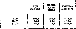
A healthy person (68 kg) contains 12.36 kg of fat (approximately 18% of their weight). In fattened animals, fat reaches up to 50% of their live weight. "That fats from food, when consumed in significant quantities, are transported to fat depots is evident from a series of experiments. Thus, when dogs and mice are fed colored pork lard, the dye passes into all fat depots, with only the liver and adrenal glands remaining uncolored; the dye also passes into milk fat (Jacobsthal); obviously, the deposition of fat in the liver is a secondary process - fat is transported there from other tissues; however, with very abundant introduction of fat, the liver fat also becomes colored. In addition, it has been established that 'foreign' fat can be deposited in fat depots if it is introduced with food for a long time and in large quantities; thus, dogs after prolonged feeding with mutton lard accumulate it in their subcutaneous tissue. The same has been noted when feeding mutton lard to goldfish and carp. Herbivores consuming green fodder have harder fat than when fed grain fodder (according to Rosenfeld, fat from green fodder is hard, fat from grains is liquid). Various fats were tested on animals - rapeseed oil, linseed oil, sesame oil, cow's fat, coconut oil, and mutton fat - and all of them could be found not only in the fat depots of the experimental animals, but also in milk, in chicken eggs, and in the uropygial gland of birds. On the other hand, each species of animal has its own characteristic fat. This does not contradict the above, since each species also has its characteristic combination of foodstuffs. Finally, large amounts of fat arise not from fats, but from carbohydrates of food or from proteins. Intermediate fat metabolism. Fatty acids, when burned in the body, undergo gradual 'stepwise' destruction, which is characterized by oxidation of the β-carbon atom, and at this point the chain breaks and a carbonyl group is formed. CHa - CH2 - CHa - CHa - CH2 - COOH cha - ch2 - ch2 - CO -/- ch2 - COOH CH2 - CH2 - CHa - COOH CH2-CO + CHa - COOH ch2 - COOH. Obviously, with each phase of oxidation, two C atoms are split off (in the form of acetic acid or its derivatives); it is also obvious that acids with an even number of C atoms must pass through the stage of a four-carbon chain - butyric acid; acids with an odd number of C atoms do not form butyric acid; on the contrary, one of the last intermediate products in them is propionic acid. Experiments on animals confirm and clarify this scheme of destruction of fatty acids in the body. Thus, Knop (Knorr), using the observations of Nentsky, Sal'kovsky, and others, showing the difficulty of burning benzene nuclei in the body, introduced phenyl derivatives into the body having various fatty side chains, which did not have time to burn completely in the body, stopping at certain intermediate products passing into the urine. In this way, it was established that fatty acids really obey the general law of β-carbon oxidation and the sequential splitting off of two-carbon segments from the chain. Thus, β-phenylpropionic acid 7 09 CH2-CH2COOH passed into benzoic acid 1 ICOOH, while phenylacetic acid I \ CH2COOH and mandelic acid CH(OH)-COOH were excreted from the body unchanged (they did not have a β-carbon in the side chain for oxidation); phenylbutyric acid CHЙ-CH2-CHЙ-COOH and its β-γ-derivatives gave phenylacetic acid. Deviations from this law exist, but they do not contradict it and are explained by special chemical conditions; for example, fatty chains with an amino group in the α-position are deaminated before oxidation, and further oxidation, according to the general rule, proceeds at this point of the chain (i.e., at α-C). The different attitude of the body to acids with even and odd numbers of C atoms was shown by Embden on the surviving liver, namely, fatty acids, when added to the fluid passed through the liver, give acetone, and the acetone-forming properties of acids with an even number of carbon atoms are significantly higher than those of acids with an odd number; in addition, it was found that with elongation of the chain, the acetone-forming properties of acids decrease. Table 15. gvo vshe-tona l Substances added to the fluid passed s "5 щ through the liver blood gSri«H Kol] formed in mg blood Norm, butyric acid CH3(CH2)2COOH » valeric acid CH3(CH2)3COOH » caproic acid CH3(CH2)4COOH » heptanoic acid CH3(CH2)5COOH 12 » octanoic acid CH3(CH2)6COOH » nonanoic acid CH3(CH2)7COOH » decanoic acid CH3(CH2)8COOH It is interesting to note here that acids with an odd number of C atoms with a straight chain, when passing through the surviving liver, not only are weak acetone-formers, but also suppress the acetone-forming properties of acids with an even number of C, if added together with them to the fluid passed through the liver (antiketogenic action). Experiments of the Embden school showed that fatty acids with an even number of C atoms, when passing through the surviving liver, serve as material for the formation of acetoacetic acid. All this series of facts allows us to conclude that fatty acids with an even number of carbon atoms pass through the stage of butyric acid during their destruction, acids with an odd number - through the stage of propionic acid. Both of these intermediate products obey the general law of β-carbon oxidation. Thus, butyric acid must turn into β-hydroxybutyric acid or acetoacetic acid, propionic acid (analogously to phenylpropionic acid) - into β-hydroxypropionic acid or β-aldehydopropionic acid and further CH8-CH2-CH3-COOH → CH3CH2COOH CH3-CO-CH2-COOH and CH3-CH(OH)-CH2-COOH g*8 CH2OH-CH2-COOH CH-CHaCOOH into acetaldehyde (the further fate of this compound see carbohydrate and protein metabolism). As for the mechanism of β-carbon oxidation itself, according to Wieland's theory, the appearance of keto-acids is preceded by the formation of an unsaturated compound with one double bond (dehydrogenation) and R-C-C-COOH ! ! K-C=C-COOH. H H dehydrogenation (α) The existence of this process is proven by the experiments of Leathes and Hartley, who showed that when phenylpropionic acid L J-CH2-CH2-COOH is introduced into the body of animals, phenyl-cinnamic acid II CH-CH.COOH can be obtained in the urine, and Dekin additionally, under the same conditions, found β-phenyl-β-hydroxypropionic acid and β-phenyl-β-ketopropionic acid SNOH-CHЙ-COOH and o- co- acid -CH2-COOH and respectively acetophenone and benzoic acid. Summing up these and other studies, Dekin believes that saturated acids pass through the stage of unsaturated acids and then go to oxy- and keto-acids, breaking at the site of the keto group. The general scheme of oxidation is as follows: H H H H -H2O | |
+H2O I | -HaO K-C-C-COOH-→R-C=C-COOH-→K-C-C-COOH-* I I
II H H
4-N2O → R-Ct^CH2-COOH → R-COOH+CHs-COOH. !i o The weak point of the scheme according to Dakin is the transformation of the oxyacid into the ketoacid. Oxyacids in the organism are generally used with some difficulty, and it is more correct to imagine that the oxyacid appears as a side reaction of the reduction of the ketoacid; indeed, when feeding benzoylacetic acid (o-phenyl-ketopropionic acid) C6H5.CO.CH2.COOH, the corresponding alcohol acid was obtained C6H5CHOH.CH2COOH. Moreover, injection of β-oxymethyl acid into a starving animal under the skin does not increase the excretion of acetoacetic acid and acetone, but such an increase occurs after the injection of butyric acid. On the other hand, it is known that β-oxymethyl acid in an isolated liver is easily oxidized to acetoacetic acid. Therefore, the most correct view (Dakin) may be considered that all oxy-, keto-, and unsaturated acids are in a state of mobile equilibrium with each other and can transform into one another. When a two-carbon segment is cleaved off at the β-carbon atom, a carboxyl group arises in the latter, while the cleaved segment probably represents acetic acid, which can either be oxidized to CO2 and H2O or pass through succinic acid (obtained as a result of the synthesis of 2 molecules of acetic acid), fumaric, malic, oxaloacetic, and pyruvic acids into lactic acid and then participate in the formation of carbohydrates. Recent works by Shaternikov and Molchanova on the study of oxygen consumption and carbon dioxide excretion by adipose tissue in a starving ram (by comparing gas exchange before and after amputation of a significant part of the fat tail) speak in favor of the transformation of fats into carbohydrates in the organism. It turned out that the respiratory coefficient (see.) in adipose tissue is 0.34-0.35; the theoretical coefficient of Chauveau, given by him in his formula for the transition of fat to carbohydrates: 2C57H110O6 + 67O2 = 16C6H12O6 + 18CO2 + 31H2O is equal to R = 0.268. II. Lipoid metabolism. The question of the metabolism of lipoids (see.) first of all encounters the complexity of the concept itself; since this concept combines a whole series of chemically different groups and compounds [phosphatides, sterols (e.g. cholesterol), cerebrosides, etc.], when considering metabolism, it is necessary to separately examine these groups. Physiology is very poor in information on the general metabolism of lipoids; the most studied body of lipoid nature is lecithin (see.), belonging to the group of phosphatides. Under the influence of the enzymatic work of the intestinal canal, lecithin-like substances can break down into their constituent parts: thus, lecithin gives choline, phosphoric acid, and glycerin. All authors note the rapid absorption of lecithin; the place of this absorption is apparently the small intestine, however, the question of whether preliminary cleavage of lecithin is necessary for absorption or not remains unresolved to this day. The paths of lecithin and other lipoids beyond the intestine are also not known with precision; from the experiments of Eichholtz it is seen that after the administration of large amounts of lecithin, its content in the blood rises by a small amount; it can therefore be assumed that lecithin goes through the v. portae to the liver, where it is retained; at least from some experiments it is seen that the blood of the v. portae is richer in lecithin than the blood of peripheral veins; Salkovsky also indicated that the liver accumulates lecithin-like bodies after their abundant introduction into the intestine; various organs react differently to the reception of lecithins: while, for example, the liver accumulates them in large quantities, other tissues, for example the brain, do not show anything similar. From the intestine, phosphatides are also absorbed by lymphatic pathways: Slovtsov managed to show an increase in lipoid phosphorus and nitrogen in the lymph 5 hours after the administration of lecithin. The question of the synthesis of phosphatides in the animal organism was raised very long ago and was resolved in different ways; it is very difficult to draw conclusions from experimental data. Here one can only dwell on the research of McCollum and others, which showed that chickens on food practically devoid of lecithin (skimmed milk and rice flour) produced 294.5 g of yolk with 36.51 g of phosphatides, of which 8.83 g accounted for lecithin. A similar observation on ducks was made by Fingerling, in whose experiments these birds synthesized lecithin at the expense of the inorganic phosphorus of the food. The metabolism of lipoids of the sterol group - see Sterols, Cholesterol. The question of the absorption of food sterols in the intestine, when resolved, encounters great difficulty due to the fact that much cholesterol is excreted from the body with bile, intestinal secretions, and shed epithelium. Part of the cholesterol that enters the intestine from food and bile is absorbed, part is destroyed by bacteria, turning into other sterols (coprosterols). In dogs (14-15 kg weight) with a fistula of the bile duct, the excretion of cholesterol in the bile was established as 0.04-0.08 g, while the content of this substance in the feces of a control dog was 0.21-0.32 g pro die. Food cholesterol is absorbed in amounts up to 54% (in dogs). An increase in fat increases the absorption of cholesterol, an increase in carbohydrates worsens its absorption. With a significant increase in cholesterol in meat, a noticeable amount of it disappeared from the intestine (almost 38%). However, it remains unclear to this day whether the disappearance of cholesterol from the intestine can be considered only as an expression of its utilization. Fürth (O. Fürth) draws attention to the possibility of partial bacterial destruction of cholesterol in the intestine. Furthermore, it was noticed that an increase in protein in the diet (and not cholesterol in it) causes an increase in cholesterol in the bile. It is also interesting to note that the breakdown of erythrocytes caused by the introduction of tolylene diamine into the organism increases the excretion of cholesterol with bile. On stationary bile fistulas, it was also established that an increase in the administration of cholesterol (with food) does not directly affect its content in the bile, but causes (temporarily) an increase in cholesterolemia, and when the latter reaches a somewhat higher level, the excretion of cholesterol with bile begins to increase. The mechanism of the passage of cholesterol through the intestinal wall is unclear; in some experiments, an increase in cholesterol in the chyle (for example, Munk-Rosenstein's on a girl with a chyle fistula) was established together with an increase in fat in the food. The idea was expressed that cholesterol 'moves along the same paths as fat.' The question of the synthesis of cholesterol in the organism is resolved positively (observations on the content of cholesterol in chicken eggs before and during their incubation). In infants on exclusively milk diet, the excretion of cholesterol exceeds its intake with food by 1½-2 times, while the amount of cholesterol increases with growth; this is seen from the following tables 16 and 17. Table 16. Cholesterol Factor Food Food Feces Female 540 » » 0.050 0.072 1200 » » 0.107 0.070 0.125 0.114 0.212 0.177 0.125 0.424 2.3 2.9 1.7 1.8 3.4 a b Table 17. Age ev « A <° ya И rt £ o к o. и b Age ev B CO E-1 ey [-£0 O A i ar R rt КОЮ child o o o o, Я» ev child rt a> » o я £ o н£ ] Ufflo Newborn 3 050 1,507 9 m. . 6 000 7,682 Ц m. . . 3 800 4,272 18 m. . 9 000 12,140 5 m. . . 5 700 5,883 If during growth the amount of cholesterol absolutely increases everywhere in tissues, as well as in the brain, then in the presence of a constant negative balance, it can be assumed that somewhere in the body its new formation is taking place. In relation to adults, more recent research also emphasizes, contrary to old data, the excess of cholesterol output over intake. Therefore, in an adult too, new formation of cholesterol in the body can be assumed. The destruction of cholesterol in the organism is still little clarified; in any case, this body is very resistant. In starving dogs that have lost up to 2/3 of their weight and a very significant part of their fat depots, the total content of cholesterol in the body was found to be very close to that of control animals; this shows that cholesterol is not destroyed in the body during starvation (Beumer) and the ratio of cholesterol to higher fatty acids strongly shifts in favor of the former. It is interesting to note that the ratio of cholesterol to phosphatides remains unchanged. From the organism, cholesterol is removed not only through bile, but also partially through the skin. Cholesterol is always present in the secretion of the sebaceous glands and in some animals is excreted in large quantities by this path. Thus, in a ram, lanolin is excreted, containing a lot of the ester of higher fatty acids and cholesterol. Of other sterols, the so-called ergosterol has been most studied in recent years. (The origin of fats from carbohydrates - see carbohydrate metabolism; the origin of fats from proteins - see Protein metabolism). b. Lavrov. III.
Lipid-carbohydrate exchange. While the conversion of carbohydrates in the organism to fats is a recognized fact (see carbohydrate exchange), the reverse conversion of fats to carbohydrates is still disputed by many authors (Tanhauser). There are only indirect proofs of the possibility of fats converting to carbohydrates through the ketone stage. Chauveau already emphasized the role of fats in the energy processes of the organism. He believed that the energy necessary for the organism is supplied by the direct combustion of carbohydrates, and these can be carbohydrates that originated from proteins and fats. This premise is increasingly confirmed by observations of exchange processes in various pathological and physiological conditions (see fat exchange). The leader of supporters of the theory of gluconeogenesis from fats, not only under pathological but also physiological conditions, is Geelmuyden. The study of lipemia, glycosuria, ketonuria, the D:N ratio, the respiratory coefficient under various conditions (diabetes, glycosuria of pregnancy, ketonuria in intractable vomiting of newborns, phosphorus and floridzin poisoning, pancreatic diabetes, fasting, anemia, physical exertion, etc.), research on the fate of a number of ketogenic and anti-ketogenic substances force one to recognize that carbohydrate and fat exchanges are interconnected. The intermediate product in the conversion of fats to carbohydrates are ketone bodies. Magnus-Levy showed that the ketones formed from the breakdown of protein in diabetic coma do not in any way cover the amount of ketones excreted in the urine, consequently the role of fats in the formation of ketones cannot be doubted. The conversion of fats through acetone bodies to carbohydrates in pathological processes is also almost universally recognized. Geelmuyden, however, believes that ketone bodies are a physiological product of exchange and that in normal conditions too, although very insignificant, gluconeogenesis from fats occurs. Ketone bodies circulate in the blood in small quantities, and only if they do not have time in the organism to convert to glycogen, they accumulate in excess and are excreted by the kidneys. Embden and his colleagues proved that the liver is the only organ where ketone bodies are formed, while their further processing can occur both in the liver and in other tissues. The amount of ketones undergoing complete transformation in the liver depends on the presence of anti-ketogenic substances in it (some amino acids, glucose and mainly glycogen). In the absence of anti-ketogenic substances in the liver, ketone bodies are carried by the blood to the periphery, where under the necessary conditions they undergo final metamorphosis. The conversion of fats to carbohydrates apparently occurs either through β-oxidation (Knopp) or through lysis (Leathes), i.e., through dehydrogenation, and then β-oxidation. The regulation of lipid-carbohydrate exchange by the nervous system apparently occurs from the floor of the third ventricle, mainly the infundibulum and tuber cinereum, the upper cervical segment of the spinal cord and the splanchnic nerve. In hormonal regulation, the main role belongs to insulin and adrenaline (Wertheimer, Raab and others); the role of the hormone of the middle lobe of the pituitary gland is less clear. The discovery of insulin, which regulates carbohydrate exchange, promotes the conversion of carbohydrates to fats and so sharply affects lipid and ketone exchange, gave new evidence of the reality of gluconeogenesis from fats. Wertheimer believes that insulin is a catalyst in the conversion of fats and ketones to carbohydrates, and in the first phase of its action only fats present in the liver convert to carbohydrates. Under the influence of large doses of insulin, glycogen disappears, under the influence of small doses it is newly formed (influence on the formation of fats or on the formation of carbohydrates). Under the influence of adrenaline injections, the neogenesis of glycogen from fat is accelerated and intensified, while nitrogenous breakdown does not increase, liver glycogen increases, and fat disappears. A number of experimental works also testify, although less convincingly, to the possibility of neogenesis of sugar from fats under the influence of pituitrin.
c. Fats. Pathology of fat exchange.Fats are supplied to the animal organism partly in ready form, partly are formed in exchange from other substances and biologically play the role of reserves entering into exchange under diverse conditions and capable of significantly influencing the general exchange with their high caloricity. The importance of fat is also evident from the fact that in a healthy state every organism contains quite a lot of fat, and in chronic diseases in most cases emaciation is observed, depending mainly on the disappearance of fat. Only extremely rarely does fat practically disappear completely, usually however a small amount of it can still be found in fat depots. The cause of fat disappearance is not always clear; sometimes emaciation can depend on the lack of food introduced (refusal to eat, inability to swallow, pyloric obstruction, etc.). The disappearance of already deposited fats is also caused by insufficient absorption of food fat. The breakdown and absorption of fats from the intestine suffer in the absence of pancreatic enzymes and the absence of bile! In these cases, severe diarrhea are often observed, which further disrupt nutrition. This same toxic or cachectic disappearance of fat occurs in most exhausting diseases. The disappearance of fat is first detected by weighing the patient and in clinical examination. It is much more difficult to detect fat loss by controlling metabolism, since the formation and breakdown of fats are slow processes requiring long-term observations, and the formed exchange products for accounting require the use of complex methodology in an inconvenient form for the patient (gas exchange). To this is added the circumstance that the disappearance of fat is accompanied by loss, and deposition-retention of water in tissues (up to 40% in adipose tissue). With insufficient fat deposition, as well as with excessive fat accumulations, the sequence of fat formation or breakdown processes does not suffer, the fat "exchange in its individual stages does not differ from normal, and nowhere in the organism is the formation of intermediate exchange products observed, nor is the appearance of such noted in the exhaled air or in the urine. There are no exact measurements that would allow one to definitely speak of excessive or insufficient fat deposition, and all questions concerning the need to fatten the patient or reduce his weight are decided on the basis of the entire clinical picture. It is noted that there are people who remain thin despite unconditional overeating. The inability to replenish fat reserves by some authors is even singled out as a special pathological form, which is associated with a disorder of the endocrine glands. More often however one has to deal with patients prone to obesity. And here disorders of internal secretion have essential significance, but the mechanism of increased fat deposition is not yet completely clear. The intermediate products of fat exchange—acetoacetic and β-oxymalic acids in normal conditions disappear almost without a trace, which is explained by their final oxidation. In cases of pathological acidosis, a significant part of ketone bodies is not oxidized, but is excreted in the urine. The cause causing an excessively large intake of ketone-forming substances into the blood consists in the impoverishment of the liver in glycogen, after which the strongest mobilization of fatty acids occurs, which leads to the formation of oxymalic and acetoacetic acids. Thus, the development of ketonuria and ketonemia occurs as a secondary phenomenon after the disappearance of glycogen from the liver cell; initially a disorder of carbohydrate exchange occurs. The antagonism between glycogen deposition in the liver and filling of the liver cell with fat is observed besides various glycosurias also in all sorts of poisonings (phosphorus, arsenic, chloroform, etc.), under the influence of bacterial toxins, especially also in periodic vomiting of children.-Among the group of lipids, cholesterol possesses the most characteristic features, the exchange of which has been studied better than others. Pathological phenomena associated with cholesterol can be distinguished as increased content of colloidal cholesterol or as deposits of this substance in an insoluble state. Probably there also exist changes concerning the ratio of cholesterol esters to free cholesterol (see Cholesterol). Disturbance of cholesterol exchange with accumulation of cholesterol in the organism and its deposition in tissues is associated with the origin of atherosclerosis of the arteries and gallstone disease. Indeed, in these diseases (as well as in diabetes mellitus and sometimes in chronic nephrosis) an increase in cholesterol content in the blood is found. Experimentally it is possible, by introducing cholesterol for a long time, to cause in some animals (mainly in rabbits and guinea pigs) deposits of it in the vessel walls, very reminiscent of the picture of human atherosclerosis.
Fromhold. Pathological anatomy of disorders of fat metabolism in the sense of excessive deposition of neutral fat - see Obesity. Deposition of cholesterol usually as a result of hypercholesterolemia manifests in arteriosclerosis (see), in the pictures of xanthelasmas and pseudoxanthomas (see Xanthoma), in the formation of cholesterol gallstones. A special pathological-anatomical picture is given by certain special diseases associated with disorders of fat metabolism substances and deposition of fat and lipoids in tissues (see Gaucher's disease, Niemann-Pick disease). Pathological anatomy of local disorders of fat metabolism of cells - see Fatty degeneration. VI. Carbohydrate metabolism. Polysaccharides (starch, glycogen) and disaccharides coming with food undergo hydrolytic cleavage under the influence of digestive enzymes to the stage of monosaccharides, which, like preformed monosaccharides that came with food, are absorbed mainly in the small intestines. Under normal conditions, disaccharides that have not undergone hydrolysis, although they are soluble in water, are not absorbed. The cellulose coming with plant food undergoes only partial hydrolysis under the influence of intestinal bacteria. - Fate of glucose after absorption. If we trace the content of glucose in the blood of the portal and hepatic veins, we will see that the content of glucose in the blood of the hepatic vein during digestion changes little; in the portal vein, however, its content during digestion sharply increases; it is also influenced by the amount of carbohydrates in food. This difference in the content of glucose in the two aforementioned veins is explained by the fact that the excess glucose entering the portal vein during absorption is retained in the liver, being deposited in its cells in the form of glycogen. The transformation of glucose in liver cells into glycogen is a fermentative process. Glycogen deposited in liver cells is a reserve carbohydrate; under the influence of enzymes present in the liver, this glycogen can again be split into molecules of glucose, which passes into the blood. In the human liver, up to 150 g of glycogen can be deposited. Approximately the same amount can be deposited in muscles. In addition, glycogen in small quantities can be deposited in almost every cell of the animal body. If there are too many di- and monosaccharides in food, then a very large amount of glucose is brought to the liver at once through the portal vein, and part of the glucose, without being converted into glycogen in the liver, passes further into the hepatic vein, which causes an increase in glucose content in the vessels of the great circulation, i.e., the so-called alimentary hyperglycemia occurs - excess sugar is excreted by the kidneys, the so-called alimentary glycosuria occurs. Alimentary hyperglycemia and glycosuria cease as soon as the delivery of simple carbohydrates with food decreases. Various carbohydrates are converted with unequal ease in the liver into glycogen and deposited in it; for example, the assimilative capacity of the liver for glucose is 100 g, i.e., in a healthy person, glycosuria occurs when more than 100 g of glucose is introduced per os at once. The assimilative capacity of the liver with respect to galactose is only 40 g. In liver diseases, especially in diseases of the hepatic parenchyma, the assimilative capacity of the liver decreases. Therefore, the determination of assimilative capacity is used for functional diagnosis of the liver. Carbohydrates are deposited as a reserve not only in the form of glycogen but can also be converted into fats, which are deposited in adipose tissue. During starvation, glycogen deposited in the liver is converted into glucose. The latter passes from the liver into the blood, and from the blood into various tissues, where it is burned. Glycogen and muscular work. During work of muscles, the content of glycogen in them, as well as in the liver, decreases. This was first established by Claude Bernard, who found that in hibernating animals there is much glycogen in muscles and in the liver; when they, after awakening from hibernation, began to move, the content of glycogen in both muscles and liver decreased. Kültz found that in the body of a dog that had been pulling a cart with a load for ten hours, there was about 1 g of glycogen per 1 kg of body weight, while in the bodies of other dogs that had not performed such work, there was about 38 g of glycogen per 1 kg of weight. Thus, during muscular work, carbohydrates are spent; if the work is short, the glycogen content in muscles decreases. With more prolonged and heavy work, the glycogen reserves also decrease in the liver. The respiratory coefficient during intense physical work equals one. Intermediate carbohydrate metabolism in muscles. The transformations of carbohydrates in muscles, associated with the release of energy, pass through a series of intermediate stages leading to the formation of final products of carbohydrate oxidation, carbonic acid and water. First of all, the glycogen present in muscles is split into molecules of glucose. If there is no glycogen in muscles, then glucose enters the muscles from the blood. Then glucose combines with phosphoric acid, and glucomonophosphoric acid is formed (see Lactacidogen). This process occurs under the influence of the enzyme phosphatase. Lactacidogen is split into phosphoric acid and an active (labile) form of glucose, easily subject to further transformations; the splitting of lactacidogen occurs under the influence of the enzyme hexosemonophosphatase. The next stage of carbohydrate metabolism is the transformation of active glucose into 2 particles of methylglyoxal, and it is possible that as an intermediate product first methylglyoxalaldol is formed. The enzyme catalyzing this process is called glycolase. CH2OH
CH2
CHa
CH3 ! CHO
CO
CO I
-OH I
I
COH I
-OH I CHO
CH
CHa
CH3 I! COH or CO COH
COH
COH
COH glucose
methylglyo
methylglyoxal xaldehyde
(2 particles) Methylglyoxal under the influence of keto-aldehyde mutase (or glyoxalase) is transformed into lactic acid, which, undergoing dehydrogenation, is then transformed into pyruvic acid: CH3
CH3
CH3 I
!
|| I
I COH O
COOH
COOH lactic pyruvic
acid acid Pyruvic acid can be formed directly from methylglyoxal as a result of dehydrogenation of its hydrate under the influence of oxidoreductase (dehydratase): CH3 -OH | CH3
CH3 +H2O CHO CO H C-OH CHO CO I COON Pyruvic acid under the influence of the enzyme carboxylase, discovered by Neuberg, catalyzing the cleavage of a CO2 particle from pyruvic acid, is transformed into acetaldehyde. Acetaldehyde is then oxidized into acetic acid, and the latter is finally oxidized to CO2 and H2O. CH I CO CH. +HOH CH CHO ~ acetaldehyde
aldehyde COOH CO2 3-H3, C4H4O2 \pyruvic acid C3H4O3 hydrate of acetaldehyde C3H6O3 Such is also the course of transformation of carbohydrates in alcoholic fermentation (see), the study of which helped greatly in elucidating the exchange of carbohydrates in the animal organism; only in alcoholic fermentation acetaldehyde is not oxidized to acetic acid, but is reduced to ethyl alcohol. In alcoholic fermentation hexose also first enters into combination with phosphoric acid, with the formation of hexosediphosphoric acid. The formation of some of the above-mentioned intermediate products (e.g. methylglyoxal, acetaldehyde) has been proven experimentally (Neuberg). Zymase (see), which is a complex of enzymes causing both alcoholic fermentation and glycolysis, can be divided into apozymase and cozymase. Some stages of carbohydrate metabolism proceed only in the presence of both components of zymase, while others can occur in the presence of only apozymase; so for example the transformation of hexose into methylglyoxal can proceed in the absence of cozymase (in the presence of only apoglycolase); the further transformation of methylglyoxal into lactic acid can proceed only in the presence of coenzyme (in the presence of apoglyoxalase and coglyoxalase). Neuberg removed cozymase or weakened its action; then he found the accumulation of methylglyoxal, since in the absence of coenzyme its further transformation did not occur. The formation of acetaldehyde was proven by Neuberg by the method of trapping acetaldehyde by combining it with sodium or calcium sulfite. If carbohydrate metabolism proceeds in the presence of sulfite, then acetaldehyde, formed from pyruvic acid, enters into combination with it; such bound acetaldehyde cannot undergo further transformations, and it accumulates as the end product of carbohydrate metabolism (alcoholic fermentation); it can be isolated and determined quantitatively. In fermentation, the oxidation of methylglyoxal to pyruvic acid and the reduction of acetaldehyde to alcohol are two opposite, interconnected processes: acetaldehyde is reduced by means of the hydrogen which is liberated during the oxidation (dehydrogenation) of the hydrate of methylglyoxal. In fermentation in the presence of sulfite, the reduction of acetaldehyde is impossible, and then the hydrogen liberated during the dehydrogenation of methylglyoxal reduces another particle of it: as a result glycerol is formed, which together with acetaldehyde (bound with sodium sulfite) and CO2 are the end products of this form of alcoholic fermentation. In its oxidation acetic acid apparently passes through the following stages: as a result of dehydrogenation of two of its molecules succinic acid is formed, which upon further dehydrogenation is converted into unsaturated fumaric acid; the latter, by adding water, is converted into malic acid, which by dehydrogenation is converted into oxaloacetic acid. From oxaloacetic acid, as a result of the splitting off of CO2, pyruvic acid is formed, from which another molecule of CO2 is then split off, and acetaldehyde is formed, which is then oxidized to acetic acid. The process proceeds further in the same order, so that molecules of acetic acid are gradually oxidized one after another to CO2 and water (the hydrogen which is liberated during the dehydrogenation of the above-mentioned intermediate products is oxidized by oxygen to water). The processes of carbohydrate metabolism are reversible reactions: they can proceed in both directions—both toward the breakdown and formation of ever simpler products, and toward the synthesis of glucose from the breakdown products of its intermediate substances. Regulation of carbohydrate metabolism. In carbohydrate metabolism two hormones play an enormous role: adrenaline and insulin. Adrenaline and insulin influence the glycogenic function of the liver and they are antagonists in this respect: adrenaline stimulates the processes of breakdown of glycogen in the liver, or in other words, its transformation into glucose; adrenaline also increases the permeability of the kidneys to sugar. Insulin stimulates the synthesis of glycogen in the liver from carbohydrates, if they are present, or from protein or fat in case of carbohydrate or complete starvation; insulin also stimulates the transfer of glucose from the blood into the tissues and the oxidation of carbohydrates in the tissues. Due to such a role of these hormones, the introduction of adrenaline into the organism causes hyperglycemia and glycosuria and is accompanied by a decrease in the content of lactacidogen in the muscles. The introduction of insulin, on the contrary, causes hypoglycemia; upon the introduction of large doses of insulin causing a strong depletion of tissues of carbohydrates, hypoglycemic convulsions may occur. Insulin, as shown by the research of Neuberg and Gottschalk, and later Ahlgren, enhances the formation of acetaldehyde in the tissues. According to some authors (Winter, Smith) insulin stimulates the transformation of blood glucose into the active form of glucose (so-called gamma-glucose). Carbohydrate metabolism and vitamins. Disorders of carbohydrate metabolism are also observed in the absence of vitamins in food. Funk pointed out that experimental polyneuritis sets in the sooner the more carbohydrates there are in the food of pigeons, and that during polyneuritis the sugar content in the blood changes. The connection between vitamins and carbohydrate metabolism has been studied in detail for experimental scurvy. It has been established (A. Palladin) that in scurvy caused by the absence of vitamin C in food, the blood sugar curve undergoes certain changes: first it rises (sometimes after a brief temporary decrease) and reaches a certain maximum, after which it begins to decrease; finally hyperglycemia gives way to hypoglycemia, progressively increasing until the death of the animal. The appearance of clinical symptoms of scurvy (bleeding gums, loosening of teeth, characteristic odor from the mouth, etc.) coincides with the transition from hyperglycemia to hypoglycemia, i.e. with the moment when the sugar content in the blood is close to normal. Therefore, when studying the sugar content in the blood in humans, i.e. at the first appearance of clinical symptoms of scurvy, it is found to be close to normal. Parallel with the change in the blood sugar curve, the content of diastase in it also changes. The content of glycogen in the liver gradually decreases as scurvy develops, and finally the liver is found to be devoid of glycogen. Such is in general the picture of changes in the blood sugar curve in experimental polyneuritis (Collazo), caused by the absence of vitamin B in the food of birds, and in feeding with food devoid of all vitamins. Thus in the absence of vitamins in the food of animals the organism loses the ability to utilize carbohydrates normally. Avitaminosis with respect to disorders of carbohydrate metabolism resembles diabetes, all the more so since the introduction of insulin in avitaminosis reduces hyperglycemia, just as it reduces hyperglycemia in diabetes.
A. Palladium. Pathology of carbohydrate metabolism. Disorders of carbohydrate metabolism are not uncommon. At the patient's bedside, they may not be accompanied by any characteristic symptoms, and their detection requires analysis of urine, respiratory exchange, blood research, etc. Considering that in normal urine under ordinary dietary conditions only traces of glucose can be found, which increase to quantitatively determinable amounts when overfed with sugars, it is possible to establish the maximum number of grams of mono- and disaccharides that does not cause such an increase in normal glycosuria and which characterizes tolerance. Increased tolerance is observed in cases of dystrophia adiposo-genitalis and in myxedema, when patients sometimes tolerate enormous amounts of carbohydrates (500 g of glucose). More often one can encounter the opposite phenomenon—decreased tolerance, which is characteristic of diabetic metabolism. The concept of 'tolerance' should be distinguished from the concept of 'assimilability' (assimilation), under which is meant the amount of carbohydrates that have entered into metabolism (destroyed or retained). If a healthy person is given glucose in an amount exceeding tolerance, not all that is given beyond tolerance will be excreted in urine, and the more glucose is given, the more will be excreted, but the more will also be consumed by the body. Disturbance of tolerance and glycosuria are sometimes observed not in sugar disease (poisonings, apoplexy, infections, liver diseases). Different kinds of sugars enter into metabolism and are destroyed with varying ease. Fructose and galactose, for example, are particularly easily excreted in urine in liver diseases. Galactose has also been found in the urine of infants with intestinal disorders. Finally, cases of the appearance of fructose and various pentoses in urine have been described. Cases of pure fructosuria, i.e., the appearance of fructose in urine without simultaneous glycosuria and provided there is no fruit sugar in the diet, are rather rare. They run like a mild diabetes, with which they may be hereditarily connected. Observations of pentosuria are interesting in that sometimes pentose is excreted in an optically inactive state, which occurs extremely rarely in animal and plant organisms. Feeding with pentosanes does not affect pentosuria. Pentoses are part of nucleic acids, and the pentosuric anomaly of metabolism is of more interest from the point of view of nucleic metabolism than from that of carbohydrate metabolism; the greatest significance is attached to those quantitative deviations of chemical transformations that occur with carbohydrates in diabetes. It should be noted that in the analysis of both tissues and all kinds of fluids and excretions of a diabetic, it is impossible to find any products of metabolism completely foreign to the normal organism. Even acetone bodies, representing such a striking feature of severe diabetic metabolism, although in much smaller quantities, are still found in normal metabolism. Thus, the diabetic organism in metabolism creates no new features in the chemical structure of substances, and all syntheses and decompositions occur with the formation of usual intermediate substances. This circumstance allows to a certain extent the data found on a healthy organism to be transferred to the diabetic. This above all refers to that important rule according to which sugars in their transformations in metabolism break down to the formation of three-carbon complexes (lactic acid, methylglyoxal), which break down in the further course of metabolism to carbon dioxide and water. On the other hand, many questions essential for normal metabolism find their resolution when considering the diabetic disorder. Such is, for example, the problem of the formation of sugar from substances of non-carbohydrate structure. If one observes the effect of feeding glucose on sugar excretion in a diabetic, it is easy to be convinced that under the influence of eaten glucose the patient excretes more sugar. However, it would be incorrect to think that the absorbed glucose does not enter into any further reactions but is directly excreted in urine. Glycosuria increases under the influence not only of glucose but also of carbohydrates of other structures, so one has to admit at least the necessity of rearrangements. The question also arises of the cause of the sharp increase in sugar content in blood, which does not occur immediately after the introduction of carbohydrate into the stomach, lasts longer than in normal cases, and speaks of a disturbance of the complex regulation of glycemia. Levulose (fructose) of food, although it increases urinary glucose, does so less than glucose. But from levulose there occurs a more abundant deposition of glycogen in the diabetic liver. On the other hand, liver glycogen also turns into urinary glucose, proof of which is the fact that a Claude-Bernardian injection is accompanied by a positive result only in a liver filled with glycogen. Thus, the question of the transition of food carbohydrates into glucose gives grounds to suppose that all food carbohydrates probably pass through the stage of glycogen, which the diabetic liver poorly retains and easily gives up in the form of glucose. One source of sugar is thus found in the carbohydrates of nutrition and liver glycogen. As for the height of sugar concentration in blood, its explanation at present encounters great difficulties. The previously prevailing view that an increase in concentration alone is sufficient for the appearance of glycosuria cannot be fully maintained at present. Thanks to the possibility of chemically analyzing the sugar content in a drop of blood, it is often possible to observe without any glycosuria great glycemia in cases of sugar diabetes, with appropriate diet, in hypertension, etc. Glycosuria is not in direct connection with hyperglycemia; there are even cases of glycosuria occurring without an increase in blood sugar (renal and floridzin diabetes). The opposite extreme—low blood sugar concentration—is observed besides cases of the use of excessively large doses of insulin also in Addison's disease, phosphorus poisoning, and experimental removal of the liver. All experimental observations with certainty indicate the dependence of the height of blood sugar on the glycogen content in the liver. The attempt to find isomers of sugar, of which only one passes through the kidney, remained fruitless. In severe cases of sugar diabetes, the excretion of sugar does not cease even when the patient for a long time feeds only on fats and proteins or even simply fasts. Since the existing glycogen reserves are quickly mobilized and lost in the form of urinary sugar, further excretion of sugar can be maintained only by the formation of sugar from other original products. In view of this, one has to admit the possibility of the transformation into sugar of proteins or fats or both of them. It is immaterial whether this sugar formation from these substances occurs directly or passes through the preliminary stage of glycogen or another substance. What is important is that a substance of non-carbohydrate structure can in one way or another turn into a carbohydrate (it is clear that we are not talking about the use of a ready carbohydrate group contained in some proteins). To solve this question, clinical observation can serve, consisting in the fact that the more protein a severe diabetic receives in food, the more sugar he excretes. To this should be added that glycosuria also increases from feeding with amino acids. For the formation of such an extra-sugar, amino acids with an unbranched straight carbon chain are suitable. All these substances in metabolism are deaminized and then, passing through the phase of oxo- or keto-acids, are used as material for the formation of sugar. The liver plays a visible role in this process. However, from all this it does not yet follow with unconditional necessity that food protein turns into sugar. Glycosuria may develop not because a large amount of original material is introduced into metabolism, but because either the general metabolism increases due to the specifically dynamic action of protein or from some other influence of eaten proteins on the course of metabolism in general. This is all the more probable that the connection between extra-sugar and food protein is expressed by a very complex, and not a simple linear, dependence. A certain connection between the amount of sugar excreted and nitrogen excreted in sugar disease exists. The coefficient ? (ratio of glucose to total urine nitrogen) somewhat changes depending on the observed case and the setting of the experiment. Its value is sometimes 2.8, sometimes 3.65, sometimes even more. It is incorrect to think that all the sugar formed passes directly into urine and that the decomposition reaction of protein quickly reaches urea. It is not surprising therefore that if calculations are made based on actually observed ?, the amount of sugar corresponding to 100 g of protein is not more than 45-58 g. Without doubt, even the relative constancy of ? deserves great attention. A huge number of works conducted to resolve the problem of the transition of protein into sugar has at present led to the resolution of this question in a positive sense. The coefficient ? was also used for their proof by supporters of the view asserting that sugar comes from fat. According to calculations, the coefficient ? in the formation of carbohydrates from protein in the best case is equal to 6.37 (Geelmuyden).
Meanwhile, various observers published figures significantly exceeding the limit value. This means that the decomposed protein that gave off N could not have provided all the sugar excreted, which in these observations could only have come from fat. But the protein molecule may not break down directly, but may pass through a series of intermediate stages, and moreover, the time required for the formation and excretion of sugar and urea may be different. The excretion curves of protein nitrogen and protein sugar would be shifted relative to each other. Another proof in favor of sugar formation from fat was derived from the respiratory coefficient in diabetics. In normal metabolism, the smallest value of this coefficient is calculated for fat and equals 0.707. Meanwhile, in cases of severe diabetes, it is quite common to encounter figures significantly lower. This indicates that the oxygen bound during respiration (the denominator of the coefficient) does not expel all the carbon of the oxidized fat through the lungs (the numerator of the RQ coefficient becomes smaller). This can happen if fats, upon oxidation, give carbohydrates that are excreted in the urine as glucose. But oxygen can also be bound and remain in the body without giving off CO2, and in the formation of acetone bodies or in other fixation of O2 in the body. Both proofs of sugar formation from fat, based on rather complex calculations, are usually countered by the well-known clinical observation that feeding fats does not increase glycosuria. This evidence, based on an extremely striking clinical fact, is disputed on the grounds that the transition of fat into carbohydrates may be very slow, and moreover, the body's fat might in metabolism give different breakdown products than dietary fat, and differ in its effect on glycosuria. Thus, the question of carbohydrate formation from fats is by no means considered resolved. It is primarily hindered by the fact that the methodology for observing fat metabolism has not yet been sufficiently developed. It is difficult to imagine the transition of fat into sugar chemically, since fat metabolism occurs through intermediate substances with a number of carbon atoms equal to 4, while in carbohydrate metabolism, substances with three carbon atoms play an important role. No intermediate products between these two types of chemical substances in metabolism have been found. Despite this, it is fully proven that the reverse path - the synthesis of fat from carbohydrates - belongs to the frequently occurring processes in metabolism. It remains to be added that the glycerol residue contained in fats can apparently serve in the body as a source of carbohydrate formation. Apart from the question of the sources of sugar, it is still necessary to determine what constitutes the essence of the diabetic disorder. Even substances that are difficult to oxidize in the body of a diabetic are destroyed just as completely as in a normal person, and there is no basis for asserting that oxidative processes in general are reduced in a diabetic patient. The disorder concerns exclusively carbohydrate metabolism. There are two assumptions for explaining the sugar disease. The first is that sugar in diabetes is oxidized more slowly than in normal conditions. Therefore, the introduction of carbohydrates into the intestine does not significantly increase the diabetic's RQ. As a result, the normal flow of glucose from the intestine and tissues (mainly the liver) does not have time to undergo timely oxidation, leading to glycosuria. But a complete loss of the ability to oxidize sugar is never observed. This would contradict the modern view of sugar as a source of muscular activity, which does not diminish in a diabetic, and moreover, muscular work even in severe diabetes increases the RQ. The slowing of sugar oxidation in diabetics is confirmed by observations of the action of insulin, under the influence of which carbohydrate oxidation in muscles is increased experimentally. The second assumption put forward to explain diabetic glycosuria speaks of enhanced sugar formation. According to it, the carbohydrate flow, the sources of which are lost in the tissues and intestine, flows more turbulently than in normal conditions, flooding the entire body with sugar, which does not have time to burn in the appropriate organs and is therefore excreted unused through the kidneys. The adaptations that should prevent carbohydrate 'flooding' (glycogen formation) do not work in a diabetic. This assumption is more difficult to prove than the slowing of glucose oxidation, although the enhanced production of sugar by a diabetic in a working condition is quite probable. There is nothing impossible in the fact that both sugar formation and sugar utilization are impaired in a diabetic patient. A peculiar deviation of carbohydrate metabolism has been found in tumor cells. Every cell, to maintain its vital needs, extracts energy from carbohydrates either with the help of oxidation processes or without the help of oxygen. In the first case, we have respiration before us, in the second, what is called fermentation. Both abilities are in a complex relationship with each other. If one experimentally studies the metabolism of normal epithelial cells occurring in the presence of oxygen, it is easy to prove their respiration. Under the same conditions, cancer cells, in addition to respiration, actively ferment glucose into lactic acid. Research on the same objects under anaerobic conditions shows that normal epithelium also has the ability to cause fermentation, but to a much lesser degree than the cancer cell. The ability to cause fermentation in the presence of oxygen is by some authors associated with the atypical growth of cancer. This feature is considered characteristic of cancer metabolism, and it has been proven that venous blood flowing from cancerous tumors is richer in lactic acid than normal blood.
E. Fromgold. Pathological anatomy of carbohydrate metabolism disorders is expressed only in glycogen deposits in tissues, which is a consequence of general disorders of carbohydrate metabolism as in diabetes (see Diabetes mellitus), and sometimes in local abnormalities of this metabolism, for example in tumors and foci of inflammation (see Glycogen infiltration). In cases of prolonged disorders of carbohydrate metabolism, the pathological anatomical picture may be complicated by signs of general nutritional disorders in the form of cachexia of varying degrees. TP. Mineral metabolism. Mineral metabolism encompasses a series of phenomena concerning the absorption of mineral elements, their journey through the body, their distribution in the tissues and fluids of the organism, and their excretion to the outside. The gross content (in percent) of mineral substances in the human body is as follows (excluding 02):' Table 18. Element % Element o/ /o Ca Na Cl Mg Fe P I S 1.5 1.0 0.35 0.25 0.15 0.15 0.05 0.004 0.00004 Zn Si Al, Br, Cu, F, Mn As, B, Pb, Ti, Co, Ni 1 thousandth J PARTS » ten-V thousandths ) parts ^ hundred-thousand-i th parts Not all elements from the indicated series belong to 'bioelements', some of them, for example Cu, Pb, Co and Ni, are not an essential part of the body, at least in higher vertebrates. Mineral elements in the body are present: 1) in the form of individual elements that are part of organic compounds, for example S in cystine, Fe in hemoglobin, I in iodothyronine; 2) in the form of mineral acids and mineral or organic salts, and they (acids and salts) can be a) free and in solution in dissociated or undissociated state, b) bound; this binding can be either weak or b. or m. strong. Electrolytes, mineral cations and anions, in their distribution throughout the body's fluids and cells create that complex system of concentration differences which can determine the course of various osmotic and diffusion processes in the organism. However, the distribution of individual electrolytes in tissues, which follows the Donnan principle, shows that their role is not limited to the creation of individual osmotic pressures; thus, it is known that the normal excitability of muscles and nerves also depends on a certain combination of electrolytes in them (compare the composition of Ringer's, Locke's and other fluids), and changes in their ratios are permissible only within very narrow limits. The mineral composition of the blood and normal tissues of animals is generally characterized by great constancy (under given conditions), which depends mainly on the action of four factors: on the intake of inorganic compounds from the outside; on the continuous destruction in tissues of organic materials in the process of catabolism, which releases mineral elements in the form of electrolytes, - increases the concentration of mineral substances in the body fluids; on the constant excretion of mineral substances by the kidneys, skin, intestines, etc.; and finally on the accumulation or release of mineral elements by the mineral depots of the body - the skeleton and probably many other tissues (depending on the concentration in the blood or under the influence of physicochemical properties, for example blood pH). The four factors mentioned represent a system where at any given moment mutually opposite actions are observed: some of them p o d n i m a- ю t the level of mineral compounds in the blood (for example the intestine during periods of absorption or depot tissues during periods of impoverishment of the blood with mineral content), others lower this level [for example excretory organs (kidneys) or various depot tissues during their replenishment with mineral compounds]. When the nutrition of the organism changes, the mineral equilibrium shifts temporarily in one direction or another until the action of the mentioned factors restores it to the previous level. With various (long-term applied) systems of nutrition i organisms may show corresponding differences in their mineral composition, especially sharp in cases of defective nutrition. The ability of the organism to create a kind of mobile equilibrium between mineral elements - salts and ions - of the blood and various organs is in turn the result of influences on the metabolism of individual organs, on the one hand, and external influences on the other. For example, the lactic acid that arises during muscle work changes the concentration of hydrogen ions, which in turn changes: the ratio between the various components of the blood buffer system (see Buffer properties); the composition of the muscle itself, depriving it among other things of potassium, which is released from the colloidal bonds of lactic acid (Burridge; 1906). The increase in H+ (acidosis) in this case also causes greater solubility of Ca, as a result of which temporary calciuria occurs. From endogenous influences on mineral metabolism, one can point to its relations with the endocrine and nervous systems. Thus, it has been established that after pancreatectomy (in dogs) hypochloremia develops, which leads to a decrease in the excretion of Cl by the urine, while its content in the organs changes in various ways: it increases in muscle tissue and decreases in the liver and skin (Meyer-Bisch, Ni; 1926, 1927), and changes in chlorine content occur completely independently of Na content. In addition, in a number of experiments it was noted that adrenaline lowers the Cl level in the lymph (retention of Cl by tissues!); opposite to this action are preparations of insulin and 'pituglandol'. There are also indications of the participation of the thyroid gland in the metabolism of Ca and P. According to recent works by Mizokami and Nishimura (Mizokami, Nishimura; 1929) in dogs there is an increase in Ca excretion after feeding the thyroid gland and a decrease after thyroidectomy. Similarly, in humans it has been established (Aub; 1929) that the administration of thyroid substance or thyroxin causes an increase in Ca and P excretion. In connection with this, in hyperthyroid conditions (Basedow's disease, adenoma of the thyroid gland) we have an increase, and in hypothyroidism a decrease in Ca and P excretion .{Figure 5}. The increased excretion of Ca and P in the described cases occurs at the expense of bone substance; in recent years this has been confirmed radiographically (in long-term Basedow's disease - greater transparency of the skeleton, in myxedema - the opposite). At the same time, it has also been established that Ca in the blood decreases in Basedow's disease and in a normal person sh Number of cases 2", "15 ( Nzh and "Fig. 5:I-illness Basedow; //-adenoma of the thyroid gland; /L-normal; IV-myxedema; V-para-thyroid tetany. after administration of thyroxin; the opposite phenomenon is observed in myxedema (Leicher; 1922). The parathyroid glands also participate in the metabolism of calcium and phosphorus. Experimental removal of them leads to significant hypocalcemia and high phosphatemia, which is associated with the simultaneously occurring phenomena of tetany. However, it should be noted that in tetany the lime content in the fully grown skeleton, as well as in muscle tissue, is not below normal (Behrendt; 1926), so that the defect in calcium metabolism does not concern the total content of this element in the body, but its transportation throughout the body. At the same time, it has been clarified that in tetany the excretion of Ca by urine is decreased (retention of Ca); similarly, the excretion of phosphorus is also decreased, so that in the presence of hyperphosphatemia one can speak of an overload of the body with phosphorus and consider the decreased calcemia a consequence of the enrichment of the blood with phosphates [experimentally shown that the administration of phosphates (humans, animals) significantly", "Figure 6: I-normal; //-postoperative tetany; III-normal, 50-5 5 units. parathyroid hormone; /V-normal, 80- 100 units of parathyroid hormone; V - hyperparathyroidism, Q, 41 g."
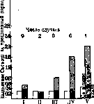
lowers serum calcium (Binger, 1917; Gyorgy, 1924; and Klercker, 1925; Gates, 1927)]. The combination of all these phenomena expresses an alkaline state of the organism, which arose as a result of a violation of the regulatory mechanism with the loss of the action of the parathyroid hormone. Conversely, the introduction of this hormone into the body causes diametrically opposite phenomena (Fig. 6). The connection of mineral metabolism with the nervous system constitutes a topic still very little developed. The influence on mineral metabolism of endocrine organs suggests this connection through sympathetic and parasympathetic innervation. A direct illustration of nervous influences on mineral metabolism can be the experiments of Alpern (1925), who showed a change in the K content in saliva after irritation of the sympathetic nerve, as well as the research of Neuschloss and Trelles (1924), who discovered a difference in the K content in muscles deprived of their innervation compared to muscles with intact nerves. Furthermore, according to experiments by a number of authors, the section of the n. splanchnici, removal of plex. coeliaci cause a decrease in Ca in the serum without tetanic phenomena, while the level of P remains unchanged; section of both nn. vagi increases the level of Ca. Absorption of mineral elements. The mineral components of food are absorbed mainly in the small intestines, where not only free salts but also weakly bound to organic compounds (e.g. with amino acids) disappear from the intestinal canal. The process of digestion contributes to the release of more firmly bound mineral components of food. For alkaline metals and chlorine, very rapid and complete absorption is noted; in a normal human stool, the content of Cl is 0.05 g pro die (Wendt). An illustration of absorption can be the experiment of Heidenhain; a loop of dog's empty small intestine isolated by two ligatures absorbed NaCl solution as follows (see table 19). Obviously, absorption proceeds faster from hypotonic solutions than from hypertonic ones; from hypotonic solutions, osmotic water is quickly absorbed, and the solution concentrates to the salt content in the plasma (about 0.65%), however, NaCl penetrates into the intestinal wall despite its concentration in the intestinal cavity being less than in the blood; the reason for this phenomenon must be sought in the fact that diffusion currents from the intestinal cavity are determined not by the blood's concentration, but by the living epithelium of the villi, the metabolic processes of which are still unknown to us. Hypertonic solutions cause osmotic secretion of water into the intestinal cavity and inhibit salt absorption. In the observed phenomena, the matter is further complicated by the fact that probably the pressure on the intestinal contents, arising from the work of the intestinal wall muscles, forces it to be pushed into the intercellular spaces of the intestinal wall ('filtration' of fluid), similarly to which a special 'pump' of the villi also acts. The transition of Cl into the blood after absorption under ordinary dietary regimes little changes the concentration of this element in the blood - from 0.36% to 0.38%. Arterial blood is richer in chlorine than venous blood. Short-term hyperchloremia (e.g. 0.407% Cl in blood against 0.364% at the beginning) is observed only after a large dose of NaCl. The general water-salt regime affects the level of Cl in the blood; prolonged low salt content in food reduces the Cl content in blood to the lower limits of its normal fluctuations. Constant consumption of very salty food increases the Cl content in blood. In the first hours of digestion, some decrease in Cl in blood is observed under the influence of increased secretion of gastric juice. Similar to chlorine, sodium and potassium after absorption, depending on their content in food, the rate of absorption and water regime, can increase their concentration in blood for some time (normal fluctuations of Na and K in plasma go within the limits of 0.28-0.35%, resp. 0.016-0.024% in humans and close to these numbers in other mammals), with a clearly expressed antagonism between K and Na being noticeable: an increase in the content of one of them in blood reduces the content of the other. The excretion of chlorine and alkaline metals from blood occurs mainly through urine, as well as by sweating; with significant sweating, the body loses not a small amount of chlorides; according to Tuteur, up to 4% of total chlorine loss goes through the skin. With very profuse sweating due to mechanical work at high temperature (e.g. when ascending a mountain in hot weather), according to Cohnheim, Tobler and others, up to 10 g NaCl was lost in a few hours. According to Aggazzotti, during a mountain trip, the loss of Cl through the skin was so great that for several days NaCl did not appear in urine at all and significant hypochloremia was observed (up to 0.43% NaCl in blood, resp. 0.17% Cl).
table 19. The question of the assimilation of calcium and phosphorus is difficult, since both these elements are excreted from the body not only by the kidneys but also by the intestines, and by the latter path very significant amounts of these elements are often removed; therefore, in ordinary studies of Ca and P assimilation, when a comparison is made of their content in stool and in food, it is impossible at all to determine what part comes from unabsorbed food salts and what part was formed as a result of the excretory function of the intestine. From experiments proving the ability of the intestine to absorb Ca and P, besides old experiments (Voit, Forster, Raudnitz; 1855-1893), one can also point to the experiments of Aristovsky, Walsh, Ivy (1925-1928) with fistula dogs; it turned out that resorption of Ca and P occurs in the ileum, and for P also in the jejunum, but 40-50% of the introduced elements leave the small intestines without being absorbed. Table 20. Food: 600 cm³ of milk In the chyme through the fistula was excreted Percentage of excreted to introduced Ca Ca Ca % 0.595 0.595 0.543 0.578 0.83 0.83 0.84 1 0.84 | 0.224 0.219 0.244 0.230 0.308 0.458 0.493 0.412 37 36 45 39 37 55 58 47 600 i meat | 1.34 1 1.34 0.083 | 0.083 ! 0.092 | 0.089 0.187 0.159 7 7 224 190 From table 20 it is seen that with calcium-poor food, a very significant loss of this element through the fistula is obtained, which illustrates the excretory ability of the intestine. Often in humans too, a significant excess of Ca in feces over the amount in food was observed. In the experiments of Aub, on average over a 3-day period, fecal Ca (in 13 individuals) was 0.60 g against 0.33 g of Ca in food. Similar observations reveal the excretory function of the intestines also with respect to P (table 21). Table 21. Authors Ttigerstedt P food |
0 R of feces 0.134 ►0.223 0.229 0.065-0.044 In experiments by R. Berg (1911), increased excretion of Ca and P by the intestine continues for some time after the intake of Ca and P in food ceases. Ca absorbed through the intestine does not cause noticeable changes in the Ca level in the blood; only very large doses of Ca (up to 30 g of anhydrous CaCl2) cause a slight increase, so that the Ca content in blood plasma is very stable (9-13 mg% in almost all mammals), which indicates the active work of calcium depots (bones), which quickly respond to changes in calcemia. As for phosphorus, its intake in food, although not always, causes an increase in P in the blood; in rabbits and small children, a gradually increasing phosphatemia has been traced after administration of phosphates, reaching a maximum (up to 180% of the original) after 2-3 hours. The fluctuations of free phosphates in blood undergo very significant variations, according to various authors from 4 to 18 mg%; general nutrition also has an effect; with a phosphorus-poor diet, inorganic phosphates in the blood lower their level. Like other elements, iron absorption occurs in the small intestine; this has been proven by various methods, including morphologically (microchemical reaction for iron). (Regarding the question of the forms in which iron is absorbed, see Iron.) As with the previous elements, the same fact very complicates the solution of the quantitative problem of iron assimilability, namely, that in cases of secondary erythropenia, normalization was achieved by the intensified introduction of iron with food (see below pathology of exchange), which speaks for the influence of dietary iron on erythropoiesis. With normal blood condition, an increased supply of food rich in iron can increase the content of this element in its depot tissues (bone marrow, spleen, liver) without affecting the Fe content in the blood; thus, when mice were fed iron, its content in their bodies increased 3 times compared with control animals. Absorbed and passed into the blood mineral substances are distributed throughout the body, pass into corresponding depot tissues, increasing their mass, replace gaps that have arisen in other tissues, and the inorganic materials that have not found application leave the body by the above-mentioned paths. Different tissues make different demands on the mineral components of the body, characterized by a more or less constant composition. It is most constant in bones, although here different studies give heterogeneous numbers and even in the same animal symmetrical bones show different content of mineral elements. Tissues Bone.........j 1780 Striated muscles........ 80 Smooth muscles . . . i 112 Brain..........100-180 white matter . . . . ! - gray matter . . . . I - Liver.........! 60 Skin (to dry matter) . ! 100-143 Spleen.......| - Blood (see Blood)
that the intestine is at the same time both an absorbing and an excretory organ for iron. It is assumed that the stomach and small intestine perform resorption, while the large intestine performs excretion of iron, however, there are actually no solid grounds for such a distribution of roles in the intestine. In addition to the intestinal epithelium, Fe is also excreted by bile, in humans up to 1 mg pro die; this value is considered constant, not depending on the influx of iron in food or parenterally, since it comes from the breakdown of red blood cells; it is possible that this iron in the small intestine is partially reabsorbed. According to Hofmeister's calculations, from the breakdown of red blood cells, 40-50 mg Fe should be released, of which 1-2 mg is excreted by urine, and the rest through the intestine. Iron absorbed in the intestine penetrates both into the blood and into the lymph; this has been traced morphologically (microchemical reactions), however, it has not been possible so far to notice enrichment of these fluids with iron even under conditions of maximum absorption; the point is that blood plasma and lymph are considered practically free of iron (according to Erben, 1 kg of blood plasma contains 23 mg Fe; 1 kg of white blood cells - 95-122 mg) and in ordinary blood samples the addition of absorbed iron cannot be accounted for. As for the amount of Fe in whole blood, it depends on the Fe content in red blood cells (see Blood) and on the number of the latter in the blood. It is difficult to trace the effect of various dietary regimes on the Fe content in the blood, only in rare cases of experimental anemia and experimen-
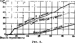
Table 22. Content of mineral elements in some tissues (according to various sources) in mg%. Na Ca Mg P Fe Cl K Bone 320 250 240 400 58-119 7 940 7,5 5 2-17 26-78 5-15 12-21 8-14 9-17 7-10 15 1 2 5-10: Cl 70 120 150 3-60/100-200 - '240-480 72 161 p 3 850 - (224) (9) (210) (92) Bone is characterized by great constancy of its composition, which at one time caused attempts to establish a certain chemical "formula" of bone substance (Hoppe-Seyler). However, according to Hofmeister's view (1911), the observed constancy of bone composition depends on the constancy of the composition of the medium nourishing it - blood fluid. Improper nutrition of the body, different types of salt starvation, can change the content of mineral compounds of bone (see below pathology). A change in blood pH in the acidic direction causes dissolution of bone minerals and increased excretion of Ca and P through urine, which is characteristic of acidosis. "Artificially" induced acidosis (administration of lactic acid, oxalic, weak sulfuric, etc.) leads to impoverishment of bones with lime. From the table it is clearly seen that bone is very rich in mineral elements and is indeed a depot tissue for these elements, especially for Ca and P. During starvation, bone tissue "melts" strongly, losing its mineral elements (decrease in bone ash) (Morel, Mouiquand et al.; 1921-1925) and in particular Ca and P (Iwabuchi; 1922). A long-term deficiency of calcium in the diet can lead to complete resorption of some bones (Lavrov, Yarusova). Of course, in addition to bone mass, mineral elements are also released from other tissues breaking down in the order of catabolism. Guided by the loss of protein in the body (by nitrogen) and assuming that this protein comes from the destruction of muscle mass, one can calculate how many mineral elements should be released in this process (knowing the mineral composition of muscles) and compare their amounts with those of urine and feces of the subject. In the table (observation by Gamble on a starving girl), each mineral element is expressed in decimilliequivalents, in other words, in equivalent amounts of cm3 p/10 solution. Table. Periods of hunger (3 days each) Na Mg o,™ £ « o o i° я io o S3 if! o м си п o B ев So й- аю art O) '<* £ Я CD E- >s) Мм K <D Од * Ив Яо да и 13,3 11,5 10,3 0,3 0,9 1,1 0,7 1,0 1,0 « 0,6 1Д 0,9 ]_ From table 23 it is seen that for K and Mg the ratio of found to calculated is very close to 1; this indicates that the muscle mass destroyed during starvation is indeed the source of these elements, whereas for Ca the found number exceeds the calculated one more than 10 times; therefore the source of Ca in this case must be another tissue; such a tissue is bone. Comparing the mineral composition of other tissues, we see that most of them are characterized by a relatively high content of potassium and poverty of calcium; in terms of iron content, the first places are occupied - still significantly inferior to bone - liver and spleen, which in relation to this element are depot tissues; significant fluctuations in Fe in the liver depend on the "delivery" of the element to this organ, and this "delivery" can be exogenous (from food) or endogenous (from other organs).- The skin has also attracted the attention of researchers due to large fluctuations in its mineral composition. The skin has made it possible to discover a certain dependence of alkali metals and chlorine on the type of food. Some authors therefore consider the skin as a depot tissue for the aforementioned elements. In addition to the elements indicated in the table, rarer elements have been found in different tissues, the appearance of which seems to be accidental, e.g. Zn (3-5 mg%), Cu and Al in muscles, Zn up to 15 mg% (human) and 34 li% (horse) in the liver, there also Cu up to 12 mg%, Sn 10 li%, Mn, As, B, F and others in fractions of li%. The significance of mineral elements in nutrition is not limited only to the absolute level of one or another element in the body or food, but also to the ratio of these elements to each other. Thus, L. Redin noted the optimal ratio of Na : K as 5:1 (on white mice). It has also been clarified that Ca has a beneficial effect on the utilization of Fe in the body: with an increase in Ca in food, it is possible to maintain iron balance at lower levels. As for the question of mutual influences of mineral elements, cases of antagonistic phenomena are related. Thus, with well-balanced diets, administration of magnesium causes loss of Ca in the body; the unfavorable effect from Mg injection disappeared after subsequent Ca injection. Ca in general can correct the disturbance of inorganic balance; in any abnormal effects caused experimentally by alkalis (Na, K, Mg), Ca acts in a restoring (balance) manner. This also includes the classic experiment by Bunge, who caused abundant excretion of Na from the body by administering a large amount of K. Based on balance observations over pregnant women and on the basis of data from analyses of human fetuses at different stages of development, the following diagrams for P and Ca could be compiled (fig.7 and 8). The ratio of mineral acids to mineral bases in the blood fluid and in the body in general. Comparing analyses of various researchers giving the mineral composition of human plasma or serum, some authors (Heubner, Kramer, Marrack and others) could compare the acid and base equivalents of the blood fluid (plasma). The difference between acids and bases presented in milliequivalents for plasma is 151-135 = 16 milliequiv. (according to other authors from 10 to 25) in favor of bases, with acids (135 milliequiv.) being fully covered by sodium (139 milliequiv.). The excess of alkalis (16 milliequiv.) is probably associated with acidic proteins, possibly also with higher fatty acids. Such a setting in the body, close to neutrality [pH=7.36-7.32 (37°)], is very important for a number of physiological intermediate metabolism processes (see Alkalosis and Acidosis). All cases that disturb this setting require a corresponding reaction from the body; first of all, the buffer system is used, then the work of the respiratory center, which by accelerated breathing evacuates CO2, then the dissolution of reserve alkalis from tissues occurs, and finally - the cleavage of ammonia from amino acids (resp. proteins) as a neutralizing base; - in this last reaction the liver plays an essential role, and in all stages of acidosis development also the kidneys, insofar as they are capable of excreting acidic urine. A priori one could imagine that food containing different ratios of mineral acids to bases is capable of producing certain shifts of the pH norm in one direction or another, and only by the compensatory work of depots and excretory organs can this norm be maintained. From this point of view, it is by no means indifferent to the body what the ratio of acids to bases is in food. This question began to be studied long ago in America in Henry Sherman's laboratory (1911-12), and in the first place the research concerned ordinary foodstuffs to establish their "acidity" and "alkalinity". For illustration, a brief table from Sherman's works is presented.
Products Products of acid elements in cm* per 10 g of acid per 100 g of product Products Corn ..... Bread ..... Rice ..... Eggs ..... Egg white Egg yolk Meat ..... Fish ..... Oat flour Rice ..... Whole wheat Wheat flour Apples ..... 6.95 7.81 9.89 11.10 5.24 26.69 0.05-13.91 9.5-16.0 12.93 7.8 ! 9.66 ! 11.6 ! Beans ...... Beets ..... Cabbage ..... Cauliflower Milk ..... Oranges . . . Peas ...... Potato. . . . Plums ...... Radish ..... Raisins ..... Turnip ..... From table 24 it is seen that acid products include meat, eggs, grains, and alkaline products include milk, fruits, vegetables. T. o. a new aspect was introduced into the evaluation of food. It was necessary to determine what the optimum ratio of acids to bases in food should be. However, to this day neither physiology nor clinic nor animal husbandry have yet provided definite solutions to this question. On the other hand, the question of human need for one or another mineral element has long been studied in physiology, and at the basis of all research lay the assumption that for a person of a given age there should be some minimum and optimum for each mineral element. Such a formulation of the question is essentially incorrect, since besides the ratio of acids to bases in food, it is necessary to study the complex tangle of dependencies of the need for mineral components of food on its organic components, on the water economy of the body, on the relationships of alkaline elements to each other, on the compensatory abilities of the human body with respect to the establishment of equilibria in the system: 'depot-tissue-blood-consuming and excretory organ', and these abilities in turn are connected with professional habits, exercises and generally with all living conditions. Therefore, all indications by authors of the level of human needs for individual mineral elements must be considered a provisional solution to the question. Thus, Sherman conducted 97 experiments on people with the aim of determining at what level of calcium (also with respect to other elements) equilibrium could be achieved [i.e. when the balance of Ca (Ca food - Ca excretions)=0]. It turned out that in different subjects the minimum level of Ca suitable for establishing equilibrium varies extremely-from 0.27 to 0.82 g per person per day. An average value was taken: 0.45 g. Similar experiments were conducted for phosphorus, where the average was 0.88 g per person per day (variations 0.52-1.20), and a small number of experiments for iron, giving variations from 6 to 16 mg per person per day; here they stopped at a rounded number-15 mg. Of course, Sherman for both phosphorus and calcium proposed increased values, namely for Ca--0.68, for P- 1.32 g per person per day. These values have been accepted in America as GIG. NORMS.
B. Lavrov.
Pathology of mineral metabolism. Mineral salts are a constituent part of tissues and fluids; their plastic role is revealed during starvation, when together with the excretion of products of the breakdown of organic components of the body, mineral salts are also excreted. Deprivation of mineral salts leads to the death of the animal. A prolonged predominance in food of some salts and the absence of others, apparently to a certain degree
can change the mineral composition of the organism (demineralization according to Noorden). Some authors believe that a certain ratio of mineral salts in food with an excess of acid or basic valences can influence the general metabolism in the organism (Bürgi), the course of disease processes, and the healing of wounds (Sauerbruch, Hermannsdörfer). Mineral salts have regulatory significance. Table 24. Excess of alkaline elements in food per 100 g of product 23.8 10.9 4.3 5.3 2.37 6.61 7.0 5.5-7.2 24.4 2.9 23.7 27.7 for maintaining the constancy of the reaction of tissues and body fluids (see Buffer properties, Reserve alkalinity). Thanks to the buffers of blood and tissues, the accumulation of acids or bases in the organism does not lead to a change in the active reaction of the blood (compensated acidosis, compensated alkalosis). A shift in the active reaction in the acidic or alkaline direction is extremely rare, usually shortly before the death of the organism. But in compensated acidosis, despite the unchanged pH of the blood, there is a change in the composition of the blood: a decrease in reserve alkalinity, a decrease in carbon dioxide tension, a decrease in the CO2 binding curve, and in addition, the amount of bases, mainly Na, decreases in the blood. Sometimes in diabetic acidosis, such losses of Na make it impossible to combat acidosis only with the help of insulin, but require the administration of NaHCO3. The reverse process occurs in alkalosis. Mineral salts regulate the osmotic state (see Osmotic pressure) of tissue fluids, especially blood. A decrease in the freezing point of blood from A=-0.56° by a few hundredths of a degree often indicates significant disturbances in the organism. A transient increase in osmotic pressure is observed with large losses of fluid (sweating when working in hot shops, in cholera), and for a long time—mainly in kidney insufficiency. The concentration of mineral salts (electrolytes) is of great importance for the stabilization of colloids and has an influence on the state of excitability of cells. Thanks to the research of Ringer and Loeb (Ringer, J. Loeb), it has been established that the correct functions of cells are maintained by a mixture of K, Na, Ca, Mg ions in certain ratios: a change in these relations can lead to excitation or paralysis. Changes in these relations are closely related to diseases of the endocrine glands and to disturbances in the activity of the nervous system. Kraus and Zondek (Kraus, Zondek) associate the action of K with the vagus nerve, the action of Ca with sympathetic innervation. A decrease in Ca and an increase in K in the blood is observed in tetany, both parathyreoprivic and idiopathic and guanidine; changes in electrolytes are accompanied simultaneously by alkalosis; a decrease in Ca is found in hyperthyroidism, an increase in hypothyroidism. Changes in the composition of mineral salts in the blood occur in allergic conditions, infections, and fever. Often it is not the absolute content of one or another ion that is important, but the relationship between two ions, for example -^ in arteriosclerosis (see Blood). Special exchange of various mineral salts and ions. Sodium chloride. The retention of sodium chloride and its excretion are closely related to water exchange (see below) (see Edema, Nephritis). In lobar pneumonia and many febrile processes, there is significant retention of NaCl: the urine contains almost no table salt; simultaneously in the blood the amount of NaCl not only does not increase, but on the contrary, its content decreases (Monakov).-Calcium. During starvation, the amount of Ca excreted in the urine increases, that excreted in the feces decreases. Parallel to the increased excretion of Ca, the excretion of Mg decreases. The opinion about demineralization, loss of Ca in t.b.c. has not been confirmed. The exchange of calcium is closely related to the exchange of phosphorus: bones are the main place of deposition of calcium phosphate. In addition to calcium phosphate, bones also contain calcium carbonate, which can be extracted from bones when there is a large amount of acids in the organism to saturate them (e.g. in diabetes). In pathological processes associated with the skeletal system and characterized by insufficient ossification or melting of bone tissue, as in rickets and osteomalacia, the amount of Ca and P in the bones decreases, but the ratio of Ca++ and PO4'" does not change. In the florid period of rickets, the excretion of Ca and P is increased (Ca and P in the blood, see Blood). Simultaneously with the increased excretion of Ca, there is retention of Mg. In osteomalacia, the excretion of Ca and P does not appear uniform; in the bones, as in rickets, there is a decrease in Ca and P, but their ratio does not change (see Blood). In various forms of calcification and ossification (myositis ossificans, calcinosis, etc.), the relationship between Ca and P and CO2 is the same as in normal bones. The deposition of lime salts is apparently a secondary process; primarily a regressive change in tissues develops. The deposition of lime salts is often found in various parts of the body damaged by infection or toxic effects (tuberculous lesions, thrombi, arteriosclerotic vessels). The excessive administration of Vigantol (vitamin D) in recent times often leads to the calcification of vessels, which may depend on the fact that vigantol changes the connective tissue in which lime salts are then deposited. Phosphorus. During starvation or insufficient nutrition, the amount of phosphates in the urine increases; by calculating the ratio of excreted N:P, it was found that the phosphates in the urine are of both organic and inorganic origin. Large losses of phosphorus occur when feeding with thyroid preparations and in Basedow's disease. Retention of phosphates is observed along with retention of chlorides in acute febrile diseases, in nephritis (see Blood). Phosphaturia is not a disease of exchange in the narrow sense; it, like oxaluria (see below), belongs to anomalies of their form of excretion. Phosphaturia is the excretion of urine in which phosphates are contained in the form of sediment. The sediment is usually amorphous [calcium phosphate, Ca3(PO4)2, calcium carbonate], partly crystalline [CaHPO4, calcium hydrogen phosphate; triple phosphate-Mg(NH4)PO4]. The urine usually has an alkaline reaction. There is no direct dependence between the formation of sediment and the total amount of phosphates in the urine. According to Sendtner, Tobler, in phosphaturia the amount of Ca in the urine is increased, the amount of phosphates is normal. Apparently the precipitation of phosphates depends on the state of the colloids of the urine (Lichtwitz). Some authors (Minkowski, Lichtwitz) consider phosphaturia as a secretory neurosis, expressed in the loss of the ability of the kidneys to excrete acidic urine.-Oxaluria is the appearance in freshly passed urine of crystals of calcium oxalate. The increased precipitation of oxalates in the sediment does not depend on the concentration of oxalic salts; the precipitation of calcium oxalate can occur in acidic, alkaline, and neutral urine. Klemperer and Fischler (Klemperer, Fischler) consider the solubility of oxalates to depend on the content of magnesium salts, but magnesium salts do not increase the solubility of oxalates, although they can to a certain extent suppress oxaluria. Lichtwitz believes that oxaluria, like phosphaturia, depends on the state of the colloids of the urine. Yu. Gefter. Path.-anat. manifestations of disturbances of mineral exchange can be twofold: either it represents a result of a decrease in the amount of salts in the tissue or it is expressed in the deposition, precipitation of salts in some tissue or in a cavity. The first concerns calcium, a decrease in the amount of which in bone tissue occurs in a number of special 'malatic' diseases of the skeletal system, giving characteristic path.-anat. pictures (see Osteomalacia, Fibrous osteitis, Rickets). The precipitation and deposition of salts can also concern calcium (see Lime deposits, metastases), as well as other salts (see Gout, Concretions). Path. anatomy of disturbances of iron exchange - see Anemia, Hemosiderin, Hemochromatosis, Iron deposition. VIII. Water exchange. O. v. water exchange is essentially the main type of intermediate exchange, since water is the main constituent of the organism. In adults, water constitutes 58-64% of their total body weight, in newborns it reaches even 74% or more. Along with this, almost all chemical processes taking place in O. v.—diffusion of soluble substances, the influence of osmotic forces, swelling and deswelling of blood and tissue proteins, the formation of secretions and excretions—take place either in aqueous solutions or with the indispensable participation of water. In individual organs and tissues, there is an extraordinary stability in the percentage content of water. Of the approximately 50 substances that make up the composition of urine and blood, only a few can be dissolved in other solvents besides water (Henderson). In addition, the water molecule, due to its dipolarity, is an excellent dielectric (see), which creates conditions for the best dissolution of electrolytes and their dissociation in it (see Electrolytic dissociation).
Water also possesses maximum (after mercury) surface tension, which explains the variety of phenomena of adsorption from aqueous solutions. Finally, the dipolarity of the water molecule and the instability of its compounds determine its dispersing effect on colloidal suspensions (see Gels). The maximum water content is found in the kidneys (82.7%), in muscles (75-80%) and in nerves (up to 82%), while the minimum is in adipose tissue (up to 29%) and in tooth enamel. In absolute terms, muscles contain about half of all water in the body (Table 25), and it remains unclear to this day whether muscles are the site of accumulation of the body's water reserves, its depot, or, what is more likely, water serves as a constant participant in the very intense biochemical processes occurring in muscles. The water content in organs is maintained with exceptional constancy, since the artificial removal or loss of 10% of the total amount of water leads to the death of organisms. Table 25. Percentage of water content in the main organs of humans and distribution of water in them. Organs and tissues Blood..... Fat...... Skin...... Bone tissue Muscles . . . . Nervous tissue Intestines..... Heart ..... Liver ..... Lung ..... Spleen . . . Kidneys..... Other..... Quantity-Percentage
Percentage of water in % of body weight 77.9-83.0 29.9 31.9-73.9 22-34 73.0-75.7 75-82 73.3-77.0 79.2-80.2 68.3-79.8 78-79 75.8-86.0 77-80 4.9 23 6.2 16 37 2.7 4.7- 6.6- 9.0- 47.7-2 3 2 1 2 0 0 11 - 9.0 ,3 -11,0 -12,5 50.8 .7 ,2 ,5 ,8 ,4 ,4 ,6 ,0 In intermediate metabolism, water may participate either as a solvent for molecular particles or as hydration water that is part of the proteins of plasma, cell protoplasm, lymph, and intercellular substance as hydrophilic colloids (see Gels). The water acting as a solvent is characterized by great mobility, 'fluidity,' and is easily taken up and given off by tissues ('intercellular' water); however, the hydration water, especially in cell protoplasm ('constitutional' water), is the main component of tissue colloids, and its loss is sometimes equivalent to the death of that tissue. At the same time, it is undoubtedly true that there is no 'free' water circulating in the body, since even the most pathological transudates are colloidal solutions that also contain some salts. In terms of its origin, the water participating in obliteration can be of different natures. Most of it is absorbed from the intestine and enters the blood, lymph, and tissues - so-called food water. Furthermore, in the process of metabolism and primarily in oxidative processes, for example the oxidation of carbohydrates and fats, water is formed within the body itself - so-called metabolic water; water is also formed as a result of neutralization reactions. Finally, tissue and blood proteins, under the influence of hydrostatic and osmotic forces, can swell (Entquellung of German authors), giving up their constitutional water. At present, all the guiding forces and paths by which intermediate water exchange occurs are far from being fully elucidated. Thus, water taken orally is not absorbed in the stomach, however, its entry into the stomach is already sufficient to cause a vasomotor skin reaction and increased skin perspiration. With the same water passing into the intestine, its absorption begins, the rate of which is determined by the ionic and molecular composition of the liquid consumed. Furthermore, the food water absorbed from the intestine does not immediately enter the general bloodstream and lymph, but is partially delayed in the liver, and the degree of this delay depends both on the nature of the solution in which the water is absorbed (water per se, saline or sugar, hyper- or hypotonic solutions) and on the state of the liver parenchyma and its circulatory system. From a physiological standpoint, it remains unclear whether this 'lock' role of the liver for water is due to the specific structure of its venous system (easy permeability of its capillaries, presence of a sphincter or venous valves at the site of exit of hepatic veins from the liver) or whether the cause is the exceptional looseness of the liver parenchyma, the ability of its cellular elements to swell, and finally the presence of a huge number of tissue spaces in the liver. A significant portion of the water absorbed from the intestine, especially after abundant drinking, passing through the liver, enters the circulatory system. However, the insignificance and short duration of hydremia (see) even after large water loads (1-1.5 liters) indicate that almost all the absorbed water is quickly removed from the blood. By means of weighing and accounting for diuresis, one can easily verify that the main mass of the water consumed passes into the tissues and lymph and is then gradually partially excreted in urine. It deserves attention that pure water is more easily removed from the blood, resp. causes less hydremia, than an isotonic solution of table salt. The further movement of water through the vascular wall and then through semipermeable cell membranes is carried out mainly by three forces: the hydrostatic pressure existing in the vascular bed, the osmotic pressure on both sides of the capillary wall, and the oncotic pressure of blood colloids and tissue fluid. It is quite obvious that all attempts to explain the exit of water from the circulatory system solely by its filtration under pressure (Ludwig) or by the influence of osmotic forces (Starling) have proved futile. Moreover, it should be taken into account that the three guiding forces of water exchange have their point of application in the capillary endothelium, resp. cell membranes, which are by no means completely subject to all those regularities that were obtained in model experiments in vitro (Loeb). While lymph, which is a filtrate of blood plasma, contains a corresponding amount of molecular particles from the blood plasma, the cerebrospinal fluid, the peritoneal serous fluid, and in pathological conditions also the edema fluid, have a quantitative salt composition very different from that of blood plasma. Physical-chemical biology explains these deviations, teaching that the active reaction of the medium on both sides of the membrane, the qualitative and quantitative composition of electrolytes determine the permeability of the mentioned 'barriers' (hemato-encephalic, peritoneal, pleural, etc.) for a number of ions and non-electrolytes, and with them for their solvent - water. Along with this, it is also noteworthy that in most organs and systems, direct water exchange between the blood and parenchymal cells is generally impossible, since everywhere between them there is a greater or lesser amount of connective tissue. The latter has the role not only of supporting tissue: the colloids of its elements have an exceptional ability to adsorb acid valences (histochemically they are acidophilic), as a result of which there is a rapid influx of fluid with positively charged H-ions into the intercellular substance and its swelling. Moreover, recent research (Hulse, Hueck) has questioned the very existence of tissue fluid in the intercellular spaces. From this point of view, the colloids of the intercellular substance (with the help of the so-called 'tissue current') themselves transport nutrients to the cellular elements and return the dissolved metabolic products back to the blood. In other words, connective tissue is the most important active physico-chemical factor of water exchange, extremely sensitive to changes in ion content in the surrounding tissues (blood or organs) and in turn influencing this environment (see also Hydrophilia, hydrophilicity). Numerous observations on the stability of erythrocytes in solutions of neutral salts and on tissue cultures show that the swelling and shriveling of cells depend primarily on the osmotic pressure of the surrounding medium. However, in this exchange with the environment, not all the water that makes up the cell participates, but only that mobile part which acts as a solvent; the constitutional water, as mentioned, is given off by the protoplasm with great difficulty and only before the death of the cellular structure. Therefore, the degree of cell swelling is not in direct proportion to the concentration of the surrounding solution (resp. tissue juices). In addition, on the surface of the cell itself there occur complex processes of adsorption and changes in surface tension, which determines the permeability of cell membranes or their boundary layers. These membranes themselves can either facilitate or hinder the implementation of osmotic processes in the cell and the exchange of water in it. Elucidation of such influences is at present difficult at least because all those physico-chemical phases and states in which the cell protoplasm and in particular its boundary layer are found are little known, as well as what forces hold water in cells. Subjected to osmotic influences, the cell protoplasm, which itself carries a certain electrical charge but is not capable of diffusion, participates in the so-called anomalous osmosis, i.e., creates unequal concentrations in the surrounding fluid and inside the cell for a number of anions (Cl, HCO3). In other words, the distribution of these ions on both sides of the cell membrane - between the tissue fluid and the cell contents - occurs unevenly according to the laws of the so-called Donnan equilibrium (see Donnan equilibrium). The water balance of the body consists of water introduced with food or formed as a result of the vital activity of organs and tissues, on the one hand, and water excreted through urination, perspiration (through lungs, skin), on the other. The introduction of water into the body is regulated by a special physiological sensation - thirst, the degree of which is determined primarily by the content of osmotically active substances (resp. molecular concentration) in the blood and tissue fluids (see Thirst). As for the excretion of water, in physiological conditions it is in strict accordance with the amount of water introduced. With abundant drinking, this regulation is carried out quickly (mainly by the kidneys). The resulting diuresis is the result of the shifts that occur in the water-salt economy of the body. These shifts are sometimes imperceptible and not always related to changes in blood composition; thus, the degree of urine excretion is not in direct dependence on the dilution of the blood.
Along with the kidneys, however, extrarenal water excretion can also have a significant influence on the water economy of the organism, which occurs mainly at high external temperatures (climate, conditions, work in a hot workshop) or during fever. The evaporation of water from a relatively large body surface plays the role of a thermoregulator, which under appropriate conditions helps maintain body temperature at a certain level. At the same time, it must be emphasized that in case of excess water in the organism, extrarenal excretion sharply increases even at normal ambient temperature, and under pathological conditions, for example in hyperthyroidism (especially experimental), the role of extrarenal excretion in water balance sharply increases, reaching up to 50% of all excreted water. However, even under normal conditions, the amount of water removed by extrarenal pathways can reach 800-900 cm3 per day in humans. Of this amount, 500-600 cm3 is excreted through skin perspiration or sweating, while the remaining amount is in the form of water vapor in exhaled air. Incidentally, the qualitative distinction made in relation to water balance between perspiration and sweating is poorly justified, since the former is carried out by the same sweat glands, and is only invisible to the naked eye. Water loss through the skin provides broader possibilities for regulating water economy than respiration, since skin perspiration is directly dependent on the extremely sensitive to temperature fluctuations, labile and amenable to fine regulatory innervation apparatus. The amount of water in exhaled air, which has a fairly constant temperature and therefore stable vapor pressure, is subject to much smaller fluctuations. The ability to maintain water economy at a certain level, resp. to equalize water balance, under physiological conditions can be studied by means of the so-called "water test." Upon introduction of 1000 cm3 of water, the latter is excreted in excess within 3-4 hours, with 80-% being eliminated renally in adults and 70-80% in children (see also metabolism in children and Kidneys, functional diagnosis). However, the results of the water test are in the closest dependence on preliminary nutrition and in particular on preliminary saturation of tissues with water and salts. Thus, in rabbits fed "dry" food (for example oats), the water test is eliminated much more slowly than in those fed water-rich carrots (Siebeck). All this applies to an even greater degree to humans, who consume extremely varying amounts of liquid and NaCl (from 5 to 20 g per day). Of the ions Na+ and Cl- making up its composition, the former undoubtedly has a water-retaining property, since other salts containing Na+ cations also promote water retention (NaHCO3, Na2HPO4). Strangely enough, the opposite (diuretic) effect is exerted by cations K and Ca, which also carry a positive charge, as well as their salts and food substances containing them (potatoes, fruits). Consequently, the nature of preliminary nutrition, not only in terms of its water content but also its mineral composition, has a decisive influence on the course of water exchange. All this merely indicates that water exchange should not be considered as a process occurring independently, in particular outside of connection with mineral exchange. Sometimes in experiments (demineralization, artificial polydipsia, overheating), as well as under pathological conditions, one can observe the development of a prolonged negative water balance; in such cases, dehydration (see.) occurs, accompanied by severe phenomena up to symptoms of severe intoxication. Cases with a positive water balance are no less severe; these are conditions that have occurred after a sharp and rapid retention of large amounts of water in the body. Such conditions can be created by introducing large amounts of fluid followed by an injection of pituitrin (Vovsi, Itsekson and Vagon). The picture observed in this case of "water poisoning" (Rowntree) is sometimes characterized by extremely alarming symptoms (tremor, restlessness, pallor, nausea, etc.). Under physiological conditions, however, in the organism, water balance is continuously and extremely rapidly equalized, creating that relative stability of water economy which is the prerequisite for the normal course of all intermediate metabolism processes. Such regulation is carried out first of all, centrally, which was experimentally confirmed by means of the so-called "water" and "salt" injections. Analogously to Claude Bernard's "sugar" injection, which is accompanied along with glycosuria also by polyuria, Meyer and Jungmann, as well as Leschke (Meyer, Jungmann, Leschke) showed that an injection into certain areas of the gray eminence and intermediate brain leads to enhanced urine excretion. It can be accompanied by simultaneous increased excretion of table salt ("salt" injection) or be limited to excretion of liquid, salt-poor urine ("water" injection). Centripetal impulses are received by these centers apparently either directly from the tissues (see Thirst) or through the hormonal apparatus, whose influence on the function of water-regulating centers is exceptionally great (see below). Of the endocrine glands, the posterior lobe of the pituitary gland, the thyroid gland, and the pancreas play a decisive role in regulating the water economy of the organism. Extracts of the posterior lobe of the pituitary gland (pituitrin and its analogs) have, as is known, a pronounced antidiuretic effect, and it remains unclear whether this effect is due to a special hormone or whether it is a property of a single pituitary secretion (see Pituitary gland). The antidiuretic effect of pituitrin is especially pronounced in cases of abundant diuresis: after water load, after extirpation of the pituitary gland in dogs or in diabetes insipidus (see Diabetes insipidus). In the blood after injection of pituitrin, a more prolonged dilution is usually noted, while in the tissues there is an accumulation of fluid (resp. an increase in body weight). Meyer and Meyer-Bisch (Meyer, Meyer-Bisch) also showed that the usually observed increased flow of lymph into the thoracic duct from the liver when introducing so-called first-order lymphagogues (see Lymph) does not occur if pituitrin is injected simultaneously with these substances. In the latter case, a small amount of thick lymph flows from the fistula of the thoracic duct. Along with this, there are numerous observations that in the kidneys themselves, the effect of pituitrin is manifested by transient but sharp constriction of renal vessels and reduction of renal blood circulation. Therefore, many researchers tend to associate the decrease in diuresis with the influence of pituitrin directly on the kidneys. In any case, the antidiuretic effect of pituitrin is not self-sufficient and is carried out through the central nervous system, not by direct action on the executive organs, since when cutting the splanchnic nerves, when destroying the gray eminences, or finally when turning off the cerebral cortex, this effect either disappears or is sharply diminished. The mechanism of action and the point of application of the thyroid gland hormone, which also participates in water or rather in water-salt exchange, are completely different. The substance of the thyroid gland (thyroidin and its analogs), as well as its hormone thyroxine, have a pronounced dehydrating effect. With prolonged oral administration of thyroidin, a decrease in body weight is usually observed with almost normal diuresis. This weight loss is caused more by increased extrarenal water excretion than by enhanced urine excretion. The dehydrating effect of thyroxine is especially effective when there is preliminary retention of water and table salt in the tissues, in particular in myxedema, in edema in decompensated cardiac patients, in nephrosis, in obesity. For this reason, there is every basis to classify the thyroid gland hormone as a water- and salt-excreting substance with a point of application in the tissues. In particular, the weight loss observed when taking thyroidin in obese people is accompanied not so much by an increase in their basal metabolism as by the removal of large amounts of salts and water from the body. The secretions of the insular apparatus of the pancreas also promote water retention in the body. The retaining influence of insulin, in contrast to the function of the pituitary gland, is manifested, however, only in cases when a certain amount of Na-salts and carbohydrates are introduced into the body along with water. Apparently under the influence of insulin, the enhancement of gluconeogenesis is accompanied by water retention in the liver. Similarly, the enhanced introduction of sodium salts, especially those with an alkaline reaction (bicarbonates, dibasic phosphates), in itself promotes water retention in tissues and even favors the formation of edema. The simultaneous introduction of insulin merely accelerates and intensifies this process. Pathology of water exchange. Under pathological conditions, disturbances of water economy occur in a variety of diseases.
Schematically, all these deviations can be reduced to conditions or symptoms with a positive or negative water balance. A negative balance is observed mainly in childhood in so-called exicosis (see), atrophies, with severe diarrhea (particularly in cholera and infantile dyspepsia), in cachexia, in the initial period of starvation, and with overheating and diaphoretic procedures. In all these cases, the cause of the negative water balance is primarily extra-renal excretion of water. Excessive loss of fluid by the body also occurs in polyurias of various origins, of which diabetes insipidus and diabetes mellitus should be placed first. In all these diseases, the elasticity and vital turgor of tissues, primarily those that serve as water depots of the body, such as connective and muscular tissues, decrease; simultaneously, the blood also becomes dehydrated. The amount of dry residue in the blood (quantity of proteins and salts) reaches high figures. The protein content of plasma reaches 10%, and NaCl reaches 650 mg%. Correspondingly, the freezing point of the blood also decreases. The sharp loss in weight observed in these conditions is due primarily to the loss of a significant amount of intercellular tissue water. However, tissue dehydration does not pass without trace for the colloids of cell protoplasm, especially of parenchymatous organs, as a result of which the enhanced protein breakdown with the excretion of excessive amounts of nitrogen in the urine (negative nitrogen balance) usually observed in the second period of dehydration occurs. Continued urination with a high percentage of salt and nitrogen in the urine creates the prerequisites for the development of the main symptom observed in negative water balance—thirst. In cases with more prolonged or sharply expressed water loss, restlessness, a feeling of heaviness in the head, sensation of heat, pains in the extremities, and sometimes mental excitement are added to the thirst. The described signs of intoxication hardly relate only to water loss, but are rather close to the very characteristic picture described by Fischler under the name of meat intoxication and observed when products of protein breakdown enter the blood. It is interesting to note that all the mentioned symptoms can also occur with the accumulation of significant amounts of fluid inside the body, i.e., with apparently positive water balance, for example, with the increase of ascites in patients with cirrhosis of the liver. In such cases, these phenomena are pathogenetically due to the same tissue dehydration, resp. thickening of the blood, which occurs here just as with significant water losses. Polyurias, which as a rule lead to a negative water balance, can occur from the most diverse causes. The most common and well-known polyurias are in diabetes mellitus and diabetes insipidus, at the beginning of renal insufficiency, in so-called primary polydipsia, etc. (see Polyuria, Nephritis, Nephrosclerosis, Polydipsia). The occurrence of a positive water balance and a sign of water retention in the body would naturally be considered oliguria. However, experience teaches us that in so-called primary oligurias, which are most often caused by nervous or endocrine influences (see Oliguria), the water balance is equalized by extra-renal excretion of water, and thus retention does not occur. Therefore, the conditions that characterize a true positive balance are only those accompanied by a rapid increase in the total weight of the patient with or without oliguria. Water retention in organs is most pronounced during the development or increase of visible or hidden edemas. The latter arise primarily due to a violation of one or several of the factors regulating the intermediate metabolism of water in the body. Thus, congestive edemas are most often associated with an increase in hydrostatic pressure in the venous system and, at the same time, a decrease in the permeability of the capillary endothelium for water, caused by its oxygen starvation (Landis). Renal edemas, especially nephrotic ones, are obviously based on a different genesis and are primarily associated with a decrease in the amount of proteins in the plasma, a change in the dispersity of their particles, a shift in the albumin-globulin coefficient, and consequently a decrease in the colloido-osmotic pressure of proteins, i.e., with a decrease in the force with which proteins retain their water. Finally, shifts in the tissue reaction, the accumulation of certain cations and anions (Na+, HCO3-) in them also lead to significant water retention in tissues (inflammatory, hungry, alimentary, salt edemas). (For more details, see Edema.) A predominantly tissue nature is also apparently possessed by the significant water retention observed in the initial period of various infectious diseases (for example, in lobar pneumonia). Leyden, on a large amount of material, showed that the loss of water in the febrile period is 2 times less than in the post-critical period. The assumption that fever plays a decisive role in this retention is hardly justified, since a similar picture is also observed after tuberculinization. Only it is certain that in these cases water retention occurs in parallel, and perhaps is caused by the sharp retention of salt and the enhancement of protein breakdown observed in these conditions. For the pathological anatomy of changes in water metabolism disorders—see Vacuolar degeneration. Edematous disease, Edema.
m. completely. IX. Metabolism in children. O. v. and energy metabolism in children, especially of early age, in many respects proceeds completely uniquely compared to the metabolism of adults. To obtain sufficiently accurate results when studying O. v. in children, it is necessary, in addition to the punctual observance of the usual general rules for studying metabolism in adults, to also consider the following main points: 1) the group of children under study should be as homogeneous as possible in terms of health status, constitution, age, and method of feeding; 2) the conditions of observation in all cases compared with each other must be completely identical; 3) the duration of observation should be at least 5-7 days; the preparatory period with the child's load on the basic diet should be sufficiently long (3-4 days); 4) it is necessary to take into account the influence of the time of year, lighting, richness of the solar spectrum with ultraviolet rays, and other meteorological moments; 5) special attention should be paid to the careful collection of urine and stool, which in children of early age presents special difficulties and requires a special technique. For this purpose, many different methods have been proposed, of which the best must be recognized as the Bendix-Finkelstein method and the Benjamin method. The position of the child and the urine collector when collecting the daily amount of urine-see vol. XIX (art. 107, fig. 4). Stool is collected on a rubber pad. For studying gas exchange in children, special chambers are available (with Benedict and Knipping apparatus). For blood research, it is mainly necessary to use micromethods. Otherwise, the study of various types of O. v. in children is usually carried out using the same methods as in adults. Energy metabolism (basal metabolism). The energy received by the child and evaluated by the caloric content of the food he consumes is spent on maintaining life on an empty stomach in a state of rest (basal metabolism), on growth and increase in body mass, and on muscular activity. Helmreich gives the following approximate distribution of energy in a child and adult with moderate food: table 26. Distribution of energy On basal metabolism ...... On growth and deposition of substance............... On specifically dynamic action ............ On work for body movement............... On losses with excretes..... In child (in %) In adult (in %) 15 0-5 15 5-10 Basal metabolism in a child depends not only on his live mass, but also on the individually varying intensity of metabolism in the cells of different subjects. In the same subject, basal metabolism differs with amazing constancy and fluctuates within a range of about 10%. There is a certain dependence between the magnitude of basal metabolism in a child and his age, weight, and body surface. To determine body surface in an infant, Lissauer's formula is suitable: S (body surface) = 10.3 × weight^2; for older children, the constant 10.3 should be replaced by others, varying depending on the child's weight; the value of the constant for boys ranges from 10 to 11.5, for girls from 10.1 to 11.1 (Talbot). Lissauer's formula is mainly suitable for normal children. More precise is du Bois' formula: body surface = weight^0.425 × height^0.725 × 71.84. Benedict and Talbot for calculating basal metabolism in newborns (weighing up to 6 kg) propose the following formula: daily calorie production = 12.65 × length (in cm) × 10.3 × weight^2 (in kg). In newborns, basal metabolism is very low and approximately proportional to their weight. According to studies by various authors (Benedict, Talbot, Bailey, Murlin), daily energy expenditure in a newborn weighing 3 1/2 kg is approximately 146 cal., or 42 cal. per 1 kg of weight; the same ratios between energy expenditure and weight are also obtained in children of the first day of life with lower and higher initial weight; by the 2-3rd day of life, energy expenditure per 1 kg of weight increases to 48 cal. and, gradually increasing further, reaches maximum figures by 1 1/2 years (50-60 cal. per 1 kg), and then begins to decrease. In boys aged 2 to 3 years, it falls to 52 cal., by 6-7 years to 42 cal., by 10-11 years to 38 cal., and by 12-13 years to 34 cal. per 1 kg of weight (Benedict). Thus, it can be considered indisputable that basal metabolism calculated per unit of weight is much higher in early age than in older children. The difference between basal metabolism in boys and girls begins to appear approximately from 1 1/2 years; from this age until the period of puberty, energy expenditure in girls is somewhat less than in boys, then the girls' curve overtakes the boys' curve and only with the end of puberty in men does the predominance of metabolism again restore. This sexual difference, one must think, depends on the smaller amount of energetically active tissue in girls, but on the other hand, a larger amount of fatty tissue. Well-nourished children, whose tissues are rich in fat and water, give lower figures of basal metabolism than lean children. During puberty, in some cases, apparently an increase in basal metabolism occurs. Age-related changes in basal metabolism in children, calculated not per age and unit of weight, but per unit of body surface, are characterized by the following figures: in a newborn about 612 cal. per 1 m^2, by the end of the year-about 1,100, by 10 years-1,000 and by 13 years-950. The respiratory coefficient (see) in the newborn period undergoes the following changes: on the first day 0.80, by the third day it falls to 0.73 and then by the 5-6th day reaches 0.81-0.82. 'Specifically dynamic' action of food in early childhood is somewhat different than in older children and adults. According to Helmreich's studies, the introduction of milk with food in small children almost does not cause an increase in basal metabolism, whereas in older children the increase in calorie consumption is quite significant and reaches approximately 10%. During puberty, according to Gottche's data, specifically dynamic action was strongly reduced in 75% of cases and in most cases (50%) was accompanied by an increase in basal metabolism. The regularity of this puberty reaction (Pubertatsreaction), as Gottche calls it, cannot be considered finally proven. There is no doubt that the state of energy metabolism in a growing organism is greatly influenced by the endocrine apparatus. Nitrogen metabolism. Food proteins are absorbed in children mainly in the form of amino acids and partly in the form of polypeptides. The passage of native proteins through a normal intestinal wall is apparently possible only in newborns, when some protein bodies can directly penetrate through an undamaged mucous membrane. Very little is known about the further fate in the child's body of the products of protein breakdown absorbed in the intestine. The process of protein absorption in the intestine, especially in early childhood, occurs very perfectly and hardly depends on the child's age and method of feeding; in breast-fed children, 70% to 90% (on average about 80%) of the introduced proteins are absorbed, in artificially fed children only slightly less. Increased introduction of proteins with food, as shown by the studies of Finkelstein and Jonas, does not impair the process of their absorption. As for the utilization of absorbed amino acids and polypeptides, according to older observations (Langstein, Meyer, Rietschel and others), the percentage of food nitrogen used varies depending on the child's age and method of feeding, while the amount of protein retained in the body depends on the child's age and hardly depends on the amount of food protein. However, the latest studies by Rominger and Meyer show that the retention of nitrogen in the body of a healthy breast-fed child also largely depends on the amount of protein introduced; the child's body, in contrast to the adult's body, has a pronounced ability to increased accumulation of protein, due to which the tissues of an artificially fed child are somewhat richer in proteins than the tissues of a child receiving only breast milk. Somewhat unique relationships are observed in newborns, according to the observations of Langstein, Niemann and Birk, negative nitrogen balance is noted, which quickly and quite completely equalizes with natural feeding with colostrum and is significantly delayed with artificial feeding. The ratio between the amount of protein in food and the amount of nitrogen excreted with feces remains more or less constant; with a normal protein load in a child, about 10% of the introduced proteins are excreted with stool.
According to Pintozzi, the amount of nitrogen in the feces can serve as a measure of absorbed nitrogen, since only a relatively small part of the stool nitrogen is due to the nitrogen of digestive juices flowing into the gastrointestinal tract, and an insignificant amount is due to the final products of protein metabolism excreted by the intestine. The main mass of the latter, as in adults, is excreted in the urine, but they are distributed in it in a completely unique way. The total amount of urine nitrogen is relatively very large in newborns, then it somewhat decreases, only to subsequently begin to increase again approximately parallel to the amount of proteins introduced with food. In breastfed children, regardless of the method of feeding, about 40-60% of the nitrogen introduced with food is again excreted in the urine. The daily amount of urine nitrogen in a newborn averages about 0.5 g, by the end of the first year of life - 1.5 g, at 6-7 years of age - 7.5 g, and by the end of the childhood period about 10 g, which when recalculated per 1 kg of body weight gives: 0.12-0.16 in an infant, 0.45 in a child of 6-7 years, and 0.31 in children during the period of puberty. A certain general idea of the distribution of nitrogen in the urine of an infant can be given by the following example from Kammerer: out of 5.52 g of total nitrogen in the urine of the child under observation, 4.8 g was urea, 0.29 g - ammonia, 0.065 g - uric acid, and 0.013 g - purine bases. According to the latter author, in a breastfed child, urea accounts for about 75-80% of total urine nitrogen, in bottle-fed children - about 84%, and in older children - from 90% to 94%; the age curve of urea excretion undergoes approximately the same fluctuations as the curve of the total amount of nitrogen. Uric acid is excreted in the newborn period approximately 5 times more than in adults and twice as much as in older children, in whom 0.018 to 0.027 g per day is excreted per 1 kg of weight; this value changes little depending on the child's age, but largely depends on the food consumed (Gopport). Purine bases are excreted parallel to the amount of uric acid (Niemann), but in the urine of newborns they are present only in traces. Due to urine ammonia, children excrete more N than adults; thus, in newborns this value is 9.5-19.5% of the total amount of urinary nitrogen (Reuss, Keller), in infancy - 8%, at 3-4 years - 7%, and at 13-14 years - 6%. The percentage ratio of ammonia nitrogen to the total amount of nitrogen, i.e., the so-called ammonia coefficient of urine, fluctuates within wide limits in children (normally from 3 to 10), changing significantly depending on the state of acid-base balance in the body, increasing in the state of acidosis and decreasing with shifts toward alkalosis, and is thus a fairly sensitive indicator of the correct course of general metabolism. Amino acid nitrogen gives high figures in the first days of life; there is especially much of it in the urine of premature infants (up to 9.5-25% according to Gebel'ro); in the infant period this value fluctuates from 2.7% to 4%, i.e., 2-2½ times more than in older children, in whom amino acids account for only about 1.6-1.8% of total urine nitrogen. The excretion of final products of nitrogen metabolism with urine, besides the child's age, is also influenced by his constitutional features (Matorina, Mats, Tur). The distribution of nitrogen in the urine depends on the state of the oxidative capacity of the child's body; as a result of its insufficiency in the newborn, there is increased excretion in the urine of a large amount of products of incomplete nitrogen metabolism, which indicates the body's inability to fully break down proteins and consequently the impossibility of sufficiently perfect utilization of food proteins. The energy of oxidative processes increases with age, reaches optimal conditions by 5-6 years, and then decreases again to the period of puberty. These features of nitrogen metabolism, as well as the basic property of the child's body - the vigorous course of growth processes, possible only in the presence of a large amount of plastic material,-make it necessary to introduce relatively large amounts of proteins into children with food. In artificially fed children, along with a significant delay in the body of introduced nitrogen, there is also increased excretion of it with urine (Rominger and Meyer), and therefore the diet of such children should be richer in proteins than for children fed only with breast milk. Due to the peculiarities of intermediate protein metabolism in children, there is increased excretion of organic acids with their urine: in premature infants and newborns - 10.1 cm³ of lactic acid per day, in infants and preschool children - 12.5, while in adults only 8.2 cm² per 1 kg of weight (Hottinger). According to the opinion of most authors, proteins should cover approximately 10% to 15% of the total calories in children. The protein requirement in a child of the first year of life averages about 2.5 g per 1 kg of weight (in breastfed from 1.5 g to 2 g, in bottle-fed from 3 g to 4 g), at 1-5 years of age it is somewhat higher and averages 3-3.5 g, by 8-10 years it falls to 2.5 g and by 12-15 years to 1.5-2 g; in the presence of certain indications, the amount of introduced proteins can be both increased and decreased within fairly wide limits without violating the normal course of metabolic processes and the correctness of general development. According to data from many German authors, a child's protein requirement is significantly less. For the correct development of the child's body, the absolute amount of introduced proteins is not as important as their completeness, determined by the content and nature of amino acids. To the vital amino acids for a child should be tryptophan, lysine, tyrosine, and cystine, to the less important but in some cases absolutely necessary - arginine, histidine, asparagine, and glutamic acid. Insufficient introduction of these amino acids inevitably leads to the development of dystrophy, and their complete absence is incompatible with life. The requirement for tryptophan in the infant period is 0.058-0.1, in older children 0.055-0.063 per 1 kg of weight. Practically, it is sufficient for about 50% of the introduced proteins to be of animal origin. For proper protein assimilation, a certain ratio (correlation) with other basic food ingredients - fats, carbohydrates, and water - is necessary. An excess of introduced proteins does not improve, and sometimes apparently even worsens nitrogen utilization, and leads to a state of relative acidosis; excessive introduction of fats, according to some authors (Freund, Neumann), worsens nitrogen absorption and retention, according to others (Pintozzi), has no effect at all; carbohydrates, according to Keller, improve nitrogen balance, while according to Pintozzi, they somewhat impair protein absorption and reduce the need for them. A moderate predominance of alkaline equivalents over acid ones in food improves protein assimilation. Insufficient introduction of water, due to the pronounced specific dynamic action of proteins, can lead to hyperthermia (Eiweissfieber). The specific dynamic action of proteins increases with the child's age, i.e., with the increase in body surface area; in small children it is less than in older children; in the latter it is less than in adults (Helmreich, A. Seifert); in boys it is greater than in girls. The maximum of the specific dynamic action of proteins is observed 3 hours after food intake and fluctuates between 9.2% and 31.4% of the fasting value. Fat metabolism in children also has a certain peculiarity. It can be considered established that even in early childhood, fat digestion already begins in the stomach under the influence of gastric juice lipase; the presence of a large amount of lipase in breast milk is of substantial importance for fat digestion in the infant. The process of fat breakdown and absorption mechanism in children basically proceeds the same as in adults. According to Orgler, Keller, E. and F. Müller, and according to the latest data from Potter, in a breastfed child about 98% is absorbed, in a bottle-fed child about 95%, in older children from 95% to 97% of fats introduced with food. The main part of the broken down and absorbed fats in the intestine, entering through the portal vein and liver into the general bloodstream, leads to the appearance of the so-called digestive hyperlipemia; the latter increases throughout the entire process of digestion, reaching a maximum with cow's milk after 3 hours, with breast milk - somewhat later, and in the first case it lasts about 5 hours and in the second about 9 hours. In the blood serum of fasting children, there are 170-200 mg% phosphatides, 140-170 mg% cholesterol, and about 400 mg% fatty acids. In the blood of a newborn, there are significantly fewer lipoids, but by 1-1½ months their amount increases by 50-75% and then slowly but gradually increases during the first years of life. At 3-11 years of age, the total amount of lipoids averages 700 mg%, which almost corresponds to their content in adults. The method of feeding the child does not significantly affect the amount of blood lipoids.
Unused fats in the amount of about 5-10% (according to Freund's observations up to 14-18%) are excreted with the stool in the form of neutral fat, water-insoluble free fatty acids, alkaline (water-soluble) and alkaline earth soaps. Their distribution in the feces of infants and artificially fed children somewhat differs. In a normal infant, the fecal fats contain 10-30% neutral fat, 10% free fatty acids, 40% alkaline soaps, 20% alkaline earth soaps, 0.8% cholesterol, and a small amount of lecithin; in an artificially fed infant, about 35% neutral fat, up to 40% free fatty acids, 10% alkaline soaps, and 13% alkaline earth soaps. In an infant, more cholesterol is excreted with the stool than is introduced with food, which indicates the possibility of its synthesis in the growing organism; however, Knauer considers this assumption unlikely. The distribution of fat in the stool changes depending on the composition of food and the state of intestinal peristalsis. Absorbed fats are partly burned in the body and thus serve as a source of energy formation, and partly, being deposited in tissues, mainly in subcutaneous fat and the mesentery, form a reserve fat depot. The fat of an infant is poor in liquid oleic acid; with age, its amount gradually increases. The growing organism can develop for a relatively long time without fats on proteins and carbohydrates alone, and the latter can be the starting material for the formation of fat deposited in tissues. However, this possibility of replacing fats with isodynamic amounts of carbohydrates only indicates the great adaptability of the child's organism, but does not give the right to the conclusion made by some authors (Pirquet, Greer) that fats are not absolutely necessary for the organism. First, the fat formed in the body from carbohydrates differs in composition from fat in normal nutrition, and second, with a fat-free but otherwise calorically sufficient diet, sooner or later in a child, symptoms of avitaminosis (Bloch, Wagner) appear, xerophthalmia develops, and the body's immune forces sharply decrease (Weigert). The normal need for fats in infancy is relatively large, usually ranging from 3 to 5 g, on average equal to 4 g per 1 kg of weight, after a year - 3.5 g, from 6 to 12 years - 3 g, and during puberty - 2-2.5 g. If, on the one hand, the child's organism can apparently develop correctly with a significantly smaller amount of introduced fats, then, on the other hand, their amount in the presence of certain indications can be significantly increased (e.g., up to 6 g or more per 1 kg of weight in infancy). However, excessive introduction of fats is not indifferent to the organism and easily leads to the development of a state of true acidosis. The need for fats is determined not only by their quantity but also by their quality; the adequacy of fats depends on the content of lipoids and vitamins in them, which should be especially taken into account in the nutrition of young children. The correct course of fat metabolism is possible only with the proper correlation of fats with other nutrients; in an infant, up to 50% of all introduced calories should be covered by fats, in older children only 25-30%. Carbohydrate metabolism. Carbohydrate metabolism in a child, generally subject to the basic laws regulating it in an adult, has a number of characteristic features. The exclusion of carbohydrates from food causes a decrease in body temperature in a child; this fact confirms the enormous importance of carbohydrates for heat production and thermoregulation in the child's organism. The question of the possibility of increasing body temperature in children under the influence of excessive sugar introduction cannot yet be considered finally resolved. Carbohydrates are absorbed mainly in the form of monosaccharides (glucose, levulose, galactose), although the possibility of absorption of dextrins is not excluded. In the blood of healthy infants, the amount of sugar on an empty stomach ranges from 0.07% to 0.09%, in older children from 0.08% to 0.1%, and finally in children 12-14 years old from 0.09% to 0.12%. During digestion, the amount of sugar in the blood increases to 0.16-0.2%; this hyperglycemia quickly equalizes, as part of the sugar is oxidized, used for purely dynamic purposes, part is deposited mainly in the liver and partly in the muscles in the form of glycogen, and finally part is used for the formation of fat. The ability of the child's organism to assimilate carbohydrates introduced with food has a certain limit; if this limit of assimilation is exceeded and the amount of sugar in the blood more or less steadily increases to 0.18-0.2%, the organism equalizes the disturbed carbohydrate balance by excreting the excess sugar with urine. The child's tolerance to carbohydrates is subject to significant individual fluctuations; it depends on the age of the child and the constitutional features of his organism, it is different for different carbohydrates and in general is much greater than in an adult. In an infant, alimentary lactosuria occurs with the introduction of 3.5-4 g of milk sugar per 1 kg of weight; in an adult, only 1 g is sufficient for this. Cane sugar has the same limit of assimilation as lactose; for grape sugar and maltose the limit of tolerance lies somewhat higher, and levulose has a particularly low limit of assimilation. According to Aschenheim, alimentary glycosuria in infants occurs with the introduction per 1 kg of weight of 4 g of galactose, 8-12 g of dextrose, 4-5 g of levulose, 6-8 g of lactose, and 6-8 g of sucrose. These absolute figures have very little value, they change greatly not only depending on the individual characteristics of the child and the state of his gastrointestinal tract, but also on the dissolving medium in which these carbohydrates are introduced. The absorption of sugar in human milk occurs faster, and the glycemic curve rises earlier and falls steeper than in whole cow's milk. An aqueous solution of sugar causes a steep (maximum in 1/2-1 hour) rise and early disappearance (after 2-3 hours) of the glycemic curve. On the glycemic curve, as shown by studies by Styrikovich, are reflected, first, the state of activity of the gastrointestinal tract and liver (the first rise of the curve) and second, the features of intermediate metabolism (the second half of the curve). Proteins, fats and carbohydrates introduced simultaneously with sugar characteristically change the glycemic curve, which depends on the resulting changes in intermediate metabolism. The specificity of glycemic reactions in early childhood indicates the intensity and mobility of carbohydrate metabolism in children and its close connection with the intermediate metabolism of proteins and fats. As one of the intermediate products of glucose metabolism in the body, lactic acid is formed. The glycolytic ability of the organism, as shown by Warburg's research, is of great importance for the growth process. According to Gyorgy, Brehme and Brahdy, in the blood of children in the first quarter of the year there is on average 18.7 mg% of lactic acid, in older infants 13.8 mg%, whereas in adults only 10.2 mg%. Part of the carbohydrates that escaped absorption undergoes fermentation under the influence of bacteria. The lower fatty acids formed in the intestine during fermentation in small amounts are useful and necessary for the organism: they stimulate intestinal peristalsis, improve nitrogen and mineral metabolism and are thus, as Klotz says, catalysts of metabolism. On the contrary, large amounts of these acids act destructively, promoting bacterial invasion into the upper parts of the intestine, suppress metabolism and worsen assimilation. What part of the carbohydrates undergoes enzymatic breakdown and absorption and what part undergoes fermentation cannot be established exactly. In the feces of a healthy infant, carbohydrates (monosaccharides) are absent; with accelerated peristalsis, sugar and starch appear in the feces. Carbohydrates, like proteins, are absolutely necessary for the growing organism and cannot be completely replaced by an isodynamic amount of fats. Even a short-term complete exclusion of them from food is hardly tolerated by the organism, especially if a large amount of proteins and fats is introduced (tendency to acidosis). The minimum amount of carbohydrates necessary for a child is probably individually different and to a large extent depends on the quantity and quality of other food ingredients received by the child. If this minimum amount of carbohydrates is absent in the child's food, the organism strives to replenish it at the expense of protein breakdown. Insufficient introduction of carbohydrates leads to a violation of water metabolism. The daily need of an infant for carbohydrates is about 10-12 g per 1 kg of weight. At the age of 1 to 5 years, on average about 10 g per 1 kg of weight, and according to some authors even less; after 10 years the daily need falls to 8-9 g. The need for carbohydrates in absolute figures is about 120-130 g by the end of the first year of life, 150-160 g at 3 years, about 200 g at 6 years, about 225-250 g by 10 years, and up to 350-450 g by 14-15 years. At first, all the needs of the child are covered by milk sugar alone, at 5 months a need for polysaccharides appears.
The ratio with other basic food ingredients in infancy is most favorable when it corresponds to that in human milk, i.e., proteins relate to fats and carbohydrates as 1:2.8:5.5, and fats to carbohydrates as 1:2. With the amount of proteins taken as 1, the ratios are as follows: 1:3.5:7. In later age these ratios can fluctuate within wider limits, but it is desirable that carbohydrates cover 35-60% of all calories. Mineral exchange in the growing organism is even more complex and in many respects less studied than the exchange of organic substances. Salts play a very important and extremely diverse role in the economy of the intensively developing young organism; prolonged absence of even one main ion is incompatible with life and proper development of the child. To a certain extent, salts determine the correct course of water, nitrogen, fat, and carbohydrate exchange; without them, the construction of new tissue and the renewal of old, dying tissue during work cannot occur. Salts maintain a certain concentration of hydrogen ions, the violation of which changes the acid-base balance; even the slightest violations of the latter adversely affect the normal course of all life processes and are often dangerous for the child's life. The preservation of osmotic pressure, the normal state of colloids, the maintenance of a certain balance in the state of excitability of the nervous system, and the normal enzymatic activity of the child's organism are directly dependent on changes in mineral exchange. Salts that participate in tissue construction are especially important for the growing organism. If mineral substances are removed from the child's food, which is fully adequate in terms of calories, there is first a delay in growth, and then a decline in the weight curve. Absorption of salts introduced with food occurs mainly in the small intestines. Absorption of salts in the intestine and their retention in the body differ somewhat depending on the child's age and method of feeding. This ability is already sufficiently well expressed in the newborn, who retains in the body, according to Michels, 170-256 mg P2O5, 181-291 mg CaO and 95 mg Cl. Long-term observations by Rominger and colleagues give the following average figures characterizing mineral exchange in children during the first year of life: Table 27 Feeding method With breast feeding With artificial feeding With mixed feeding Introduced per day Retained in absolute amount per day 1.07-1.5 0.37-0.72 2.46-5.8 0.83-3.8 2.33-3.9 1.12-1.3 33-48 24-65 33-48 The previously widespread view that the retention of salts in the child's body does not depend at all or depends only to a very small extent on the intake of salts into the body, but is regulated only by the needs of the body itself, in particular its growth, can now be considered more or less refuted. From Rominger's figures above, it can be seen that in breast-fed children and children on mixed feeding, the absolute amount of salts retained in the body is significantly greater than in children receiving only breast milk. In an artificially fed child, especially large amounts of sodium and potassium salts are retained. The percentage utilization of salts in some cases is apparently somewhat less in breast-fed children than in bottle-fed children. However, the increased retention of mineral salts in artificial feeding does not lead to hypermineralization of tissues, because periods of increased salt retention are followed by periods of their enhanced excretion and possibly even brief periods of negative balance. The balance of individual salts shows significant fluctuations depending on the child's age and food and partly on the time of year. Part of the absorbed salts (Na, K, hydrochloric and phosphoric acids) is excreted by the kidneys and removed with urine, another part (salts of Ca, Mg and Fe), after passing through the blood, is excreted back into the intestine and removed from the body with stool. The latter circumstance makes the study of mineral exchange particularly difficult. The growing organism's need for calcium varies depending on the child's age, the nature of the food consumed, and the time of year. In healthy, naturally fed children during the first year of life, the daily need for CaO ranges from 0.13 to 0.21 g, averaging about 0.174 g (Orgler); for a child of 6-7 years, 0.3-0.5 g of CaO must be introduced daily to ensure normal bone growth, of which about 30% is retained in the body (Herbst); a child of 14 years retains about 0.45 g of CaO daily, which is possible with a daily intake of Ca with food in an amount of at least 1.35 g. E. and F. Müller give the following average figures for the necessary daily intake of calcium with food for older children: at age 6-8 years - 0.57-0.68 g, 10-12 years - 0.71-0.92 g, 13-15 years - 0.79-1.02 g CaO. The amount of calcium absorbed in the intestine cannot be accounted for; the amount of calcium retained in the body varies depending on the child's age and food; with breast feeding, according to Wang, up to 63% is retained, with artificial feeding up to 47.5%, and finally in older children - about 30% (Herbst). About 5-10% of the calcium introduced with food is excreted with urine, the rest is removed with stool. In blood serum, calcium is contained at about 10.5 mg%, of which 2 mg in ionized form, the rest accounts for calcium of organic compounds (about 25-30%) and for undissociated salts (65-75%). Excessive intake of proteins and fats with food apparently somewhat impairs calcium exchange. The growing organism's need for phosphorus with artificial feeding is significantly greater than with natural feeding; in the first case, 1-2 g P2O5 should be introduced daily with food, in the second - 0.2-0.3 g; in a breast-fed child, 0.1-0.17 g is retained (about 70%), in a bottle-fed child 0.23-0.5 g (about 30%). In children 6-8 years old, the daily retention of P2O5 per 1 kg of weight is 0.04 g, at age 10-12 years - 0.034 g and at 13-15 years - 0.031 g (E. and F. Müller), which is approximately 32-35% of the total amount of phosphates introduced with food. Excess introduced phosphorus compounds are excreted with urine and stool. In blood serum, phosphorus compounds are contained in the form of inorganic (averaging about 5.4 mg% P2O5) and organic P; the decrease in phosphates in blood in early spring and the increase in late summer depend on the richness of the solar spectrum with ultraviolet rays. Magnesium exchange in children is very little studied; in infants, 37.2% of MgO is retained, in bottle-fed children - 11.5% (Langstein, Meyer), in older children - from 14% to 49% (Herbst's observations on 4 children). According to observations by Schloss on rachitic children, there seems to be an antagonism between calcium and magnesium exchange. Most of the unabsorbed magnesium is excreted with urine, a smaller part - with the large intestines. Magnesium reduces the excitability of the nervous system. Sodium and potassium. In infants (2-4 months), K2O is retained per day - 0.17-0.19 g, in bottle-fed children - 0.21-0.28 g; Na2O in the same age with breast milk feeding is retained 0.21 g, in a child receiving cow's milk - 0.22-0.44 g (Langstein-Meyer); in older children, 2-12.2% of the total amount of K2O introduced with food is retained, and Na2O - 19.2-23%, which when recalculated per 1 kg of weight gives 0.002-0.009 K2O and 0.027-0.032 Na2O (Herbst). Unused alkalis in healthy children are excreted mainly with urine, in sick children their excretion with stool significantly increases. Chlorine. In whole blood, chlorides are contained at 450-550 mg%, in plasma - 570-620 mg%; but these figures are subject to significant fluctuations depending on the state of the secretory ability of the stomach. At present, there are no exact data characterizing chloride exchange in children of different ages. In older children, the need does not exceed 1-2 g. Excretion of chlorine occurs almost exclusively with urine; the daily amount of chlorides in urine in infants is 0.1-0.14 g per 1 kg of weight, in the second year - 0.45, at 6 years - 0.66, and after 10 years - 0.5-0.57 (Shanyavsky). The amount of chlorides in urine fluctuates depending on the constitutional peculiarities of the child (Mats). Iron. In human milk there is about 1.4 mg%, in cow's milk - 0.4-0.7 mg% Fe2O3, with breast feeding iron is absorbed up to 80.2%, with artificial feeding - 28-33% (Krasnogorsky). There are no data on iron exchange in older children. Iron is necessary for the formation of hemoglobin and, in addition, according to Warburg, plays the role of a catalyst in oxidative processes. The organ of excretion of iron is the large intestines, in urine it is found only in traces. Sulfur enters the organism mainly in the form of cystine and in smaller amounts in the form of inorganic sulfate salts.
The need for sulfates in the nursing period is 0.022, in the second year of life - 0.056, in the third year - 0.075 and 0.06-0.07 per 1 kg of weight in subsequent years. According to Schwarz, a five-year-old child, from 0.103 g SO3 introduced per day, excreted 0.008 with the stool, 0.063 with urine, and retained 0.032 g, which is 31% of the intake. Sulfur is mainly excreted with urine in the form of inorganic sulfates. Water exchange. The importance of water exchange is especially great for the growing organism of a child. The tissues of a child are very rich in water: in a 6-week-old fetus, 97.5% of the total body weight is water, in a 5-month-old it decreases to 90%, in a 7-month-old to 82.6% (Feling) and finally at the time of birth the water content in tissues falls to 68-70% (in an adult about 60%). Throughout the further life of the child, starting from the age of 3-4 months, there is a gradual drying out of his tissues. The tissues of a child are not only rich in water, but in addition, the water in them is in a state of extremely unstable equilibrium (Rominger, Langstein and Meyer). This hydro-lability, inherent in the child in general, is especially sharply expressed in children with constitutional anomalies-exudatics, pastous, lymphatics. The amount of water necessary for the proper development of a child is subject to significant fluctuations depending on age, work performed, food, temperature and humidity of the air, constitution and sex. The need for water in the first weeks of life, according to Meyer and Widmer, reaches 150-170 g, at 6 months - 110 g and at the end of the year about 90 g per 1 kg of weight. Approximately the same figures are given by Panteleyeva; according to Shabanova, the need for water in older children gradually decreases: from 95 g per 1 kg of weight in the second year of life to 40 g by 13 years. A one-year-old child should receive about 800 g of water per day (water contained not only in liquid but also in solid food is taken into account) (Czerny), a child 2-4 years old should receive 957 g, 5-6 years old - 1,200 g, 7-10 years old - 1,333 g, 11-14 years old - 1,510 g. The need for water in girls is somewhat less than in boys. The absorption of water from the intestine in infants is 3 times greater than in adults, the passage of water through the gastrointestinal tract is significantly accelerated, the hydremic reaction during the water test occurs faster in children, water balance after dry eating in adults equalizes only after 5 hours, and in children after 11/2 hours (Rominger). All this speaks of an extremely intense water exchange in children; Meyer and Nassau indicate that each particle of water during its passage through the child's body makes the path: intestine-blood vessels-intestine at least 5 times. According to Kammerer, in an infant 59-60% of water is removed by the kidneys, 33% by the skin and lungs, 6% with the stool and only 1-2% is retained in the body. The distribution of excreted water can greatly change in the same child depending on external conditions and the food they take. Extrarenal excretion of water (through the skin and lungs), or so-called perspiratio insensibilis, is relatively greater in children than in adults, and is only slightly reduced in the neonatal period. According to Birk and Edelstein, a newborn excretes 21.7-37.5 g per 1 kg of weight per day by this method; a normal infant excretes 32.2-38.3 g per 1 kg of weight (pro die), a rachitic child - 44.4 g (Heubner, Rubner). According to Kammerer, by the method of perspiratio insensibilis, in the first days of a child's life about 80 g is excreted per day, by the end of the 1st week - 100-110 g, on the 4th week - 130 g, on the 10th - 180 g, by half a year - about 300-400 g and by the end of the year - about 500 g. After the first year, extrarenal excretion increases slowly, amounting to 550-650 g at 10 years of age, and by 11-11/2 years - 680-780 g. Perspiratio insensibilis is greater during wakefulness than during sleep, greater during restlessness and crying than at rest, greater in a vertical position than in a horizontal position, and finally in dystrophic children (27-40%) greater than in eutrophic children (33-60%) (Lokshina and Alexandrova). Water exchange is closely related to carbohydrate and mineral exchange. The exclusion of carbohydrates from a child's diet causes a decrease in weight, and conversely, their introduction contributes to water retention. Calcium and potassium enhance the release of water by tissues, NaCl - in some cases promotes water retention in the body, in other cases it can enhance its excretion. With concentrated food, diuresis decreases, and extrarenal excretion increases (Bosh). Water exchange to a large extent depends on the state of the child's endocrine system. Limiting the amount of water introduced leads to phenomena of exicosis: depression of the fontanelle, fall of tissue turgor, dryness of mucous membranes, brain phenomena, concentrated urine and dry dark stool. Restriction of water with sufficient protein intake causes even more severe phenomena resembling the picture of food intoxication, and is accompanied by an increase in body temperature (Müller, Riehel, Finkelstein). Changes in metabolism in pathological conditions (metabolic diseases) and constitutional anomalies - see the corresponding words.
a. Turk. Pigment metabolism, see Pigments.
Related articles
Mentioned in
Cite this page
“Obliteration.” Soviet Medical Encyclopedia. English translation of Bolshaya Meditsinskaya Entsiklopediya, 1st ed. (Moscow, 1928–1936), ed. N. A. Semashko. https://sovietmedicalencyclopedia.pages.dev/article/obliteration/