Hydrocele
Historical document, translated for reference. It reflects medical knowledge of the 1920s–30s and is not medical advice.
Summary
Hydrocele is the accumulation of serous fluid between the layers of the tunica vaginalis of the testicle, either congenital or acquired. The article describes various forms of hydrocele, their clinical manifestations, diagnosis, treatment approaches, and complications.
Encyclopedia article (1928–1936)
HYDROCELE, hydrocele (from Greek hydor- water and cele- tumor, hernia), "water on the testicle," accumulation of serous fluid between the layers of the tunica vaginalis of the testicle, of congenital or acquired nature. Some authors associate H. with physical labor, more often with horse riding (jockeys) and driving automobiles (drivers). H. may be limited to the area of the testicle itself, in which case it takes the form of an egg-shaped tumor. With increasing H., the tunica vaginalis may also be involved, and the tumor becomes pear-shaped, with the acute end directed toward the inguinal canal. In the presence of scar constrictions on the cord (of congenital or acquired nature), H. takes the form of an hourglass ("hydrocele bilocularis extraabdominalis"): one sac in the scrotum, another under the skin in the area of the inguinal canal. In hydrocele bilocularis intraabdominalis, the inner sac is located in the abdominal cavity, communicating with the outer one through the unobliterated processus vaginalis peritonei. In the presence of inflammatory adhesions between the two layers of the serous membrane, a multilocular H. is formed. In congenital H. (hydrocele communicans), there is no obliteration of the processus vaginalis peritonei, and fluid can freely move from the scrotum into the abdominal cavity and back. In fresh cases of H., the tunica vaginalis is smooth, shiny, thinned; in later stages it thickens due to fibrous changes and sometimes turns into skin-like tissue: fibrous plaques form in places with hyalinization reaching cartilaginous density, often with lime deposits in them (periorchitis plastica). Sometimes villous proliferations (periorchitis proliferans) can be observed; sometimes fibrinous clots deposited on the surface of the serous membranes of H., under the influence of trauma and other causes, detach and float freely in the fluid in the form of rice bodies. The testicle is usually pressed back by the fluid. In the testicle, no sharply expressed atrophic changes are observed macroscopically, however, with prolonged existence and large H., microscopically, atrophy of the epithelium of the seminiferous tubules can be found in places. The amount of fluid varies: from several cubic cm to several liters. The color of the fluid in chronic cases is amber-greenish-yellow, sometimes with an admixture of white flakes. The reaction is neutral; specific gravity 1.012-1.028. Refractive index 1.34012-1.35316; protein from 2 to 9%; globulins from 1:10 to 1:40. Microscopically, endothelium, single leukocytes, many cholesterol crystals are found. In some cases, a large number of sperm threads (H. spermatica) are found in the fluid, indicating a violation of the integrity of the testicular tissue or rupture of the so-called spermatocele. In acute cases, the fluid is turbid, sometimes with a bloody tinge, and under the microscope contains a significant number of leukocytes and erythrocytes; sometimes various microorganisms can also be found. The pathogenesis of the disease is not sufficiently clarified. Löffler, by introducing anthrax culture into the peritoneum and under the skin of guinea pigs, obtained primary lesions of the serous membranes of the testicle in them, sometimes with the development of H. Similar changes were also obtained by Calavielle with intraperitoneal injection of tubercle bacillus cultures. Consequently, the possibility of primary disease of the serous membranes cannot be excluded. More often H. is a secondary phenomenon as a result of some primary process in the epididymis or in the testicle. Therefore, a rarer primary H. is distinguished, when no changes can be found in the epididymis or testicle, and secondary H. - with lesions of these organs. Monod and Ferrillon anatomically confirmed the connection of the lymphatic system of the epididymis and the serous membranes of the testicle and see the cause of secondary H. in the difficulty of outflow of serous fluid from the cavity of the testicular membranes as a result of past or existing changes in the epididymis. Campbell, in 502 cases of H., had in 36.6% in the anamnesis gonorrheal diseases of the epididymis, in 7.7% - trauma to the scrotal organs; in addition, an etiological factor may be inflammatory (usually tbc) and neoplastic processes in the epididymis and testicle. However, in a number of cases, the etiological factor cannot be established, and then they speak of idiopathic H. Two forms of H. should be distinguished - acute and chronic. Acute H., otherwise acute periorchitis, develops within several days and remains in one position for 1½-2 weeks in order to then disappear without a trace or pass into a chronic form. Clinically, there is a tumor of half the scrotum, with diffuse redness of the skin with signs of edema. The tumor is elastic, more or less distinctly fluctuates; percussively gives a dull sound; transilluminates; very sensitive. It is impossible to palpate the testicle, pressed back. Acute H. is accompanied by a significant increase in temperature and is observed as a result of traumatic or acute-inflammatory processes in the epididymis or testicle. - Chronic H. usually develops insidiously, bothering the patient little; having reached large sizes, it causes pain in the groin, discomfort during sexual intercourse, and often during urination. The retraction of the penis into the tumor can change the stream of urine, which, moistening the scrotum, causes eczema. Clinically, there is a pear-shaped tumor. The upper border of the tumor is well defined. The skin in small H. shows no changes, in large ones it is thinned but mobile. Depending on the amount of fluid, the tumor can be sharply tense, in the form of a dense elastic formation, with fluctuation, and it is impossible to palpate the testicle and epididymis; sometimes the tension of the fluid is so insignificant that the testicle and epididymis are palpated quite well. Percussively, the tumor gives a dull sound and transilluminates. In hydrocele communicans, which also has a chronic course, the tumor appears only when walking and moving; in the lying position it can disappear completely or partially. A characteristic symptom of H. is transillumination, which is performed with the help of a pocket electric flashlight, tightly applied to the surface of the tumor opposite the stethoscope placed on the tumor; the passage of light rays excludes haematocele (see), orchitis, tumors of the testicle, which differ from H. also by their unevenness. However, with a thickened serous membrane, transillumination may also be absent. Large H., when the tunica vaginalis is also involved, must be differentiated from a hernia. The stability of the configuration of the tumor in the lying position of the patient and during coughing, dull sound (can also be with omental hernia), the characteristic constriction at the site of the inguinal canal and transillumination speak for H. By puncture, the nature of H. and presumptive etiology can be established using the Lisbonne reaction. To 20 cubic cm of hydrocele fluid, add 5 cubic cm of chloroform, stir with a glass rod and watch for the formation of flakes. Rapid coagulation of the fluid indicates the presence of a tumor, syphilis, and tbc in the testicle. Coagulation not earlier than after ½ hour speaks for the presence of chronic gonorrheal epididymitis. Absence of coagulation speaks for idiopathic hydrocele. The prognosis in acute H. is favorable. In chronic H., one cannot count on spontaneous recovery. In hydrocele communicans in children, subsequent obliteration of the processus vaginalis peritonei with complete recovery may sometimes occur. - Complications: 1) infection of the fluid endogenous and exogenous (as a result of puncture), 2) hemorrhage into the cavity of the membranes (see Haematocele), 3) rupture of the membranes due to trauma (spontaneous ruptures are also sometimes observed). The cause of them is the loss of elasticity of the membranes due to chronic inflammation. There is no definite localization of the rupture. Experiments (Charpy) indicate that rapid increase in H. often contributes to rupture. - Prevention: wearing a suspensory with any disease of the genitourinary sphere. Conservative treatment is indicated in acute and operative - in chronic cases. - Conservative treatment comes down to the application of warming compresses on the scrotum and to autoserotherapy. With a syringe, 10-15 cubic cm of fluid are obtained and 2-5 cubic cm are injected intramuscularly or subcutaneously. Injections are repeated with an interval of 2-5 days. The more acute H., the better the result. According to Caforio, in 42% complete recovery is obtained. With severe pain, puncture of H. with a thick needle or a thin trocar is indicated. The needle is inserted, remembering the location of the testicle, from front bottom obliquely upward; the tumor is slightly squeezed with the left hand and emptied

FIG. 1.
of fluid (see figure 1). In cases of chronic H., puncture is symptomatic treatment and can be combined with sequential injection of irritating fluids - tincture of iodine, 2% carbolic acid, Lugol's solution, with the aim
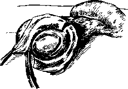
Figure 2.
cause adhesive inflammation of the serous membranes of the testicle and obliteration of the cavity. - Technique. After removing the fluid by puncture, following anesthesia of the cavity with 10-20 cubic cm of a 1% solution of novocaine, 5-10 cubic cm of T-rae Iodi are introduced and after 3-5 minutes the solution is released back. According to older authors (Vendliny), the recurrence after such treatment varies between 4 and 10%. This method can be indicated for hydrocele in elderly patients with signs of severe weakening of the general condition. Radical operations. 1. According to Bergman (Bergmann): a longitudinal incision through all layers of the scrotum to the serous membrane of the testicle, without cutting it. The testicle is dislocated into the wound. Bleeding is controlled. The testicle is placed back in position. Mattress sutures. This method is recommended for thick membranes. 2. According to Jaboulay-Winkelmann: an incision along the anterior outer surface of the scrotum through all layers to the serous membrane; dislocation of the testicle into the wound. The serous membrane is incised upward and downward to the tail of the epididymis. Sutures are placed on the everted membranes behind the epididymis (see Figure 3). Mattress sutures on the skin. Recurrences in 1-2%. This method, which causes almost no complications, is popular; it removes the patient from work for a short time. 3. Alferov's method: incision of all membranes, evacuation of fluid. For the purpose of permanent drainage, the serous membrane is sutured to the subcutaneous tissue of the scrotum. Mattress suture of the skin. Dressing. Lit.: - Khol'tsov V., Private Urology, vol. 4, L., 1927; Morris H., Diseases and injuries of the sexual and urinary organs, SPB, 1901; Weinler E., Die Erkrankungen des Hodens, des Samenstranges u. der Schleimhaute (Handbuch der Urologie, hrsg. von A. Lichtenberg, F. Voelker, H. Wildbolz, B. V, T. 3, Berlin, 1926); Demel R., Chirurgie des Hodens u. des Samenstranges, Stuttgart, 1926; Monod Ch. et Ferrillon O., Traite des maladies du testicule et de ses annexes, P., 1889; Thorek M., The human testis, Philadelphia-L., 1924; Campbell M., Hydrocele of the tunica vaginalis, Surgery, gynecology a. obstetrics, v. XLV, 1927. A. Vasil'ev.
Figure 3.
The testicle is placed back in position. Mattress sutures. This method is recommended for thick membranes. 2. According to Jaboulay-Winkelmann: an incision along the anterior outer surface of the scrotum through all layers to the serous membrane; dislocation of the testicle into the wound. The serous membrane is incised upward and downward to the tail of the epididymis. Sutures are placed on the everted membranes behind the epididymis (see Figure 3). Mattress sutures on the skin. Recurrences in 1-2%. This method, which causes almost no complications, is popular; it removes the patient from work for a short time. 3. Alferov's method: incision of all membranes, evacuation of fluid. For the purpose of permanent drainage, the serous membrane is sutured to the subcutaneous tissue of the scrotum. Mattress suture of the skin. Dressing. Lit.: - Khol'tsov V., Private Urology, vol. 4, L., 1927; Morris H., Diseases and injuries of the sexual and urinary organs, SPB, 1901; Weinler E., Die Erkrankungen des Hodens, des Samenstranges u. der Schleimhaute (Handbuch der Urologie, hrsg. von A. Lichtenberg, F. Voelker, H. Wildbolz, B. V, T. 3, Berlin, 1926); Demel R., Chirurgie des Hodens u. des Samenstranges, Stuttgart, 1926; Monod Ch. et Ferrillon O., Traite des maladies du testicule et de ses annexes, P., 1889; Thorek M., The human testis, Philadelphia-L., 1924; Campbell M., Hydrocele of the tunica vaginalis, Surgery, gynecology a. obstetrics, v. XLV, 1927. A. Vasil'ev.
Related articles
Mentioned in
Cite this page
“Hydrocele.” Soviet Medical Encyclopedia. English translation of Bolshaya Meditsinskaya Entsiklopediya, 1st ed. (Moscow, 1928–1936), ed. N. A. Semashko. https://sovietmedicalencyclopedia.pages.dev/article/hydrocele/