Testicle
Historical document, translated for reference. It reflects medical knowledge of the 1920s–30s and is not medical advice.
Summary
The testicle is the male gonad in animals where male sex cells-spermatozoa-are formed. In vertebrates, it is a compact paired organ closely connected to the excretory system, developing from a fold in the body cavity wall near the primary kidney.
Encyclopedia article (1928–1936)
Testicle, testis (testis, testiculus), the male gonad of animals, in which the formation of male sex cells-spermatozoa-occurs. In invertebrates, it sometimes has a tubular or sac-like structure and possesses its own excretory ducts. In secondary coelom animals, the testes develop in the wall of the body cavity as dense organs. Mature sexual products fall into the body cavity, and then are expelled from it outward through the excretory or separate sexual canals (ringed worms). In vertebrate animals, the testis is a rather compact paired organ, always showing a close connection with the excretory system. The testis develops, like the ovary, in the form of a paired fold in the wall of the embryo's body cavity, on the sides of the dorsal mesentery, in the area of the primary kidney. The primary sex cells, which form much earlier, migrate toward these rudiments and implant themselves in the sexual folds. The latter consist of an outer layer of thickened "rudimentary" epithelium and an inner mass of embryonic connective tissue. From the rudimentary epithelium, cell cords containing primary sex cells grow into the connective tissue, subsequently forming the seminiferous tubules and ampullae of the testis. From the Malpighian bodies of the primary kidney, epithelial cords also grow into the rudiment of the sex gland, connecting with the seminiferous tubules and forming the efferent ducts (vasa efferentia). The latter pass directly into the tubules of the primary kidney. Thus, in vertebrates, the characteristic connection of the male sex gland with the primary kidney is established, through which the mature sexual products are expelled outward through the primary kidney (Wolffian) duct. This connection develops partly through the cords of the rudimentary epithelium of the gland, and partly through cords growing from the epithelium of the excretory tubules, and in various vertebrates the significance of each of these components varies. In lower vertebrates, the testis sometimes has a complex system of internal tubules developing from cords growing from the primary kidney, while the rudimentary epithelium forms only ampullae opening into these tubules, in which the formation of spermatozoa takes place. In mammals, all the tubules of the testis itself develop from the rudimentary epithelium, while the tubules of the epididymis (epididymis) consist of the convoluted tubules of the primary kidney and their primary kidney (Wolffian) duct. In lower vertebrates (fish and amphibians), the semen is conducted through the aforementioned connecting ducts (vasa efferentia) into a certain number of primary kidney tubules and then into the primary kidney duct, which consequently simultaneously serves as the urinary duct and the vas deferens. However, in many fish and some amphibians, a certain differentiation occurs within the primary kidney, and only certain (usually anterior) tubules then perform the sexual function, usually losing their excretory function at the same time. Similarly, sometimes the vas deferens becomes separated from the urinary duct. In all higher vertebrates, starting from reptiles, the secondary, pelvic kidney develops as a permanent excretory organ, with its own separate ureter, while only the sexual division of the primary embryo's kidney remains, consisting of a greater or lesser number of tubules, and the primary kidney duct, which now serves exclusively as the vas deferens (ductus deferens). These ducts become highly convoluted and lie in the form of a rather large mass near the testis, forming its epididymis (epididymis). The testes develop in mammals, as in lower vertebrates, in the abdominal cavity at the level of the primary kidney, however, later in most mammals they move backward (downward) into protrusions of the abdominal cavity (processus vaginalis), growing into the scrotum, located externally behind the copulatory organ (descensus testiculorum). In insectivores and some rodents, the testes descend into the scrotum only during the breeding period, after which they are retracted back into the abdominal cavity.
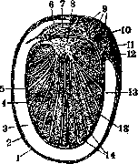
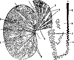
p- Shialgausen. The human testis consists of the testis (testis, testiculus, orchis, didymis) and the epididymis (epididymis, paratestis, parastata cirsoides, s. varicosa). The testis has the size of a walnut, somewhat compressed from the sides. In it, 2 surfaces are distinguished-facies lateralis et medians (flattened),-2 edges (margo anterior et posterior) and 2 ends, poles (extremitas superior et inferior). The size and weight of the organ vary: in an adult, the longitudinal size is 4-5.5 cm, the sagittal-2-3.5 cm and the transverse-1.8-2.4 cm; weight 25-30 g, with the left and right testis usually having different weights. When standing, the upper pole is slightly tilted forward and outward, and the lateral surface is turned slightly backward. Usually the left testis hangs lower than the right. The lower pole and anterior edge of the testis are free, in the posterior part of the upper edge the excretory paths of the sex gland begin (ductuli efferentes), and the entire posterior edge of the testis is occupied by the epididymis located here. The surface of the testis is smooth and shiny, like a mirror, whitish, because it is covered by a serous sheet (tunica vaginalis propria), belonging to the protrusion of the abdominal cavity; through it the tunica albuginea shines through. Vascularization, A. spermatica int. begins from the abdominal aorta near the site of the sex gland's development; it usually anastomoses with a. deferentialis. A. spermat. ext. (from a. epigastrica) and a. pudenda ext. (from a. femoralis) supply only the coverings of the testis. The veins collect in plex. pampiniformis, from which the left ones through vv. spermaticae sin. by means of v. renal, sin., and the right ones directly flow into v. cava inf. Lymph, vessels from the testis, epididymis and d. deferens pass through the inguinal canal to lgl. lumbales (aorticae).-Nerves of the testis from Dx; sympathetic and parasympathetic fibers pass through nn. splanchnici minores, plex. coeliacus, aorticus and renalis. n. carotinus. Microscopic structure. The surface of the testis is covered by the tunica albuginea, with which the visceral sheet of the serous membrane of the testis (tunica vaginalis propria) is fused, which does not exist after birth; 4-tunica vaginalis communis; 2-tunica vag. propria, outer sheet; 3-tunica vaginalis propria, inner sheet, connecting with albuginea (5); 6-tunica albuginea; 7-epididymis (epididymis) (fig. 1). On the posterior edge, the tunica albuginea forms a thickening, protruding into the testis 6-9 mm in the form of a vertically standing sheet 2-2.7 cm long, the so-called mediastinum testis (mediastinum testis) or Highmore's body (corpus Highmori); from it into the testis go partitions (septula testis), dividing it into conical lobules (lobuli testis), numbering 100-250. Inside each lobule are placed the convoluted seminiferous tubules (tubuli contorti seminiferi) from 1 to 3, which, dividing repeatedly and winding, fill its cavity; in the pointed ring of the lobule they pass into the straight tubules (tub. seminiferi recti), which, penetrating into Highmore's body, form there the network of Haller (rete testis, s. Halleri). From the upper end of this network emerge 7-15 efferent ducts (ductuli efferentes, s. Graafiani), which pierce the tunica albuginea and pass into the epididymis (fig. 2). The tunica albuginea, 0.4-0.6 mm thick, consists of dense connective tissue, bundles
Figure 1. Cross-section of the testis and its coverings (according to Keliker): 1-tunica vaginalis communis; 2-tunica vag. propria, outer sheet; 3-tunica vag. propria, inner sheet, connecting with albuginea (5); 6-tunica albuginea; 7-epididymis (epididymis); 8-branches of a. spermaticae; 9-epididymis (epididymis); 10-ductus deferens; 11-lobules of the testis; 12-vas deferens; 13-epididymis (epididymis). On the posterior edge, the tunica albuginea forms a thickening, protruding into the testis 6-9 mm in the form of a vertically standing sheet 2-2.7 cm long, the so-called mediastinum testis (mediastinum testis) or Highmore's body (corpus Highmori); from it into the testis go partitions (septula testis), dividing it into conical lobules (lobuli testis), numbering 100-250. Inside each lobule are placed the convoluted seminiferous tubules (tubuli contorti seminiferi) from 1 to 3, which, dividing repeatedly and winding, fill its cavity; in the pointed ring of the lobule they pass into the straight tubules (tub. seminiferi recti), which, penetrating into Highmore's body, form there the network of Haller (rete testis, s. Halleri). From the upper end of this network emerge 7-15 efferent ducts (ductuli efferentes, s. Graafiani), which pierce the tunica albuginea and pass into the epididymis (fig. 2). The tunica albuginea, 0.4-0.6 mm thick, consists of dense connective tissue, bundles
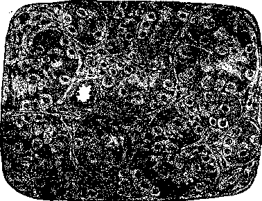
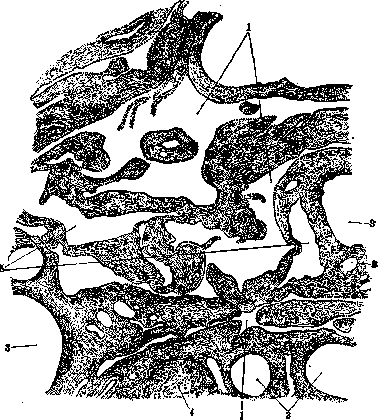
Figure 5. Section of the testis of an 8-month fetus (according to Prenant): in the convoluted tubules round, light sex cells and dark follicular (Sertoli) cells; between the tubules groups of interstitial cells are visible.
Figure 2. Scheme of the tubules of the testis (according to Ebert): 1-lobules of the testis, bounded by connective tissue partitions going from the tunica albuginea to Highmore's body (2); in it are placed the straight tubules (3) and Highmore's network (4); 5-efferent ducts; 6-vascular cones of the epididymis; 7-epididymal canal; 8-vas deferens; 9-organ of Giraldes; 10-lower aberrant tubule.
of collagen fibers with a small number of elastic fibers and cells (fibroblasts, smooth muscle cells). The mediastinum testis is a dense connective tissue with a large number of elastic fibers and vessels. The lobules of the testis contain convoluted seminiferous tubules (tubuli seminiferi contorti), 30-70 cm long, 150-250 μ in diameter, consisting of a basement membrane and a multilayered epithelium. The epithelium consists of supporting cells (Sertoli cells) and spermatogenic cells. The supporting cells are large, with a clear nucleus, located on the basement membrane. The spermatogenic cells are located in different stages of development, forming spermatogenic series. In the convoluted tubules, spermatogenesis occurs. The straight tubules (tubuli seminiferi recti) are short, straight tubules leading from the convoluted tubules to the rete testis. The rete testis is a network of channels in the mediastinum testis, formed by the straight tubules. From the rete testis, 7-15 efferent ducts (ductuli efferentes) emerge, which pierce the tunica albuginea and enter the epididymis. The efferent ducts are lined with a ciliated epithelium and have a diameter of about 100-150 μ. The epididymis is an elongated, flattened body attached to the posterior edge of the testis. It consists of the head (caput epididymidis), body (corpus epididymidis), and tail (cauda epididymidis). The epididymis is formed by the efferent ducts, which anastomose to form a single duct, the ductus epididymidis. The ductus epididymidis is a highly convoluted tube, 4-6 m long, with a diameter of 0.1-0.4 mm. It is lined with a pseudostratified epithelium with stereocilia. The ductus epididymidis continues into the ductus deferens (vas deferens). The ductus deferens is a muscular tube that transports sperm from the epididymis to the urethra. It has a thick muscular wall and a narrow lumen. The ductus deferens ends in the ampulla ductus deferentis, which joins the duct of the seminal vesicle to form the ejaculatory duct.
The testis is covered by the tunica vaginalis, which is a serous membrane derived from the peritoneum. It consists of two layers: the parietal layer, which lines the inner surface of the scrotum, and the visceral layer, which covers the testis and epididymis. Between these two layers is a small amount of serous fluid that allows the testis to move freely. The tunica albuginea is a dense, white fibrous membrane that covers the testis beneath the tunica vaginalis. It is continuous with the septula testis, which extend into the testis and divide it into lobules. The mediastinum testis is a thickened area of the tunica albuginea at the posterior edge of the testis, from which the septula testis extend. The lobules of the testis contain the seminiferous tubules, which are the sites of spermatogenesis. Each lobule contains 1-3 seminiferous tubules, which are convoluted and filled with spermatogenic cells. The straight tubules connect the convoluted tubules to the rete testis. The rete testis is a network of channels in the mediastinum testis, which collects sperm from the straight tubules. From the rete testis, the efferent ducts emerge and pierce the tunica albuginea to enter the epididymis. The epididymis is an elongated structure attached to the posterior edge of the testis, consisting of the head, body, and tail. It stores sperm and allows them to mature. The ductus deferens is a muscular tube that transports sperm from the epididymis to the urethra. The blood supply to the testis is provided by the testicular artery, which is a branch of the abdominal aorta. The veins of the testis form the pampiniform plexus, which helps regulate the temperature of the testis. The lymphatic vessels drain to the lumbar lymph nodes. The nerves of the testis are derived from the sympathetic and parasympathetic nervous systems.
The testis develops from the genital ridge, which is a thickening of the mesoderm on the posterior abdominal wall. The genital ridge contains the primordial germ cells, which migrate from the yolk sac. The primordial germ cells proliferate and form the sex cords, which are surrounded by mesenchyme. The mesenchyme differentiates into the interstitial cells, which produce testosterone. The sex cords canalize to form the seminiferous tubules. The efferent ducts develop from the mesonephric tubules. The epididymis and ductus deferens develop from the mesonephric duct. The descent of the testis from the abdomen to the scrotum occurs during fetal development and is completed shortly before birth. This descent is regulated by hormones and involves the formation of the processus vaginalis, which is an outpouching of the peritoneum that precedes the testis into the scrotum. The gubernaculum testis is a fibrous cord that attaches to the testis and helps guide it into the scrotum. After descent, the processus vaginalis obliterates, leaving the tunica vaginalis as a covering for the testis. The temperature of the scrotum is lower than that of the abdomen, which is necessary for spermatogenesis. The testis is sensitive to temperature changes, and exposure to high temperatures can impair sperm production. The testis also has a rich blood supply, which helps regulate its temperature. The pampiniform plexus, a network of veins in the spermatic cord, acts as a countercurrent heat exchange system, cooling the arterial blood as it enters the testis.
The testis is susceptible to various pathological conditions. Testicular torsion is a medical emergency in which the spermatic cord twists, cutting off blood supply to the testis. This can lead to ischemia and necrosis of the testicular tissue if not treated promptly. Epididymitis is inflammation of the epididymis, usually caused by infection. It can be acute or chronic and is often associated with urinary tract infections or sexually transmitted infections. Testicular cancer is relatively rare but is the most common cancer in young men. It usually presents as a painless lump in the testis. The most common type is seminoma, which arises from the germ cells. Varicocele is an enlargement of the veins within the scrotum, similar to a varicose vein in the leg. It can cause pain and may affect fertility. Hydrocele is a collection of fluid in the tunica vaginalis, causing painless swelling of the scrotum. Orchitis is inflammation of the testis, which can be caused by infection or autoimmune conditions. Cryptorchidism is a condition in which one or both testes fail to descend into the scrotum. It increases the risk of infertility and testicular cancer. Testicular trauma can result from injury to the scrotum and may cause pain, swelling, and damage to the testicular tissue. Microlithiasis is a condition in which calcium deposits form in the seminiferous tubules. It is usually asymptomatic but may increase the risk of testicular cancer.
The function of the testis is to produce sperm and testosterone. Spermatogenesis is the process by which sperm are produced. It occurs in the seminiferous tubules and involves the division and differentiation of spermatogenic cells. The process takes approximately 64 days and is regulated by hormones, including follicle-stimulating hormone (FSH) and luteinizing hormone (LH). Sertoli cells provide support and nourishment for the developing sperm. They also produce androgen-binding protein, which concentrates testosterone in the seminiferous tubules. Leydig cells, located in the interstitial tissue, produce testosterone in response to LH. Testosterone is responsible for the development of male secondary sexual characteristics, such as facial hair, deepening of the voice, and muscle mass. It also plays a role in spermatogenesis and libido. The testis is also involved in the production of inhibin, a hormone that regulates FSH secretion. The blood-testis barrier is formed by tight junctions between Sertoli cells, which create a microenvironment necessary for spermatogenesis. It protects developing sperm from the immune system and maintains the proper concentration of hormones and nutrients.
The testis can be examined using various diagnostic techniques. Physical examination involves palpation of the testis to detect any lumps, swelling, or tenderness. Ultrasound is a non-invasive imaging technique that can visualize the testis and detect abnormalities such as tumors, cysts, or torsion. Color Doppler ultrasound can assess blood flow to the testis, which is useful in diagnosing torsion or epididymitis. MRI is not commonly used for routine evaluation of the testis but may be helpful in complex cases. Biopsy involves taking a small sample of testicular tissue for microscopic examination. It is used to diagnose conditions such as infertility or to evaluate testicular masses. Laboratory tests may include hormone levels (testosterone, FSH, LH), semen analysis, and tests for sexually transmitted infections. Treatment of testicular disorders depends on the underlying condition. Testicular torsion requires emergency surgery to untwist the spermatic cord and save the testis. Epididymitis is usually treated with antibiotics. Testicular cancer is typically treated with surgery, radiation therapy, or chemotherapy, depending on the type and stage of the cancer. Varicocele may be treated with surgery or embolization to block the affected veins. Hydrocele may be drained or surgically removed if it causes symptoms. Orchitis is treated with antibiotics if caused by infection, or anti-inflammatory medications if due to autoimmune conditions. Cryptorchidism is usually treated with surgery to bring the testis into the scrotum. Testicular trauma may require surgery to repair damaged tissue. Microlithiasis does not usually require treatment but may necessitate regular monitoring.
The testis is an important organ in the male reproductive system, and its proper function is essential for fertility and overall health. Regular self-examination of the testis is recommended to detect any abnormalities early. If any lumps, swelling, or pain are noticed, medical attention should be sought promptly. Maintaining good overall health, including a balanced diet and regular exercise, can help support testicular function. Avoiding exposure to high temperatures, such as hot tubs or saunas, can help prevent damage to sperm. Safe sexual practices can reduce the risk of sexually transmitted infections that may affect the testis. Regular medical check-ups, including examination of the testis, are important for early detection and treatment of any potential problems. In cases of infertility, evaluation of the testis may be necessary to determine the underlying cause and appropriate treatment. Advances in medical science have improved the diagnosis and treatment of testicular disorders, leading to better outcomes for patients. Research continues to enhance our understanding of testicular function and the development of new treatments for testicular diseases.
The testis has been studied extensively throughout medical history. Ancient Greek and Roman physicians described the anatomy and function of the testis. The Roman physician Celsus provided detailed descriptions of testicular conditions and treatments. In the Middle Ages, Islamic scholars made significant contributions to the understanding of anatomy, including the testis. During the Renaissance, anatomists such as Andreas Vesalius provided more accurate descriptions of testicular anatomy. In the 19th century, advances in microscopy allowed for better understanding of spermatogenesis. The 20th century saw significant progress in the diagnosis and treatment of testicular disorders, including the development of surgical techniques and chemotherapy for testicular cancer. The study of the testis continues to evolve, with ongoing research into its development, function, and diseases. Understanding the testis is essential for addressing issues related to male infertility, sexual dysfunction, and testicular cancer. The historical study of the testis has laid the foundation for modern urology and andrology. The testis remains a subject of interest in medical research due to its complex structure and function. Advances in genetic and molecular biology have provided new insights into the mechanisms of spermatogenesis and testicular development. The testis also serves as a model for studying stem cells and regenerative medicine. The historical perspective on testicular research highlights the importance of anatomical knowledge in medical practice. The study of the testis has contributed to our understanding of hormonal regulation and reproductive biology. The testis continues to be an important focus of medical research and clinical practice.
The testis is a complex organ with a rich history of study and clinical importance. Its structure and function have been described in detail in medical literature throughout history. The testis plays a crucial role in male reproduction and overall health. Understanding its anatomy, development, and function is essential for diagnosing and treating various disorders. Advances in medical science have improved our ability to manage testicular conditions effectively. The testis remains a subject of ongoing research, with new discoveries continually enhancing our knowledge. Historical studies of the testis have provided a foundation for modern medical practice. The testis is a vital organ that continues to be of great interest to medical professionals and researchers alike.
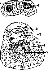
in which fibers are interwoven in various directions parallel to the surface; between them are networks of elastic fibers; the inner layer of the tunica albuginea, containing a large number of blood vessels, is distinguished by some authors as a special vascular tunic (tunica vasculosa). The proper vaginal tunic (tunica vaginalis propria) is closely fused with the tunica albuginea and cannot be separated from it; it is a fold of peritoneum covering the testicle during its development and is covered on its surface by endothelium. The body of the epididymis is formed by a thickening of the tunica albuginea and is also built from dense fibrous connective tissue, as well as the septa. On the contrary, the connective tissue lying inside the lobules between the convoluted tubules has a loose character, contains many capillaries, lymphatic vessels, and along with ordinary stable connective tissue cells, special interstitial cells (syn. Leydig cells, Waldeyer's plasma cells) (fig. 3). These are angular cells, 14-21 μ in size, with a round nucleus, near which is located a granular sphere with a centrosome; in the protoplasm, inclusions of lipoid and fatty nature are noticeable, larger around the nucleus, as well as pigment; on fixed preparations, it has a reticular structure. These cells are located in strands or groups around the vessels, under the capsule and between the tubules. In humans, special protein crystals in the form of rods, described by F. Reinke, Lubarsch and others (fig. 4), are found in the interstitial cells. These cells were formerly considered connective tissue, then a gland with internal secretion (see Interstitial gland); recently, their glandular nature is disputed (Stieve). In homosexuals, Steinae found Leydig cells of particularly large size. The parenchyma of the testicle is formed by convoluted seminiferous tubules 0.13-0.28 mm in diameter, which form a large number of bends, sometimes branch and connect with each other; the length of the tubule in 1 on macerated preparations is determined as 70-80 μ (Sappey). The tubule membrane (membrana propria) in humans is quite thick (5-11 μ according to Kölliker), has a layered structure and consists of plates of connective tissue, the bundles of which go mainly circularly and contain thin elastic fibers; the innermost membrane represents a thin elastic film; between the layers of the membrane are flat endothelial-like cells. The lumen of the tubule is lined with two types of cells: 1) follicular epithelium or Sertoli cells (Sertoli, 1865) and 2) germinal or sperm cells. Before the onset of sexual maturity, there are primary germ cells in the form of round light cells, which are located in small numbers between the follicular epithelium (fig. 3); with the beginning of spermatogenesis (see), the number of germ cells increases, and they are arranged in several rows, producing spermatozoa, while Sertoli cells are located among them in the form of columns. Fat inclusions appear in their protoplasm in the form of small droplets, which is especially clearly noticeable when examining the convoluted tubule in the fresh state. In old age, with the cessation of spermatogenesis and the disappearance of germ cells, the walls of the tubule are lined only with Sertoli cells, which take on the appearance of cylindrical epithelium. The convoluted seminiferous tubules pass into the straight tubules and through them into the network of Haller, which passes into the vas deferens.
The straight tubules, 0.02-0.09 mm in diameter, are lined with low cylindrical, cuboidal, or flattened epithelium, often with polymorphic nuclei, which sharply differs from the follicular epithelium of the ends of the convoluted tubules; the same epithelium lines the network of Haller. The latter has no capsule of its own, but represents tubules and lacunae of various diameters (from 3-4 μ to 0.1-0.3 mm), of irregular shape, hollowed out in the dense connective tissue of the body of the epididymis (fig. 5); it contains smooth muscle fibers and dense networks of elastic fibers, which pass into the elastic networks of the septa and the tunica albuginea.
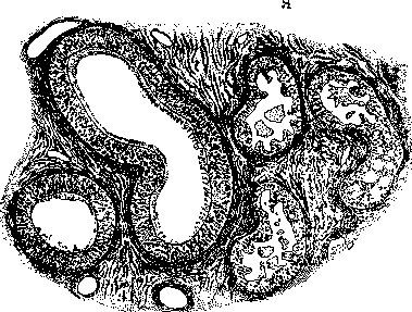
The epididymis (epididymis) (fig. 2), forming the main mass of the excretory ducts, consists of the head (caput) and tail (cauda) and closely adjoins the body of the epididymis and the posterior edge of the testicle. It originates from the efferent ducts (ductuli efferentes), which emerge from the network of Haller in its upper part, numbering from 7 to 15; each of them first goes straight, then begins to make bends of ever-increasing size, which in their aggregate form a conical lobule of the epididymis (lobulus epid.) (conus vasculosus Halleri); after this, each efferent duct opens at a right angle into the duct of the epididymis (ductus epididymis), which curves around the outer edge of the head of the epididymis. This duct, descending, begins to twist itself, forming the tail of the epididymis, and passes into the vas deferens. The epididymis is covered with a dense connective tissue capsule, which gives off septa between the conical lobules; inside the lobules, between the tubules, loose connective tissue. According to histological structure, the efferent ducts (tubules of the head) sharply differ from the duct of the epididymis and can be easily distinguished on sections (fig. 6). The epithelium of the efferent ducts has an uneven contour and is provided with bay-like indentations, with the protruding parts formed by cylindrical cells with a ciliated covering, strongly staining with eosin, the indentations lower, cuboidal in shape, without cilia and light; both contain lipoid granules and pigment; according to Schaffer, the indentations represent intraepithelial glands. In contrast to this, the epithelium of the duct of the epididymis is high (45-50 μ), double-rowed, with long non-ciliated hairs, which are glued into bundles (stereocilia). Both types of tubules have their own membrane and a circular layer of smooth muscles, thicker in the duct of the epididymis.
Associated with the epididymis are rudimentary organs, representing remnants of the Wolffian body ducts (see) (fig. 2). These include: 1) aberrant ducts (ductuli aberrantes), ducts connected with the duct of the epididymis and ending blindly, and 2) the organ of Giraldes (Giraldes, 1857), syn. parepididymis (Henle) or paradidymis (Waldeyer), representing one or more ducts, blind at the ends, which are twisted into balls located between the head of the epididymis and the vas deferens. The ducts are covered with a connective tissue capsule with smooth muscles and lined with ciliated epithelium. The third rudimentary organ is the remnant of the Müllerian duct (see Morgagni's hydatids) (appendix testis)
Fig. 6. Section of the epididymis (according to Schaffer) on the right a section of the efferent ducts, on the left - the duct of the epididymis.
abundantly supplied with blood vessels, blood and lymphatic, of which the former form capillary networks around the convoluted tubules, and the latter form a plexus under the tunica albuginea.
Karpov - Pathological anatomy and clinic. Atrophy of the testicle is a frequent phenomenon, usually arising from secondary causes, from debilitating diseases, from disruption of endocrine correlations, diseases of the thyroid gland (dysthyroidism, cretinism), pituitary gland (acromegaly, nanosomia), lesions of the base of the diencephalon, as well as the peripheral nervous system (p. spermatici, plexus mesentericus). Histologically, such testicles are characterized by the predominance of connective tissue over the tubules. The latter in this case are already. Spermatogenesis in hypoplastic testicles occurs later. - Bilateral hyperplasia of the testicle does not possess characteristic anatomical signs, although clinically this condition is recognized. Some authors consider premature maturity in diseases of the gl. pinealis as a phenomenon of hyperplasia. Unilateral hypertrophy is observed as vicarious when one testicle atrophies or is removed at an early age. The hypertrophied testicle can reach a volume several times larger than normal. Histologically, such testicles are characterized by wide lumens of their tubules (255-270 μ). - Abnormal displacement of the testicle is called ectopia. Depending on the position, femoral, perineal ectopia and a very rare form of ectopia testis transversa are distinguished, when both testicles are on one side of the scrotum with separate appendages and spermatic ducts or a common v. deferens (Merkel). In all cases of ectopia transversa, the testicles were found in the right side of the scrotum. The most common cause of circulatory disorders in the testicle is twisting of the spermatic cord, associated with abnormal structure and position of the testicle. Such testicles are poorly fixed and can rotate around their horizontal and vertical axes. With vertical rotation by 90-180°, twisting of the spermatic cord occurs, compression of vessels and the associated hemorrhagic infarction and necrosis of the testicle. Twisting is more common when the testicle is in the inguinal position, but can also occur when it is in the scrotum. Less commonly, hemorrhagic infarction of the testicle occurs from thrombosis of the plexus pampiniformis, after trauma, after varicocele surgery, from incarceration of the testicle in the inguinal canal, and from unknown causes. Hemorrhagic infarction should be distinguished from hemorrhages that occur in the testicle in scurvy, hemophilia, infectious and septic diseases, as well as from birth trauma in newborns, especially in breech position. - Disorders of metabolism greatly affect the testicle. Avitaminosis causes degeneration of the seminal epithelium in rats. The same condition is caused by unilateral nutrition. Feeding with cholesterol, extirpation of the adrenal glands, accompanied by disruption of fat metabolism, cause strong changes in spermatogenesis, breakdown of seminal filaments, spermatids and spermatocytes. Disorders of salt exchange act in the same direction. Introduction of iodine in animals causes degenerative changes in the seminiferous tubules. In severe anemias, hemochromatosis, bronze diabetes, the iron-containing pigment is deposited in the seminiferous tubules and in the interstitial tissue. Spermiogenesis suffers in this case. Prolonged intoxications with morphine, nicotine, alcohol are accompanied by atrophic processes in the seminiferous tubules. The seminal epithelium is strongly affected by X-rays. In guinea pigs, under the influence of X-ray irradiation, the seminal epithelium disappears, leaving only the walls of the tubules with Sertoli cells. With the cessation of the effect of the rays, the epithelium quickly regenerates. With repeated and prolonged exposure to X-rays, atrophy of the testicle occurs (Kyrle). Psychological factors influence spermatogenesis. Under the influence of severe emotional shocks in completely healthy men, the formation of spermatozoa may cease. Acute infectious diseases - typhus, relapsing fever, typhoid fever, rheumatism, etc. - are accompanied by the appearance in the tubules of abnormally shaped seminal filaments, and subsequently their disappearance. Spermatids, and in more severe cases spermatocytes, disintegrate, only Sertoli cells and spermatogonia remain in the tubules. The breakdown of the seminal epithelium in the lumen of the tubules is accompanied by the appearance of eosinophilic cells with pyknotic nuclei and peculiar forms of giant cells. At the height of the breakdown, the latter fills the lumens of the tubules of the rete testis and epididymis. Among the breakdown products, cells of the macrophage type containing sperm (spermiophagy) are found. Particularly devastating are the frequent orchitis in smallpox (necrosis of parenchyma, infiltrates, etc.) - see also Orchitis. In gonorrhea, thickening of the epididymis is observed. In acute cases, a serous exudate containing leukocytes and desquamated epithelial cells of the tunics appears between the tunics of the testicle. In chronic cases, the leaves of the testicle may be fused together over a considerable extent. Lime is often deposited in these adhesions. Histologically, in acute cases, the testicle may not participate in the gonorrheal process at all or participate very weakly. The process is predominantly localized in the excretory ducts of the testicle. If the testicle participates in the process, usually the part adjacent to the rete testis and the tunica albuginea is affected. In these places, there is proliferation of interstitial tissue and in the tubules - breakdown of the seminal epithelium (see Orchitis). All without exception chronic infectious diseases and intoxications cause changes in the testicle in the form of proliferation of connective tissue, death of the seminal epithelium, with subsequent hyalinization of the tubules and fibrosis of the interstitial tissue (spermatoangiitis fibrosa obliterans). The initial moment can be both inflammatory processes in the interstitial tissue and degenerative changes in the seminal epithelium. This morphological condition is called fibrosis of the testicle, regardless of the cause that caused it. Fibrosis of the testicle (fibrosis testis) is often found in syphilis. Unlike other processes in syphilis, there is diffuse proliferation of connective tissue with round-cell infiltration in the testicle in the absence of similar changes in the epididymis. However, for more accurate diagnosis, it is necessary to find syphilitic changes in other organs as well. Fibrosis of the testicle is characterized by the presence of grayish-white strands penetrating the entire organ on cross-section. The scars can occupy the entire organ or only one part of the testicle. On microscopic examination, some tubules are completely obliterated, while in others the epithelium from indifferent cells is still preserved. Near hyalinized tubules in the interstitial tissue, foci of Leydig cells. The interstitial connective tissue is increased in amount, the elastic tissue in the walls of the tubules is preserved for a long time. Among the fibrous tissue, there may be areas with normal spermatogenesis. TB of the testicle usually originates from a lesion of the epididymis. The latter turns into a sausage-shaped formation, on cross-section filled with a curdled mass. When the process spreads to the testicle, nodules first appear in the corpus cavernosum, but can be scattered over the entire surface of the testicle. The testicle can also be affected primarily by metastatic means. In the latter case, miliary forms of TB are more common. Morphologically, intratubular and interstitial forms of TB are distinguished. In the first form, the epithelium of the tubules proliferates intensely with the formation of giant cells. The contents of the tubules then undergo necrosis, i The process passes from one tubule to another. The elastic tissue of the walls of the tubules is preserved for a long time, indicating the boundaries of the former tubules. In metastatic TB, the process is initially localized in the interstitial tissue, but later also spreads to the tubules (Baumgarten). - Syphilis of the testicle rarely spreads to the epididymis. In the testicle, syphilis occurs in two forms: as interstitial fibrous and as gummatous orchitis. In the first form, the testicles are small and dense. In the interstitial tissue, there is a diffuse inflammatory process in the form of proliferation of connective tissue; the tubules are compressed and the seminal epithelium undergoes necrobiosis. Subsequently, fibrosis of the testicle (fibrosis testis) is obtained with thickening and hyalinization of the walls of the tubules up to their obliteration. In gummatous orchitis, the development of gummas is observed in the form of single or multiple yellowish nodules with a necrotic center and a small-cell infiltrate of lymphocytes, plasma cells and giant cells at the periphery. - In leprosy, lepra bacilli are excreted from the blood into the seminiferous tubules, producing a destructive effect on the seminal epithelium, therefore in leprosy spermatogenesis ceases early. Lepra cells appear in the seminiferous tubules and in the interstitial tissue. In far-advanced cases, a picture of fibrosis of the testicle with deposition of hemosiderin develops. Along the course of the testicle and spermatic cord, single and multilocular cysts filled with transparent or milky contents, in which sperm are often found, are frequently encountered. In the latter case, they are called spermatoceles (see Spermatocele). Serous cysts are more common in the epididymis than in the testicle. They are lined with low epithelium, but in some cases with ciliated or squamous epithelium, arising on the basis of metaplasia of the cells of the covering epithelium (Mayer). These cysts lie between the serous and albugineous tunics. Their occurrence is associated with inflammatory processes. - Tumors rarely affect the testicle and occur in childhood as well.
Fibromas and lipomas are rare. Chondromas of the testicle are most often in combination with elements of other tissues. They apparently belong to teratoblastomas. Myomas and rhabdomyomas occur rarely, the latter more often as part of mixed tumors. In general, mixed tumors are found in the testicle not infrequently. They often contain myxomatous tissue. Many mixed tumors are essentially teratoblastomas, developing unilaterally. Sarcomas of the testicle can be round-cell, polymorphous-cell, spindle-shaped, often in the form of a mixed tumor. Cancers usually originate from the epithelium of the tubules and rete testis. Cancers consisting of small round cells (so-called seminomas) are common, while adenocarcinoma and scirrhous types of cancer are rarer. Dermoid formations in the testicle are comparatively rare; one mainly deals with teratoblastomas (malignant teratoma) of the testicle. Teratoblastomas of the testicle are more often on the right side than on the left, and they prefer a certain age-from 20 to 40 years. Macroscopically, teratoblastomas in the initial stages appear as a small tumor of dense consistency, on the section containing cysts of various sizes. The cavities of the cysts are lined with either cylindrical or cubic epithelium, while in places multilayered squamous epithelium is also found. Around these cavities, smooth muscle fibers, hyaline cartilage, sometimes nerve bundles and ganglion cells are encountered. The tumor soon infiltrates the tissue of the testicle. Metastases appear very early, first in the retroperitoneal glands, then in the lungs, brain, and other organs.
3. Morgenstern. Congenital abnormalities. Monorchidism (syn. monorchism), unilateral cryptorchidism (see). Anorchidism (syn. aplasia), absence of both testicles. More often, both testicles do not descend into the scrotum and are located in the abdominal cavity, usually atrophied; at the same time, the external genitalia are often underdeveloped (see Eunuchoidism). Poly-orchidism (syn. polyorchidia), excessive number of testicles. This phenomenon is extremely rare. The supposed extra testicle turned out to be either an encysted hydrocele, funiculocele, or a cyst, or an omental hernia. Synorchidia (fusion of testicles with each other, usually inside the abdominal cavity), an extremely rare anomaly. Inversio testis, abnormal position of the testicle: 1) horizontal, in which its upper end is at the bottom, so that the vas deferens, in these cases shorter, originates from the epididymis, the tail of which is at the top; 2) vertical, in which the testicle is rotated around its longitudinal axis. Inversion can be complete or incomplete. The cause is unknown. Retention of testicle (retentio, ectopia testis) can occur on the path from the lower pole of the kidney to the scrotum and can be unilateral or bilateral. In persons under 10 years of age, there are no subjective sensations. Later, painful sensations usually appear, especially with sharp movements, gymnastics, etc. On palpation, the patient feels pain at the site of retention of the testicle, and sometimes an underdeveloped testicle that has not descended into the scrotum can be felt, whose absence in the latter is easily determined. Along with a number of methods for descent and fixation of the testicle (see Cryptorchidism), operations by Nikoladoni and Longard are also recommended. The latter, after separating and descending the testicle, fixed it by suturing the lower part of the tunics surrounding the testicle to the skin of the perineum. Longard for the same purpose applies prolonged traction of the testicle, connecting its tunica albuginea with nodal sutures to the skin of the scrotum. These threads are left long and for the purpose of traction are attached with adhesive plaster to the inner surface of the thigh for 8-10 days. Torsion of the testicle, rotation around the longitudinal axis, occurs when the spermatic cord twists. This rare condition usually occurs in persons with a retained testicle, more often on the right side. In all cases, the epididymis is more enlarged than the testicle, which is sometimes little changed, and sometimes can be dark and gangrenous. With torsion of the testicle, pain and a tumor suddenly appear in the corresponding groin, often accompanied by vomiting. The diagnosis is based on a tumor of the spermatic cord, located between its healthy part and the testicle, and on the position of the epididymis in front of the testicle. Treatment with early recognition consists in unwinding the testicle, with late recognition - operation, more often in the form of removal of the gangrenous mass. Spermatic cyst, spermatocele, a cystic tumor of the testicle, filled with semen. The spermatic cyst arises from the obstruction of the seminiferous tubule as a result of one or another, more often inflammatory process, leading to narrowing or even complete obstruction of it. Continuing semen secretion expands the seminiferous tubules, forming tumors from retention of secretion. The starting point of their formation is predominantly either the place where the tubules of the testicle flow into rete testis, or the rete testis itself, or the place where the vasa efferentia flow into the epididymal canal. Spermatic cysts are more often found in persons under 40 years of age. On corpses, they are found significantly more often than in living persons, due to the absence of any clinical phenomena (according to Hochenegg, they are found in 20% of autopsies, more often on the right than on the left). Depending on their location outside or inside the proper tunic of the testicle, spermatic cysts are divided into extravaginal and intravaginal. The walls of the cyst consist mainly of connective tissue elements, covered with flat, and in young people cylindrical, epithelium. The fluid inside the cyst is transparent or slightly turbid, yellowish-green in color, and often contains spermatozoa. From a clinical point of view, the spermatic cyst represents a uniform, slowly developing formation, the first symptoms of which in the form of a tumor and unpleasant sensations usually appear in old age. Their size is usually from a pea to a cherry, occasionally to a small apple. The consistency is soft and elastic. Treatment consists of puncture followed by a pressure bandage. Koher recommends the injection of tincture of iodine. The most rational treatment is the radical removal of the spermatic cyst by surgery. Hydrocele, cystic expansion of the accessory formations of the testicle, sitting either directly on it or on a stalk. They are usually of small size. Seminoma, a malignant cancerous tumor of the testicle, previously considered a sarcoma, consists of cells morphologically similar to germ cells. On section, it appears grayish-white with often encountered necrotic and softened areas. Seminoma primarily affects the testicle, subsequently spreading to the epididymis. As it grows, it penetrates the tunica albuginea and other tunics of the testicle. Subsequently, it perforates the skin or spreads upward along the spermatic cord, metastasizing not only to the glands but also to distant organs (brain, liver, etc.). Seminomas are predominantly found in young age, during the period of sexual maturity. On palpation, there is a dense, rapidly growing tumor, and after some time completely replacing the parenchyma of the testicle. It is necessary to differentiate it from a benign tumor, tuberculosis and syphilis of the testicle. The sluggish course of the process and other general phenomena distinguish these conditions from seminoma (see below syphilis of the testicle). The prognosis, even with early removal of the testicle, is not always favorable. Treatment is surgical followed by X-ray irradiation of the corresponding lymph glands. "Fungus testis benignus" is observed after spontaneous rupture of an abscess of the testicle outward with the formation of a fistula, around which later granulation and even fungoid-like growths form (see Orchitis). Stones of the scrotum are formed by the deposition of salts from urine that has penetrated into the scrotum during the rupture of an abscess, or by the deposition of salts along the fistulous tract, or, forming in the urethra, subsequently penetrate into the scrotum. They consist of uric acid or calcium phosphate. Stones of the scrotum, due to the continued deposition of salts from urine, increase in size, sometimes reaching large dimensions. Graefe reported a case of a stone weighing 26 ounces, which spontaneously ruptured through the scrotum outward. Stones of the scrotum cause pain during urination, trauma, and even on touch. Treatment is surgical, consisting not only in the removal of stones but also in eliminating the cause that caused them. Syphilis of the testicle (orchitis luética) can be of congenital or acquired nature. The latter is observed in the secondary and tertiary stages, manifesting itself mainly in two main forms: in the form of gummatous nodules (orchitis gummosa) or in the form of interstitial fibrous orchitis (fibrosis testis syphilitica, s. orchitis fibrosa). Congenital syphilis of the testicle can be observed already in the first days after the birth of a boy or manifests itself up to 2-3 years of age. The gummatous form is characterized by the appearance of initially small nodules in the parenchyma of the testicle, which, partially merging with each other, increase the testicle often to the size of an adult man's fist. Gums cause a nodular surface of the testicle. On section, its outer tunic is dense, grayish or yellowish in color, and inside there is often caseous degeneration and disintegration. Interstitial fibrous orchitis is a chronic inflammation of the interstitial connective tissue. It is characterized by the presence of dense callous thickenings, especially in the corpus cavernosum, from which they fan out in the direction of the surface of the tunica albuginea. Often, formations of a similar nature are present in the parenchyma of the testicle. Progressing, the inflammatory process turns into fibrous tissue, which, contracting, gives scar degeneration, wrinkling and finally atrophy of the organ. The clinical picture comes down to a slow gradual increase in the size of the testicle. It has a round or pear-shaped form, sometimes smooth, more often a nodular surface, painless on pressure, increased in weight. The scrotum is usually not affected except in cases where a hernia of the testicle forms. The vas deferens and epididymis of the testicle almost always remain unaffected by the process. A slight hydrocele is often observed. Sometimes one testicle, sometimes both are affected, either sequentially or, more rarely, simultaneously. The diagnosis of syphilis of the testicle is based on the corresponding anamnesis, general phenomena of syphilis, positive Wassermann reaction, presence of specific changes in the cerebrospinal fluid, as well as local changes in the testicle. Differential diagnosis with acute orchitis, hydrocele, hematocele and even with tuberculous lesions does not present particular difficulties. Great importance is attached to the differential diagnosis with malignant neoplasm of the testicle. In case of difficulty in diagnosis, specific treatment is recommended. Regression of the process speaks for syphilis, in doubtful cases a biopsy should be performed, and in extreme cases the affected testicle should be removed. Treatment consists in the combined use of mercuric iodide and salvarsan; a fistula is treated according to the rules of surgery. Tb of the testicle.
(tuberculous orchitis) occurs rarely in isolation from the epididymis. Usually the process extends from the affected tuberculous epididymis to the testicle. Etiology, routes of penetration see Tuberculous epididymitis. Tuberculous foci in the testicle, beginning with the formation of tubercular tissue, later undergo caseous degeneration, first in the center, then at the periphery. With further development of the process, fistulous tracts appear. Tubercles in the form of millet grains are found on the surface of the testicle even in the initial stage of the disease. The course of the disease is usually slow, chronic, without severe subjective disturbances, and only pain from accidental trauma or pressure may attract the patient's attention and compel him to consult a physician. Often with tuberculous lesions of the testicle, there are foci in the vas deferens, seminal vesicles, and prostate. Treatment see Tuberculous epididymis. - Orchovasoanastomosis, anastomosis of the vas deferens with the testicle or seminal ducts; proposed by Scaduto and Razumovsky. It is used after the operation of removing the epididymis to restore patency for spermatozoa. According to Razumovsky, the healthy vas deferens is split and its flattened wall is sutured with fine catgut in the area of the rete testis. Above this site, the tunica albuginea of the testicle is sutured, so that the implanted vas deferens lies in a groove. When only the tail of the epididymis is removed, the vas deferens can be implanted into the head of the epididymis. Pascal, in cases of a tuberculous vas deferens, recommends making an anastomosis between both testicles. For this purpose, he excises the partition of the scrotum, incises the tunica albuginea of both testicles, and sutures them together so that the parenchyma of both testicles come into contact with each other. Testicle transplantation was first successfully applied by Berthold on roosters. The effect produced by transplantation does not last equally long. According to Voronov, the transplant is preserved for 14 months. He transplants the transplant into the scrotum, other authors transplant it onto scarified muscle of the abdominal walls (Haberlandt). The best results are obtained with pieces of healthy testicular tissue 1-2 cm in size. Transplantation is more successful in young animals. With human material, the results are much worse. The transplant is resorbed. Histologically, the process proceeds as follows: first the germinal epithelium dies off, while the Sertoli cells prove more resistant. They proliferate with the formation of giant cells. The walls of the tubules subsequently undergo hyalinization and the entire transplant is transformed into fibrous tissue.
M. Zaigraev.
Related articles
Mentioned in
Cite this page
“Testicle.” Soviet Medical Encyclopedia. English translation of Bolshaya Meditsinskaya Entsiklopediya, 1st ed. (Moscow, 1928–1936), ed. N. A. Semashko. https://sovietmedicalencyclopedia.pages.dev/article/testicle/