Spermatogenesis
Historical document, translated for reference. It reflects medical knowledge of the 1920s–30s and is not medical advice.
Summary
This article details the complex process of sperm development within the seminiferous tubules, describing the cellular stages from spermatogonia to mature spermatozoa and the role of Sertoli cells.
Encyclopedia article (1928–1936)
SPERMATOGENESIS (spermiogenesis, sperma-topoiesis), the process of the development of spermatozoa or spermatids takes place in the convoluted tubules of the testis, the walls of which are lined with cellular elements arranged in several layers (so-called seminiferous epithelium) (Fig. 1). Among them
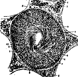
Figure 1. Cross-section of a seminiferous tubule of a rat: 1-interstitial cells; 2-vessels; 3-fatty and staining granules; 4-heads of spermatids; 5-spermatogonia; 6 and 7-membrana propria; 7-nuclei of Sertoli cells; 9-spermatocytes; 10-spermatids. (According to Kolliker.)
one can distinguish three types of cells: 1) germ cells, or primordial, which give rise to spermatids; they have a rounded shape and form the main mass of the layer; 2) Sertoli nourishing cells, the bases of which adhere to the membrana propria of the tubule, and the body in the form of a cylindrical column passes through all layers of the germ cells; 3) in certain areas, spermatids, with their heads immersed in the lobules of Sertoli cells, and with their tails directed into the lumen of the tubule. S. gives a very complex and tangled picture. 1. The spermatogenic series consists of three main cellular forms in the following sequence: a) spermatogonia (spermi-), b) spermatocytes (spermi-), c) spermatids (spermatids). Spermatogonia, descendants of maternal germ cells (see Testis, development), lie along the wall of the tubule on the expanded feet of Sertoli cells; they have a small size, round or oval nuclei (6-11 μm) and a thin rim of protoplasm. According to the character of the nucleus, two types of spermatogonia are distinguished: with a thin, finely granular chromatin network, a 'powdery nucleus' (sp. a noyau poussiereux, Regaud) and with a coarser network, a 'crust-like nucleus' (sp. a noyau croustillé); the former are the initial form, from which, by karyokinesis, spermatogonia with a coarse-grained nucleus are formed, part of which remains as a reserve. Spermatogonia with a coarse-grained nucleus in turn divide, and the products of their division are first-order spermatocytes; at first they have a small size, but then gradually grow, reaching a size of up to 25 μm with nuclei of 12-13 μm (rat; Ebner). This period, lasting quite a long time, is called the 'growth period'; during it, preparation for division occurs in the nucleus, the prophase of karyokinesis, which proceeds in a special way and ends with the formation of special chromosomal groups, so-called tetrads (see Reductional division). Tetrads (quartets) have the appearance of rings or four short sticks arranged in two rows. The subsequent 'maturation period' consists of two rapidly following each other reductional divisions. After the first division, which is characterized by a large karyokinetic figure, two smaller cells arise—second-order spermatocytes. In many animals their nuclei do not come to a state of rest, and second-order spermatocytes, barely formed, begin to divide; in others (rat; Ebner) they exist for some time as resting cells. The division figures of second-order spermatocytes are distinguished by a smaller size and a spindle occupying the entire cell; the result of the division is spermatids, which receive a reduced half amount of chromosomes. These are rounded, small cells with a delicate chromatin network, a nucleolus, and an idiosome in the cell body. At this moment, Sertoli cells come into action; protoplasmic outgrowths are formed on them, which absorb spermatids (copulation). The formation of spermatids takes place in Sertoli cells; at the height of the process, Sertoli cells acquire the appearance of candelabras with protruding sperm tails; there are 8-12 in each cell. After the disintegration of the lobules, spermatids are released. According to Ebner's calculations, the full cycle of development lasts 21 days, of which the growth period occupies 8-9 days; from the formation of spermatids to the expulsion of spermatozoa, about 10 days pass. 2. The topography of S. in mammals is very complex and was elucidated in detail only in rodents by the studies of Ebner (rat), Lenhossek (mouse), and Regaud (guinea pig). A certain number of adjacent cells pass through the cycle of development simultaneously and give an exactly identical picture; these are so-called isogenic groups. On any section of the tubule, several isogenic groups, superimposed on each other, are always visible, most often in four floors: in the very lowest place are spermatogonia, in the uppermost—developed spermatids; but these groups belong to different moments of S., between which there are gaps, which is why it is impossible to study the entire spermatogenic series on a section of one tubule and a series of consecutive sections is required. The scheme of Fig. 2 shows the distribution of groups in eight consecutive sections, exhaustively covering all the diversity of S. pictures; the successive elements of the series are denoted by numbers. In general, the process of S. spreads along the length of the tubule in a wave-like manner (Ebner's spermatogenic waves), starting from the rete testis. Such a regular course, in all tubules at once, has been described only in rodents; in man and bull, along with active areas, there are those in which S. stops. In general, the process of S. depends on the general condition of the organism and is easily interrupted by phenomena of degenerative disintegration of germ cells, for example, with strong fatigue, exhaustion, psychic shocks, alcohol poisoning (Stieve), infectious diseases, especially chronic ones, etc. Complete cessation of the process causes ligation of the spermatic duct; after final degeneration of germ cells, only Sertoli cells remain, which turn into a proper cylindrical epithelium. With age, the size of active areas decreases, but sperm production can continue until 70-80 years. 3. Spermatohistogenesis, the process of transformation of a spermatid into a mature spermatozoon, has been studied in detail in rodents and in man (Lenhossek, Duesberg, Meves). A developed spermatozoon (see Spermatozoa) consists of a head with a sheath, or acrosome, a neck, a connecting or intermediate part, and a tail. The head is formed from the nucleus of the spermatid by a corresponding change of shape, accompanied by the release of nuclear sap and condensation of the chromatin. The sheath is formed with the participation of the idiosome, inside of which a bubble arises and in it a body, staining with acidic dyes (acrosome); the bubble emerges from the idiosome and adheres to one end of the nucleus in the form of a crescent; the acrosome gives rise to the sheath. Complex changes are undergone by the centrosome, also emerging from the composition of the idiosome; it approaches the membrane of the spermatid and divides into two parts: proximal and distal; from the distal one a flagellum, the rudiment of the tail, grows out, then both are directed towards the nucleus; the proximal, adhering to the nuclear membrane, gives rise to the two upper centrosomes of the spermatozoon; the space between the proximal and distal centrosomes forms the neck. Even earlier, the distal centrosome acquires the shape of a cone,
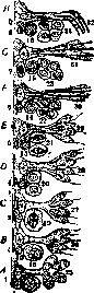
with its wide base directed towards the tail; the peripheral part of the cone separates in the form of a ring, which slides along the tail to the cell membrane, marking the lower boundary of the intermediate part. The formation of the spiral thread, winding around the axial thread of the intermediate part, occurs at the expense of mitochondria (Benda). The protoplasm of the spermatid disappears over a greater extent and remains around the neck and the intermediate part, surrounding them in the form of a drop; in this form the spermatozoon is expelled and its final development occurs already in the lumen of the tubule.
Karpov.
Related articles
Mentioned in
Cite this page
“Spermatogenesis.” Soviet Medical Encyclopedia. English translation of Bolshaya Meditsinskaya Entsiklopediya, 1st ed. (Moscow, 1928–1936), ed. N. A. Semashko. https://sovietmedicalencyclopedia.pages.dev/article/spermatogenesis/