Sexual Organs
Historical document, translated for reference. It reflects medical knowledge of the 1920s–30s and is not medical advice.
Summary
This article describes the anatomy and development of sexual organs in animals, including gonads, reproductive tracts, and external genitalia, with particular focus on vertebrates and mammals.
Encyclopedia article (1928–1936)
SEXUAL ORGANS (genitalia), organs of sexual reproduction, consisting in animals of the sex glands, which produce sex cells (eggs and spermatozoa), of the sexual passages, serving for the removal of sexual products to the outside, and in the female often also as the source of formation of the egg's protective coverings, the site of fertilization and sometimes the site of embryo development, and finally of the external genitalia, or copulatory organs, ensuring in certain animals the introduction of sperm into the female sexual passages. The sex glands of vertebrates develop as paired thickenings of the body cavity wall on either side of the dorsal mesentery. These thickenings consist of folds of epithelium with an internal mass of connective tissue and are called sex or rudimentary ridges. Subsequently, the rudimentary ridges grow deeper into the body cavity, separate from its wall, and thus become suspended inside the body cavity on their mesentery. The internal development of the glands proceeds differently in the two sexes. From the rudimentary epithelium, "Pflüger's" cell cords grow into the connective tissue stroma, in which there then occurs in the female differentiation into separate, larger primary egg cells and surrounding smaller follicular cells. Later, the cords gradually break up into separate primary follicles (see Ovaries). In the males of lower vertebrates, the rudimentary epithelial cords also break up into separate follicular vesicles, in the walls of which numerous sex cells develop. In higher vertebrates, the rudimentary epithelial cords are directly transformed into the convoluted seminiferous tubules (see Testis). From the Malpighian bodies of the primary kidney, epithelial "Hoffmann's" cords also grow into the gonad, participating in the formation of the ovarian medulla. In the male, these cords transform into a network and straight tubules of the testis. They remain connected with the tubules of the primary kidney, from which they originated. These tubules give rise to the efferent ductules (vasa efferentia), which empty into the excretory duct of the primary kidney, which now receives the name of the vas deferens ( see vol. XIX, art. 209, fig. 4). The initial portion of the latter is strongly convoluted and together with the aforementioned ducts forms the epididymis (epididymis). The remaining ductules of the primary kidney are preserved in higher vertebrates only in the form of insignificant remnants (paradidymis). In the female, alongside the ovary, insignificant remnants of the primary kidney ductules are also preserved in the form of a rudimentary appendage (epoophoron and paroophoron). In mammals, the sex glands, which initially lie in the lumbar region, subsequently move in a caudal direction. This applies partly to the ovary (descensus ovariorum), but especially to the testis (descensus testiculorum), which in many mammals penetrates through the inguinal canals into the scrotum, developing from the sex ridges.
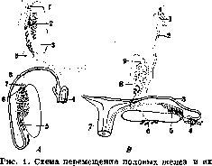
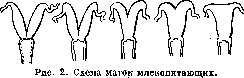
In vertebrates, the sexual passages consist of the ducts of the excretory system (see Urinogenital organs) and specifically in the male the excretory duct of the primary kidney—the Wolffian duct, and in the female a duct developing in connection with it (Müllerian duct) (fig. 1). In males, only the upper ends of the Müllerian duct are preserved in the form of a pedunculated hydatid (hydatida Morgagni) and the lower ends, connected with each other, in the form of a "male uterus" (uterus masculinus, s. sinus prostaticus). In male mammals, the lower portion of the vas deferens forms the ejaculatory duct (ductus ejaculatorius), and in connection with it, the glandular seminal vesicles (vesiculae seminales) develop. The vas deferens opens in lower vertebrates into the cloaca, but in mammals into the urogenital sinus, which is drawn out in all viviparous mammals into a long urethra. The oviducts of lower mammals also connect directly with the cloaca, but in viviparous mammals, the aforementioned urogenital sinus separates from it, remaining in the female as a very short vestibule of the vagina. The oviducts themselves in their terminal portions fuse together, forming an unpaired vagina (vagina). In connection with the development of viviparity in all typical mammals, the main part of the oviducts forms more or less voluminous uteri (uterus), in which the embryo develops. These uteri (see Uterus) may remain paired or, beginning from the cervix, more or less completely fuse, forming in various mammals a double, bipartite, bicornuate, or finally a simple uterus (the latter in bats, monkeys, and man) (fig. 2). The distal part of the oviducts forms the proper oviducts, or Fallopian tubes (see).
The external genitalia, generally more complexly arranged in males (copulatory organs), can have the most diverse origins, structures, and developments. Even in vertebrates, the copulatory organs are developed very differently. Appearing only in forms with internal fertilization, they can in individual groups reach a high degree of development. In cartilaginous fishes (sharks and rays), modified parts of the pelvic fins serve as copulatory organs, while in bony fishes sometimes the anal fin performs this function. In terrestrial vertebrates, parts of the cloaca serve for copulation. In lizards and snakes, the copulatory organs have the form of paired sacs lying under the skin at the posterior edge of the cloaca and can be protruded through it to the outside. In turtles, crocodiles, and some birds (in ostriches, ratites, and some gallinaceous birds), an unpaired copulatory organ develops on the anterior wall of the cloaca, supported by fibrous tissue (corpus fibrosum). Along the organ runs a longitudinal groove for the passage of semen, bounded by erectile cavernous tissue (corpus spongiosum). The distal end of the organ can be protruded from the cloaca by the action of special musculature. In monotreme mammals, the copulatory organ is still located inside the cloaca, surrounded by a fold of mucous membrane (vagina) (fig. 3). Compared with reptiles and birds, this organ is perfected in that the longitudinal groove is closed into a canal. This canal begins with an opening at the base of the penis in the urogenital sinus and ends at the apex. When the opening leading from the sinus into the cloaca is closed, the semen passes not into the cloaca but along the canal of the copulatory organ.
In viviparous mammals, the penis (see) reaches a higher development (fig. 3). The cloaca disappears in them, and between the urogenital sinus and the canal of the copulatory organ there occurs a permanent and continuous communication, so that the latter, being a continuation of the sinus, serves not only for the removal of semen but also for urination. The penis is located externally, surrounded by a fold of skin forming its prepuce. With the progressive development of the perineum in higher mammals, the penis moves progressively farther forward. The copulatory organ of mammals is supported by a paired or sometimes unpaired erectile fibrous body (corpus fibrosum, s. corpus cavernosum penis) and also by an unpaired erectile cavernous body surrounding the urethra (corpus spongiosum, s. corpus cavernosum urethrae) and forming at the end of the penis more or less swollen glans penis of diverse shape. The anterior part of the fibrous body is usually supported by a cartilaginous or bony axis (os penis). The latter is absent in ungulates, in
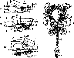
Figure 3.
Figure 4-
Figure 3. Diagram of the structure of the male sexual apparatus: I - of monotremes, II - of marsupials, III - of placental mammals; 1 - rectum; 2 - anus; 3 - cavernous bodies of the penis; 4 - Cooper's gland; 5 - pubic bone; 6 - abdominal muscles; 7 - urinary bladder; 8 - vas deferens; 9 - ureter; 10 - perineum; 11 - cavernous body of the canal; 12 - testicle; 13 - prostate; 14 - seminal vesicle. Figure 4. Sexual organs with their accessory glands in the male hedgehog (Erinaceus europaeus): 1 - kidney; 2 - seminal gland; 3 - urinary bladder; 4 - testicle; 5 - prostate; 6 - penis; 7 - glans penis; 8 - crus penis; 9 - vas deferens; 10 - ureter. In whales and in humans. In connection with the urogenital canal of mammals, a number of accessory glands develop, of which the most constant are the prostate gland (see) and the paired Cooper's glands (fig. 4). In the mammalian embryo, the external genitalia develop in the region of the anterior edge of the cloaca in the form of a small protrusion at first - the cloacal tubercle. In the anterior part of the cloacal tubercle, the genital tubercle develops, and on the lower surface of the latter, a longitudinal genital groove is incised, bounded on the sides by genital folds (fig. 5). Around all these parts, a ring-shaped fold - the genital swelling - develops. These indifferent parts of the sexual apparatus then develop differently in both sexes (fig. 5). In the male, the genital tubercle grows strongly and turns into the penis. The genital f folds fuse with each other, so that the genital groove turns into the urethra. The genital swellings also fuse with each other along the midline and form the scrotum. In the female, the genital tubercle gives rise to the clitoris, the genital folds - the labia minora, well developed in many mammals, and the genital

Figure 5. Development of external genitalia in humans: A - indifferent stage: 1 - phallus; 2 - genital swelling; 3 - primary urogenital opening; 4 - anus; B - in female embryo: 1 - glans clitoridis; 2 - labium majus; 3 - primary urogenital opening; 4 - anus; C - in male embryo: 1 - glans penis; 2 - scrotum; 3 - primary urogenital opening; 4 - anus.
swellings - labia majora, characteristic of some monkeys and of humans. In some insectivores, in many rodents, and in prosimians, the female's clitoris, like the male's penis, contains the urethra, opening outward at its end.
I. Shmal'gauzen. Parasites of the sexual organs. P. o. can be a habitat for various parasites. Among the protozoa, flagellates - Trichomonas vaginalis - are found in the vagina. Among parasitic worms, the penetration of pinworms (Enterobius vermicularis) into the vagina is observed. Pinworms can also penetrate into the pathologically altered wall of the uterus, move into its cavity and enter the Fallopian tubes. In the walls of the uterus, vagina, and in the tissues of external P. o., eggs of Schistosomum haematobium are found in bladder bilharziasis; in the genital cleft in this same disease, papillomatous growths can develop. The uterus and ovary can be affected by echinococcus. A case of a well-developed ascaris in the ovary is known. Of particular interest is the possibility of parasites penetrating from the blood vessels of the uterus into the body of the fetus through the placenta. Such intrauterine infection is possible with malaria parasites, sleeping sickness (Trypanosoma gambiense), the causative agent of Chagas' disease (Trypanosoma Cruzi), leishmaniasis (kala-azar), liver flukes (in sheep, a child?), Schistosomum japonicum, Taenia solium, Dirofilaria immitis (in dogs), hookworms, ascarids (especially in dogs), etc. (see Intrauterine invasion). The external P. o. of women can greatly enlarge in elephantiasis (filariasis). - In the P. o. of men, various parasites can also be found. Trichomonas vaginalis is rare in the urethra; dysentery amebas and plerocercoids of Piphyllobotrium mansoni, previously described under the name Sparganum mansoni, can also be found here. In bladder bilharziasis, scrotal fistulas sometimes form. One form of filariasis is lymphoscrotum, which in some cases leads to the development of elephantiasis of the scrotum (larvae of Wuchereria bancrofti are found in the lymph). Echinococcus can develop in the testicle. Dysentery amebas sometimes parasitize in the epididymis. In some forms of elephantiasis, the penis enlarges. In the pubic hair of men and women, pubic lice (Phthirius inguinalis) can parasitize.
Related articles
Mentioned in
Cite this page
“Sexual Organs.” Soviet Medical Encyclopedia. English translation of Bolshaya Meditsinskaya Entsiklopediya, 1st ed. (Moscow, 1928–1936), ed. N. A. Semashko. https://sovietmedicalencyclopedia.pages.dev/article/sexual-organs/