Ovaries
Historical document, translated for reference. It reflects medical knowledge of the 1920s–30s and is not medical advice.
Summary
The ovaries are the female reproductive organs where eggs are formed and mature. This article describes their anatomical structure, development, and variations across different animal species.
Encyclopedia article (1928–1936)
OVARIES (ovarium, oophoron, ovarion), the female reproductive glands in which the formation and maturation of eggs occurs. In lower invertebrate animals, ovaries may be absent altogether (sponges) or may exist only as temporary accumulations of sex cells (some coelenterates) at various stages of their formation. In the vast majority of animals, ovaries are completely isolated, permanent organs. Two basic types of ovarian structure are distinguished: 1) sac-like ovaries, where egg formation occurs in the germinal epithelium lining their internal cavity; mature eggs fill this cavity and are then expelled outward through special ducts; 2) compact ovaries with internal connective tissue stroma and an outer lining of germinal epithelium (which also grows to a greater or lesser degree into the stroma), in which egg formation occurs; in this case, mature eggs are released through rupture of the outer ovarian wall into the general body cavity, from where they are then expelled outward through the excretory tubules or at least completely independent of the ovaries (annelid worms, vertebrate animals). In lower worms, ovaries exist as sac-like organs, sometimes elongated into long tubes. The difference between the sac-like ovaries of acoelomate and primary coelomate animals and the compact ovaries of secondary coelomate animals is explained by the origin of the secondary body cavity through the expansion of the cavities of the sac-type sex glands (the "gonocelium" theory). However, even in secondary coelomate animals, ovaries sometimes acquire a sac-like structure through the isolation of part of the general body cavity (arthropods, mollusks, bony fishes). Generally, ovaries are usually paired organs of various shapes, the essential part of which is the aforementioned germinal epithelium. In the germinal epithelium, in addition to sex cells, which sometimes differentiate into actual eggs and various types of nutritive cells, there are also somatic cells arranged in a continuous layer around developing eggs and receiving the name follicular cells. The latter also participate in the nutrition of the egg, and often also in the formation of egg membranes. The ovaries of vertebrate animals, always paired in origin, sometimes become unpaired due to the fusion of both ovaries (in some fishes) or due to the underdevelopment of one ovary (the right one in birds). It develops from paired swellings of the body wall cavity on either side of the dorsal mesentery. These swellings consist of a fold of thickened epithelium with an internal mass of connective tissue and are called sex or germinal folds. The primary sex cells, which separate from the embryo much earlier, independently of the rudiment of the sex gland and even independently of the middle germ layer, then migrate toward the dorsal wall of the body cavity and penetrate into the sex folds. From the Malpighian bodies of the primary kidney, epithelial "Hoffmann's" cords grow into the rudiment of the sex gland, participating in the formation of the ovarian core. The sex folds then project deeper into the body cavity, separate from its wall, and thus become suspended inside the body on their mesentery. The germinal epithelium grows into the connective tissue stroma of the ovary in the form of multicellular cords (called in mammals Pfuger's), containing both primary sex cells and follicular cells. Subsequently, these cords break down into rounded complexes of cells—follicles—each consisting of one large female cell (oocyte) surrounded by a layer of many follicular cells. The maturing follicle moves toward the surface of the ovary and then
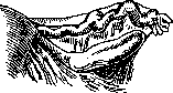
Figure 1. Anatomical preparation of the right ovary, posterior view: tube and right angle of the uterus.
the mature egg falls into the body cavity through rupture of its wall. In mammals, the follicle wall is multilayered and inside it, a cavity filled with fluid develops eccentrically. Such a follicle receives the name of Graafian follicle. In place of the follicle that ruptures when the mature egg is expelled, a scar forms from degenerated follicular cells, the so-called yellow body (corpus luteum), which is a temporary organ of internal secretion. In mammals, the ovary somewhat shifts from its original position at the level of the primary kidney backward into the area of the small pelvis (descensus Ovariorum).
I. Shmalgauzon. In the bodies of animals and humans, the ovaries undergo significant and frequent changes. The size, shape, general appearance, and structure of the ovaries change not only according to the age of the woman, but also depending on various physiological states of her body: sexual maturation, sexual maturity, pregnancy, climacterium, menopause, etc. Sharp changes in the ovaries also occur in connection with menstrual cycles. The shape of the ovaries is very variable, especially during their embryonic development and in early childhood. If the internal sexual organs of a newborn or a girl of the first years of life are removed from the pelvis and made into an anatomical preparation, the ovary will appear as (fig. 1) an almost three-sided, prismatic or spindle-shaped body with rounded sides and a smooth surface. It is attached by a short duplication of the peritoneum—mesovarium (mesentery)—to the posterior leaf of the broad ligament, with its more pointed pole, facing the uterus (extremitas uterina), connecting to the angle of the uterus by a ligament called lig. ovarii proprium; it attaches to the angle of the uterus behind and somewhat below the exit of the Fallopian tube. The blunter pole of the ovary, facing the fimbriae of the tube (extremitas tubaria), is also fixed by a special ligament—lig. suspensorium ovarii (lig. infundibulo-pelvicum)—to the fascia covering the m. psoas, i.e., to the lateral surface of the pelvis. Some authors describe another ligament of the right ovary—lig. appendiculo-ovaricum (Clado), however, the existence of such a ligament is questionable. One of the fimbriae of the tube reaches the extremitas tubaria ovarii along the outer edge of the mesovarium and is called fimbria ovarica. This frill has the shape of a groove. Some authors attributed to it an important role in the process of the egg passing from the ovary to the tube. Mesovarium attaches to the upper edge of the ovary—margo mesovaricus (rectus)—which has the shape of an almost straight line (fig. 2). The ovary is covered by a special, so-called germinal, epithelium, mor
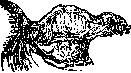
Figure 2. Anatomical preparation of the right ovary: tube and right angle of the uterus. Lig. ovarii proprium and lig. suspensorium, s. infundibulo-pelvicum. Margo liber, s. inferior. Margo mesovaricus, s. rectus. Histologically distinct from the endothelium covering the peritoneum. Mesovarium serves as the place of entry for vessels and nerves into the ovary from the broad ligament (hilus ovarii). The lower edge of the ovary has a convex shape and is called margo liber (s. inferior), hanging freely in a semicircle into the abdominal cavity. The size of the O. in a sexually mature woman varies approximately within the following limits: length between 25 and 50 mm, width 15-35 mm, thickness 5-15 mm. The right O. is usually somewhat larger than the left. During the climacteric period, the size of the O. somewhat decreases (involutio senilis). Waldeyer gives the following figures for the weight of the O.: in newborns-0.5 g, in children-2-3 g, in adults-6-8 g, in old women-1-2 g. In a sexually mature woman, the O. has an elliptical, somewhat flattened shape, sometimes spindle-shaped and even spherical. The surface of the O. during sexual maturity is slightly uneven, and at times contains folds or grooves that penetrate only shallowly into the tissue of the organ. In old women, these grooves often cover the entire O. In such cases, one speaks of ovarium gyratum. The grooves can apparently also be congenital. Basically, they form as a result of atresia of follicles and the formation of scars after ovulations. The position of the O. in the pelvis has great clinical significance. Fixed to the broad ligament by the mesentery (mesovarium) and two lateral ligaments (lig. ovarii proprium and lig. infundibulo-pelvicum), the O. in most cases has a definite, though changing within certain limits, position. It is necessary, however, to remember that the position of the O. in the pelvis in relation to the uterus differs sharply from the position that we observe on anatomical preparations. Since the uterus is deviated forward by its fundus, the tubes and O. lie somewhat behind it. In this case, the O. are located in well-defined depressions in the peritoneum (fossa ovarica), located in the area of the branching of a. iliaca com. into a. iliaca externa and a. hypogastrica. In many animals, the O. is located in a special fold of the peritoneum, forming a kind of hood over it (bursa ovarica) or even a completely closed sac (in horses), whereby the O. is as if extraperitonealized. The anterior surface of the O., looking forward on the anatomical preparation, in a living woman looks not forward, but to the side, and therefore is called facies lateralis, while the posterior surface is turned toward the median line and is called facies medialis. In this case, its extremitas uterina becomes the lower pole, and extremitas tubaria the upper pole. A number of pathological processes can change the normal position of the O., causing its prolapse or, what happens more often, an upward displacement. The position of the O. changes especially during pregnancy, when the growing uterus pulls it upward. By the end of pregnancy, the O. rises to the level of the navel (Ols-hausen), and lies not to the side of the uterus, but next to it, at its edges. The O. is supplied with blood from two arteries- a. ovarica, penetrating to the O. through lig. suspensorium (infundibulo-pelvicum), and branches of a. uterinae. From the anastomosis formed by these arteries, numerous branches originate, entering the hilus ovarii and forming numerous branches throughout the gland. Corkscrew-like twisting in the area of the hilus and the medulla layer, these arteries are accompanied by wide numerous venous trunks, which form in the mesovarium the so-called plexus ovarii pampiniformis. The O. has a wide network of lymphatic pathways. Around primordial follicles and in the area of the albuginea, lymphatic vessels have not yet been discovered, whereas in the walls of maturing follicles they are found in the form of an expressed network, covering also the corpus luteum. The O. is supplied with a system of sympathetic nerves, entering it together with the vessels. Microscopic structure of the O. When studying the microscopic structure of the O., it is necessary to take into account both the age and the physiological states during which the material for research is taken, as well as certain anamnestic data: the number of previous births and abortions, their timing, the time of menstruation, etc. It is best to study the O. on cross-sections. Even with the naked eye, one can note that the O. consists of two layers: cortical (substantia corticalis) and medullary (substantia medullaris). Examining sections from the O. even at low magnification, we can easily distinguish these two layers on them, especially on stained preparations. The cortical, denser layer horseshoe-shaped surrounds the second-medullary layer, with the absence of cortex noted toward the hilus ovarii. The thickness of the cortical layer and the power of the medullary layer depend on the age of the individual. In the child's ovary or in newborn girls, the cortical layer occupies almost all the space of the section, leaving little room for the medullary layer from the side of the hilus, whereas in old women, on the contrary, the cortical layer is thin, and the medullary layer occupies the larger part of the O. With a more detailed study of the microscopic picture of the O., one finds its surface covered with a low cylindrical or cubic epithelium, the so-called germinal or rudimentary epithelium-Keimepithel (Waldeyer), the cells of which differ in shape and size from the adjacent endothelium covering the peritoneum. The transition of the germinal epithelium from the O. to the peritoneum of the mesovarium, by which the O. is attached to the broad ligament, occurs not gradually, but rather quickly, thanks to which at its mesovarial edge (margo mesovaricus) the so-called Farr-Waldeyer line is formed. The germinal epithelium does not cover the entire surface of the O. One can assume that such a partial absence of covering epithelium on the surface of the O. is not a simple result (as was explained earlier) of mechanical reasons related to the preparation of the specimen. Another explanation is more likely: with the rapid growth of follicles and the stretching of the surface of the O. in the corresponding areas, the covering epithelium atrophies, and when the follicle ruptures, it even disintegrates (J. Miller). Under the layer of germinal epithelium in the O. of humans and most animals is located tunica albuginea, consisting of denser bundles of the main connective tissue of the O. The thickness of this layer does not exceed 0.1 mm, and it passes without a sharp boundary into the underlying cortical layer. Directly under the layer of t. albugineae lies the cortical layer of the O., consisting of 2 main tissues: the connective tissue basis-stroma (stroma ovarii)-and the parenchyma-epithelial elements of the O. The stroma of the cortical layer consists of spindle-shaped connective tissue cells with a large number of nuclei, which makes this tissue resemble sarcomatous. Directly extending in the direction toward the hilus, the stroma of the cortical layer passes into the connective tissue mass forming the medullary layer of the O. The parenchyma of the cortical layer consists of a large number of so-called primordial or primary follicles (folliculus primarius), microscopic, invisible to the naked eye formations of the O. Each of such follicles is filled with one large round cell-an egg, surrounded in follicles at rest by several barely noticeable, and that not on all sections, flat crescent-shaped cells, surrounding the egg and called granulosa cells-membrana granulosa, or stratum granulosum (Fig. 3).
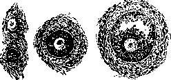
Figure 3. Primordial follicle; beginning of follicle growth; formation of liquor folliculi and its shell-theca folliculi.
The number of follicles in the ovaries varies within the widest limits: while in a newborn girl they are so numerous that they lie almost directly against each other, being separated by only the thinnest layers of connective tissue, in mature women and especially those approaching the climacteric period they are very few, and sometimes one must examine a whole series of sections to find one or two follicles. In old women, follicles almost completely disappear from the ovaries. Thus it becomes understandable why attempts to count the follicles in the ovaries have led to different conclusions. Henle found in an 18-year-old girl in one ovary 36,000 eggs, Sappey counted in a 2-3-year-old girl in both ovaries approx. 600,000 and even 1,150,000 follicles. Hoggestrom in a 22-year-old healthy unmarried woman counts in both ovaries approx. 400,000 follicles. Not infrequently in one follicle one can observe two or more egg cells, each containing two nuclei. This is especially frequently observed in childhood. The question of the origin of the egg cell, of oogenesis (see), until recently can hardly be considered resolved. Previously it was thought that the germinal epithelium covering the ovaries, growing into the connective tissue stroma of the ovary, forms the so-called Pflüger's cords, or Valentin-Pflüger's tubes, from the cells of which the parenchyma of the ovary differentiates. Due to the ingrowth of connective tissue, the entire epithelial mass, as was thought, divides into separate small cells—primordial, or primary follicles. At the present time, it can be stated with greater certainty that the germinal epithithelium does not participate in the formation of eggs and that eggs differentiate from the so-called indifferent sexual material, which forms on the inner surface of the primary kidney (Wolffian body), perhaps by migration here from other parts of the embryo's body, especially its caudal end (Rubashkin). As most researchers think, the eggs lying in the primordial follicle are already oocytes, not oogonia, since they are incapable of reproduction, but can only by the secretion of directive bodies give rise to the following generations of oocytes. They are oogonia, i.e., cells capable of division, only during the embryonic period of development. Thus, until recently, the possibility of reproduction in women, or otherwise, the formation of new eggs in the postembryonic period was denied. However, this opinion, which has fundamental biological significance, is disputed by some authors, at least for some species of animals (Paladino). Winiwarter and Semon point out that in cats after childbirth all eggs in the ovaries die and that after this eggs are newly formed. According to Kingery, in white mice oogenesis begins only 2-3 days after birth and continues until sexual maturity is reached. A whole series of authors (Allen, Sun) assert that the formation of eggs in sexually mature mice occurs periodically, depending on the estrus. However, this question is far from being resolved. Follicle maturation. Even in young age and even in sexually immature animals in the deeper parts of the cortical layer, more developed follicles are found, differing both in size and shape. In sexually mature women such a follicle can reach a large size and as a result of the maturation process becomes a Graafian follicle [folliculus oophorus (Graafi)]. The process of maturation of the primordial follicle consists of the following: the flat crescent-shaped cells of the granulosa in the maturing follicle gradually take on a cubic shape and, multiplying by mitosis, first cover the egg with one continuous dense layer, and then with two to three and many layers of follicular epithelium (fig. 3). With the accumulation of granulosa, some cells and their groups die; disintegrating, they in places give rise in the thickness of the granulosa to islands with tissue defects, filled with liquid content. Such islands later merge and lead to the formation of one common cavity filled with transparent fluid. The fluid accumulating in the follicle stretches the follicle to a large size and pushes the egg with the surrounding granulosa to one of the poles of the follicle. This fluid is called follicular fluid (liquor folliculi). The follicle containing it is called a Graafian follicle (fig. 4). The process of maturation of primordial follicles usually affects those located in the deeper layers of the ovarian cortex, and only one follicle out of all those in the ovary, more rarely two to three or more. As the maturing follicle increases in size, it occupies more and more space in the cortical layer and with its pole facing the surface of the ovary reaches its level and even protrudes slightly outward, thinning the tunica albuginea. This place is called the stigma folliculi—the place where it will rupture and through which the egg will be expelled into the abdominal cavity. The structure of a mature, ready-to-rupture follicle is very complex (fig. 4). The wall of such a follicle consists of a thick capsule (theca folliculi), built from the connective tissue stroma of the ovary. Even at the beginning of follicle maturation, two layers can be clearly distinguished in its wall: inner and outer. The inner layer (tunica propria, s. vascu-losa, s. interna) consists of relatively large polyhedral cells, which appear lighter in preparations. The protoplasm of these cells contains a special lipoid substance, apparently the same as in the lutein cells of the corpus luteum (see below). These cells are called theca-lutein cells (fig. 4). The outer layer of the theca folliculi is called the tunica fibrosa, or externa. It has no special physiological significance and consists of compacted connective tissue. However, Winiwarter found muscle elements in this layer, which is not without significance in the emptying of the follicle. The inner surface of the tunicae vasculosae, to which the granulosa cells adhere, is covered with a transparent, consisting of delicate fibrils membrane, which was formerly considered structureless and to which the name boundary layer (Höegbom) is assigned. This layer separates the granulosa from the theca folliculi proper. The entire large cavity of the follicle (fig. 4) is occupied by liquor folliculi. This fluid contains paralbumin (Waldeyer) and according to a whole series of recent researchers (Allen, Zondek, Aschheim and others) also a special sex hormone, produced by the cells of the thecae internae. In the developed follicle, the granulosa, located on the inner surface of the Graafian follicle in the form of a one- or two-layered epithelium (fig. 4), accumulates at one pole of the follicle, forming the so-called discus or cumulus oophorus, or proligerus. In the center of this accumulation of granulosa is located the largest cell of the body—the egg, surrounded by cells of the granulosa arranged around it in rays (corona radiata) and enclosed in a shiny radially striated membrane (zona pellucida) (fig. 5). The zona pellucida in places separates from the protoplasm of the egg, giving rise to the formation between the egg itself and its shiny membrane of the so-called perivitelline (perivitelline) spaces. The human egg in its developed form in the Graafian follicle reaches 0.20 mm in diameter. In its protoplasm are contained granular accumulations of yolk, and somewhat eccentrically lies the nucleus (vesicula germinativa), containing a nucleolus (macula germinativa) and a thin network of chromatin. The fate of the maturing follicle is different: the follicle, having reached its maturity, either ruptures, and the egg, carried away by the current of liquor folliculi, is expelled from the ovary (ovulation), or the follicle, without completing its development, undergoes regressive changes and gradually dies by the so-called atresia of the follicle. In the first case, after ovulation, in the cavity formed in place of the former follicle, a temporary gland of internal secretion develops—the corpus luteum (see Corpus luteum)—which quickly goes through its progressive and regressive phases of development depending on whether this body develops during the menstrual cycle or during pregnancy (corpus luteum menstruale and corpus luteum graviditatis). If ovulation does not occur, the Graafian follicle undergoes the process of atresia and turns into a corpus atreticum. The egg in such a follicle and its granulosa die, the liquor folliculi is absorbed, the boundaries that were clearly defined become smoothed over. The most important role in replacing the space formed by the death of the egg and granulosa belongs to
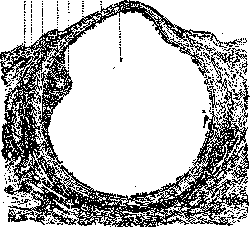
Fig. 4. 1-germinal epithelium, 2-albuginea, 3-theca folliculi interna-vasculosa (tunica vascu-losa); 4-theca folliculi externa fibrosa (tunica fibrosa); 5-discus oophorus; 6-granulosa; 7-stigma ovarii; 8-liquor folliculi; 9-boundary layer of Höegbom (Grenz faserschicht).
the connective tissue, which fills the space left after the atresia of the follicle. The corpus atreticum is thus a structure consisting of connective tissue, in the center of which are found the remains of the follicle: the collapsed theca, the degenerated granulosa, and the dead egg. The corpus atreticum is gradually absorbed and replaced by connective tissue. The fate of the egg after ovulation is also different: if fertilization does not occur, it dies and is absorbed; if fertilization occurs, it develops further (see Fertilization). The process of ovulation is accompanied by certain changes in the ovary: the stigma folliculi becomes more pronounced, the follicle wall at this place thins, and the follicle ruptures. The egg, together with a part of the granulosa (corona radiata), is expelled into the abdominal cavity, from where it is captured by the fimbriated end of the fallopian tube. The liquor folliculi is also expelled and is absorbed by the peritoneum. After the rupture of the follicle, the cavity formed is filled with blood, which is later absorbed. From the cells of the theca interna and granulosa, a temporary endocrine gland develops—the corpus luteum (see Corpus luteum).
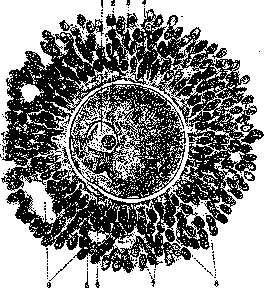
Fig. 5. Egg of a mature follicle: 1-germinal vesicle; 2-germinal spot; 3-zona pellucida; 4-perivitelline space; 5-protoplasm; 6-deutoplasm; 7 and 8-cells of corona radiata; 9-follicular fluid.
with atresia of the theca interna folliculi, i.e., cells of connective tissue character. The egg's resistant zona pellucida remains for a long time. The German border layer (see above) due to swelling, thickening and hyalinization forms a structureless, coiling into a ball, so-called glassy 772 membrane (Glasmembran). The formation called corpus atreticum is preserved in the Ovaries for a long time, but in the end it is also replaced by connective tissue. Some researchers call corpora atretica 'interstitial gland', attributing to it an internal secretory function. In some pathological cases, e.g., in hydatidiform mole and chorioepithelioma, an especially vigorous growth of cells of the thecae internae occurs in atretic follicles, which can lead to the formation of voluminous tumors, so-called lutein cysts, sometimes exceeding the size of a grown man's fist. The Ovaries belong to glands possessing double secretion. It produces a secretion which is the egg, and an incretion, or internal secretion, entering the organism through the blood and lymphatic systems and having undoubtedly immense significance in the general economy of the organism. A whole series of experimental and clinical observations speaks for this. The influence of the Ovaries on the sexual organs is especially great. Upon removal of the Ovaries, especially in childhood, the sexual sphere stops in its development, and in adults it comes into a state of atrophy. For a long time researchers, for the purpose of studying the physiological role of the ovaries, have made attempts at various kinds of ovarian transplantations to animals (Preobrazhensky, Rubinstein and others). Numerous experiments conducted in this direction allow one to come to the conclusion that autotransplantation of the Ovaries (transplantation of one's own Ovaries) in most cases ends successfully, i.e., the Ovaries not only take but the transplanted one can continue its functional work, up to follicular maturation and ovulation. Homiotransplantation, in which transplantation of the Ovaries of the same species to an animal is performed, especially in young animals and animals of the same litter, can also end in complete success. Almost negative results are obtained in the so-called heterotransplantation - transplantation of Ovaries of other species to animals. In recent times Steinach, and then a whole series of experimenters (Zavadovsky) have successfully performed a series of so-called heterosexual homiotransplantations, transplanting to animals and hens sexual glands, although of the same species of animals, but of the opposite sex. Such transplantation succeeds mainly in young animals, if previously the animal to which the sexual glands are transplanted has had its own gonads removed. With this transplantation one can masculinize a female animal and feminize a male one. These experiments once again teach us that the sexual gland, by influencing the development of secondary sexual characteristics, can accelerate sexual maturation (pubertas praecox) or retard it (infantilismus genitalis) and perhaps even pervert it (false hermaphroditism, malformations). The uterus and especially its mucous membrane - 'the slave of the ovary', as Schroeder (Schroder) says. Removal of the Ovaries leads to the cessation of menstrual function, to artificial menopause, even in young women. In recent years thanks to brilliant works of American (Allen, Doisy) and German researchers (Zondek, Aschheim and others) our knowledge in the field of physiology of the Ovaries has significantly deepened. According to the research of Zondek and Aschheim the Ovaries are entirely under the influence of the internal secretory activity of the anterior lobe of the hypophysis, producing hormones that make the Ovaries function (Prolan A and B). Under the influence of these hormones even the Ovaries of immature animals begin to function: follicles mature in them, ovulation occurs, follicles luteinize, yellow bodies develop. These changes in the Ovaries lead to the formation in them of special ovarian hormones: hormone produced by elements of follicles, and hormones of the yellow body. These hormones determine the complex periodic changes in the uterus, especially in its mucous membrane (menstrual cycle). The hormone of growing follicles (folliculin, menformon, folliculin) has now been obtained in pure form. The secretion of the yellow body, discovered earlier by Seitz, Wintz, Fingerhut, is more complex in structure. The mentioned authors divide it into two substances: 1) lipamin, occurring mainly in young yellow bodies and possessing hyperemic property, and 2) luteolipoid, occurring in large quantity in mature yellow bodies and possessing distinct hemostatic properties. Latest data have shown that the hormone secreted by the follicular apparatus of the Ovaries leads to proliferation of the uterine mucous membrane, while the incretion of the yellow body leads to the secretory phase of the menstrual cycle (see Menstruation). The activity of the ovarian hormone can be proven experimentally. By introducing to a castrated sexually mature female with ceased after operation estrus or even to an immature animal the hormone, one can cause in them estrus phenomena, which can easily be detected upon examination of a scraping from the vagina, due to the separation during estrus in animals of the upper, non-nucleated keratinized layers of the vaginal epithelium (Schollen), very characteristic in appearance and easily detectable in the vaginal secretion (method of Allen and Doisy). The minimal amount of hormone causing estrus in an immature animal is accepted to be called a hormone unit. Naturally, these units are different for various animals. Most often the mouse unit or rat, rabbit and others are used. Pathology. Being a complex formation in its physiological role and in its anatomical and histological structure, the Ovaries are also subject to various diseases. In it a number of developmental defects are found up to complete absence of this gland (see Anovaria). Along with rudimentary state of the Ovaries, Ovaries are also found that significantly exceed in size the norm, or the so-called accessory Ovaries (ovarium accessorium). Incorrect positions of the Ovaries are also observed, especially often their descent (descensus ovarii) up to formation of ovarian hernias, in which the ovary descends through the inguinal or femoral canal into the thickness of the labia majora (hernia ovarica). In these cases the pains accompanying the descent of the Ovaries sometimes force the surgeon to resort even to operative treatment, to the operation of suturing the Ovaries to the lateral surface of the pelvis or to the upper edge of the broad ligament (ovariopexia). On the soil of infection, penetrating to the Ovaries most often from the tubes, as well as by hematogenous or lymphogenous route, inflammatory processes (oophoritis) develop in the Ovaries (see Oophoritis) both in acute and chronic form, which sometimes pass into abscess of the Ovaries (abscessus ovarii). Previously inflammatory diseases also included the change of the Ovaries, bearing the name of microcystic degeneration of it, in which in the Ovaries many small cyst-like cavities are found, originating from growing follicles. There is no doubt that microcystic degeneration of the Ovaries can be a consequence of an inflammatory process, however it is also certain that this change in the Ovaries can be observed in perfectly healthy women, with regular menstrual cycles, and is a consequence of simultaneous maturation of a whole series of follicles (Schroeder). Usually upon examination in the cavity of such follicles granulosa is completely absent or it can be found only in the form of scanty remnants. From microcystic degeneration it is necessary to distinguish the state to which the name folliculus persistens is assigned. In these cases in the Ovaries usually one, rarely two or more follicles in various stages of development, with developed granulosa, are found, while yellow bodies are completely absent. Usually such state, according to Schroeder, leads to disturbance of the functional work of the Ovaries, accompanied by bleedings of the type of metropathies. In rare cases on the surface of the Ovaries papillary growths of various size and extent can be observed. Most often both Ovaries are covered with such growths. Papillary formations on the Ovaries may originate exclusively from its surface, having no relation to the so-called papillary cysts. In the formation of such superficial papillae the germinal epithelium and the stroma of the Ovaries growing into the papilla participate. The causes of formation of these papillae have not yet been clarified. It is hardly a matter here of simply an inflammatory reaction, especially of gonorrheal nature (Gottschalk). On the other hand it is difficult to agree with those authors (Olshausen, Ruge, R. Meyer and others) who deny the possibility of existence of superficial papillae of the Ovaries, if in this Ovary papillary cysts are absent. The Ovaries are especially rich in tumors, which are encountered in this organ significantly more often than in others and are of extremely diverse character. To this time there is no such classification of these tumors that could completely satisfy us. The most widespread classification of tumors of the Ovaries can be considered the classification proposed by Pfannenstiel and somewhat modified by Franke (v. Franque). According to this classification all tumors encountered in the ovary are divided into: a) non-proliferating and b) proliferating (blastomas).
Non-proliferating tumors include simple follicular retention cysts, lutein cysts, about which mention was made above, and so-called chocolate cysts, which are a variety of lutein cysts and differ from them by their dark coloration due to the admixture of blood to the fluid contained in the cyst. In essence, non-proliferating tumors of the O. are such only conditionally, and it is more correct to isolate them into a special group of cysts of the O. To proliferating tumors, true blastomas, belongs a significantly larger number of tumors found by us in the O. According to Franke, these tumors can be divided into the following groups. A. Epithelial tumors: 1) benign-adenomas: a) cystadenoma pseudomucinosum, b) cystadenoma serosum (ciliatum, papillare); 2) malignant-carcinomas. B. Connective tissue tumors: 1) benign: fibromas, fibromyomas and even osteomas, chondromas and myxomas; 2) malignant: sarcomas and endotheliomas. C. Tumors built from many germ layers, derivatives of the egg: 1) benign: dermoids (see) and 2) malignant: true teratomas (see). Of the listed tumors, the most frequently encountered, and therefore have particularly important clinical significance, are adenomas - pseudomucinous and serous. These tumors differ from each other not only in their appearance and size, but mainly in their histological structure. While pseudomucinous cysts are built according to the type of glandular tumors, serous cysts are characterized by their papillary structure. The epithelium of pseudomucinous cysts is high, cylindrical, very similar to the epithelium of the cervical canal of the uterus. The epithelium of serous cysts, having a stronger tendency to proliferation and a more cubic shape, does not secrete mucus and in structure is closer to the epithelium of the mucous membranes of the uterine cavity and tubes. The thick mucous substance contained in pseudomucinous cysts contains pseudomucin, which in its chemical nature and relation to some reagents (acetic acid) differs from mucin. The histological origin of adenocysts has not been clarified until recently. The old view of the origin of these tumors from the epithelium of primordial follicles or even Graafian vesicles (Pfannenstiel) does not correspond to modern views. More probable is the theory recently proposed by Kermauner, according to which the parenchyma of the O., like the epithelium of the uterine and tubal mucosa (otherwise the epithelium of Müllerian ducts, from which the genital tract develops), originates from the same coelom-epithelium by differentiation in various directions. One can imagine that if in the area of the rudiment of the O. its parenchyma differentiates in the direction of cervical epithelium, conditions are created in the O. for the formation of pseudomucinous cysts, and conversely, if the differentiation of ovarian epithelium proceeds along the type of the epithelium of the uterine cavity or tubes, serous cysts will develop. Tumors of the O., especially cyst-adenomas, as they grow, lead to the disappearance of all ovarian tissue and as they grow often pull the mesovarium (lig. ovarii proprium and lig. suspensorium ovarii) into a longer or shorter stalk, along which pass vessels bringing blood to the tumor and carrying it away. Tumors on a stalk possess great mobility. Often however, growing in the direction of the mesovarium, tumors of the O. split the broad ligament and become interligamentary, intraligamentary, immobile. A mobile, equipped with a long stalk tumor can easily twist on its axis and lead to torsion of the cyst, and consequently of its stalk. At this time, the vessels carrying blood away from the tumor are compressed, the tumor swells, becomes crimson-red due to hemorrhages into its wall. Twisting of the cyst is accompanied by a stormy clinical picture of collapse and irritation of the peritoneum. The twisted cyst causes, as a foreign body from the side of the adjacent peritoneum, a reactive aseptic inflammation, which leads to the formation of adhesions of the tumor with neighboring organs and the growth of new vessels through these adhesions into the wall of the cyst for the nutrition of the tumor. A special variety of pseudomucinous cystomas can be considered the neoplasm called pseudomyxoma ovarii. These cysts are very thin-walled, usually multilocular. Their contents are gelatinous and transparent. The thin wall of such a cyst easily ruptures, and the contents spill into the abdominal cavity, covering the mesentery, intestines and all other organs of the abdominal cavity. In such cases, the so-called pseudomyxoma peritonaei (false mucous tumor of the peritoneum) develops. Carcinoma ovarii can arise in various ways. There are primary carcinomas developing in ovarian tissue as independent diseases, cases of carcinomas are observed as a result of cancerous degeneration of cystic formations of the O., and finally a whole series of carcinomas of the O. of metastatic nature, transferred here from the stomach, intestines and other organs. Both primary and secondary cancers of the O. are very often bilateral. To the primary carcinomas of the O. some authors refer the tumor that has attracted attention only in the last decade, the so-called tumor of granulosa epithelium (Granulosazelltumor). In these tumors, accumulations of cells can be found, which in their radial arrangement and general appearance are completely similar to the cells of granulosa in the Graafian follicle. This anatomical similarity with the ovarian parenchyma also attracts special attention because often in women having such tumors, changes are observed in the uterus, resembling premenstrual ones, and in old women already in menopause-bleedings similar to menstruation. Also worthy of attention is the circumstance that part of these tumors clinically proceed as benign tumors, while another part is distinguished by particularly pronounced malignancy. Until recently, the histogenesis of these tumors was unclear. Most authors see in them derivatives of follicular epithelium and even the egg, assigning them the name of folliculoma or oophoroma of the O., others consider these tumors to be ordinary carcinomas without a genetic connection with the parenchyma of the ovarian follicle. To the carcinomas of the O. must also be attributed the tumor first described by Krukenberg under the name fibrosarcoma ovarii mucocellulare (carcinomatodes). Characteristic for this tumor are large signet-ring cells, lying sometimes in groups, and in other cases separately, and resembling in shape an egg. In the overwhelming majority of cases, these tumors develop in the O. as metastases of carcinoma, brought here from the stomach, more rarely from the intestine or gallbladder. Tumors of connective tissue nature in the O. are encountered significantly less frequently than epithelial ones. Besides small usually fibromas, here are also relatively rarely encountered sarcomas, endotheliomas and peritheliomas, sometimes hardly distinguishable even at microscopic examination from carcinomas. The origin of the third group of tumors-dermoids and teratomas-teratoblastomas, also called embryomas-remains unclear. Recently Bonnet expressed the assumption that these tumors originate from fertilized directive bodies of the egg or from blastomeres detached from the cleaving after fertilization egg. Dermoids grow extremely slowly and according to their clinical course should be attributed to the most benign tumors of the O., while teratomas differ both by their rapid growth and their malignancy. Among the tumors of the O. are also encountered tumors, histologically resembling the structure of the thyroid gland-struma ovarii. These neoplasms can be observed either as inclusions in dermoid cysts or as independent tumors of the O. Clinically these tumors proceed malignantly. The origin of these tumors also remains to this day controversial: while most authors consider struma ovarii an undoubted tissue of the thyroid gland and consider this tumor as a teratoma with predominant development of thyroid tissue, others (Bauer, Kaufmann) attribute this tumor to ordinary adenocystomas. At present, thanks to a series of works by R. Meyer, among the various ovarian tumors of the carcinoma type, tumors of a special type begin to be identified, different from ordinary carcinomas both in their morphological structure and often in their ability to secrete hormones of the sex glands. To this group of tumors should first be attributed the so-called granulosa cell tumors. Meyer genetically explains their origin from still undifferentiated germ-epithelium, having a tendency to develop in the direction of the female sex gland, and precisely in the direction of the granulosa of the Graafian follicle. Accordingly, these tumors, developing in girls, contribute to their early sexual maturity, in old women- the appearance of menstruation, and in general possess the ability to feminize the organism. There is also a type of tumors originating also from the germinal epithelium, but with a tendency to differentiate in the direction of the parenchyma of the male sex glands and possessing the properties to masculinize the affected organism. They are assigned the name arrhenoblastoma. Finally the third group of ovarian tumors, distinguished by R.
According to Meyer, it develops, in his opinion, from undifferentiated germ cells, devoid of hormonal properties and the ability to differentiate, i.e., dysgerminal cells, which is why they are called dysgerminomas. Despite the deep location of the Ovaries in the abdominal cavity, they (albeit rarely) are the site of development of parasitic tumors. Among parasites, the ray fungus, echinococcus, and Bilharzia haematobia are found in the Ovaries. Actinomycosis and echinococcus are rare and usually occur in the form of secondary tumor-like formations. Such tumor-like formation is extremely characteristic of actinomycosis. In some cases, swelling of the Ovaries is unilateral, more often it is found in the right Ovary. The process develops uncontrollably further, the tumor suppurates, and the pus seeks an outlet to the surface of the body, sometimes through the abdominal wall or through the vaults. In the pus that is discharged, characteristic grains of actinomycosis visible to the naked eye are found. The opened abscess has no tendency to heal, but leads to the formation of chronic fistulas. The disease of the female genital organs with echinococcus generally belongs to rare pathological forms; echinococcus of the Ovaries belongs to particularly rare diseases. The existence of primary echinococcus in the Ovaries is questionable. Clinical diagnosis of echinococcal cysts of the Ovaries encounters great difficulties, and one must remember that puncture for diagnostic purposes in echinococcosis can lead to the dissemination of the latter and therefore should not be used if echinococcosis is suspected. As an auxiliary method for the diagnosis of echinococcosis, a number of biological reactions have been proposed. The only therapeutic agent remains its surgical removal. Finally, mention can be made here of another parasite, bearing the name Bilharzia haematobia, especially frequently observed in Egypt and in Japan. This parasite is almost never encountered in Europe. Most often, it affects the external genitalia and vagina, although several cases of affection of the Ovaries have been described. Tumors caused by this parasite have the form of papillary or polypoid growths and are formed not by the parasite itself, but by its eggs. k. Svrobansky.
Related articles
Mentioned in
Cite this page
“Ovaries.” Soviet Medical Encyclopedia. English translation of Bolshaya Meditsinskaya Entsiklopediya, 1st ed. (Moscow, 1928–1936), ed. N. A. Semashko. https://sovietmedicalencyclopedia.pages.dev/article/ovaries/